AFib, Flutter, AVNRT
 Call Kai
 Learn
Learn Practice Test
 Spaced Repetition
 Match
Match Flashcards
 Knowt Play
1/22

Earn XP
Description and Tags
Includes some mapping tips and some info about the arrythmias
Name | Mastery | Learn | Test | Matching | Spaced | Call with Kai | Chat |
|---|
No analytics yet
Send a link to your students to track their progress
23 Terms
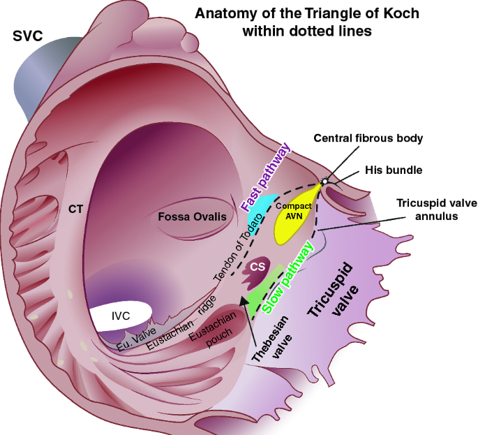
Slow pathway area
Triangle of kock
usually between cs and tricupsid leaflet
AH Jump
Confirms dual nodal physiology (30 % ppl have it)
Further testing determines if its AVNRT or AVRT
AH > 50 ms
What signals are you looking for AVNRT?
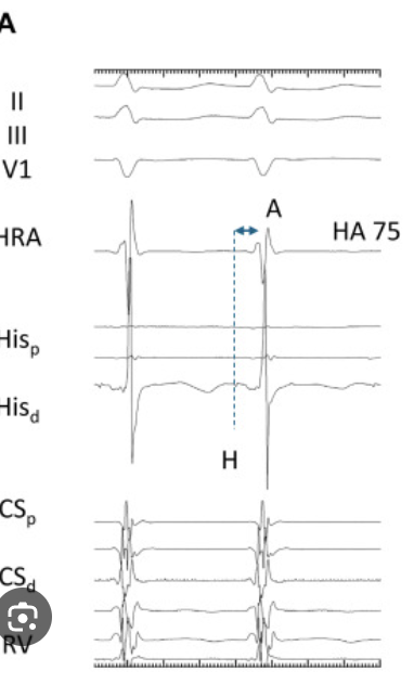
Junctionals
A and Vs happening at same time → overlapping
Mechanism for AVRNT
Premature beat occurs → fast pathway is in RP but slow pathway with fast RP → signal travels down slow pathway and hits fast pathway at the end of its RP → causes reentrant loop

What are the 2 things necessary for a reentrant circuit?
Premature beat
Two pathways with different conduction speed and different RP
Slow conduction has faster RP
Fast conduction has slower RP
What type of signals are you looking for for the slow pathway?
Crunchy W signals
Signals are crunchy and low amplitude because of the slow conduction in the slow pathway

What are you using as a reference pt for AVNRT?
BS
System can’t discern A between V since they’re overlapped
An A for every V
SVTs can start with CS ref but then switch to BS for AVNRT
What are you using as a reference pt for flutter?
CS
Can see more of what is happening in the A (more As than V)
Big A small V (far field V)
What are you looking out for during AVNRT ablation?
Fast junctionals (burning his) → heart block
You want slow junctionals (400-600 ms)
PR elongation → heart is struggling to transmit signals from the A to the V (burn AV node)
See an A but no corresponding V (heart block = bad)
Catheter moves (don’t want to hurt his)
Electrogram of Flutter
Sawtooth
Concentric → probably right sided (CS9-10 down to CS1-2)
Multiple As for every V
I, II, avF need to be opposite in sign to V1 = right sided
Make sure to look at same point on electrogram not the wave
Entrainment
Pace at a slightly faster speed than tach CL → measure from post pacing spike to next natural A signal in same pacing channel → ±30 ms of tach CL = pacing site in the circuit
LAT Histogram Flutter
Full cycle length should fill the histogram
Ensure plenty of pts are taken
What anatomy is gathered for flutter?
RA, IVC, SVC, TVA, His
Can mark annulus points when V signal is larger than A
Bi directional block Endpoint
Pace CS and measure signal on mapping cath that is lateral (near IVC)
This interval should be longer than what it was at the beginning
Also do this process from lateral to medial (MAP cath to cs cath)
Bipolar and Unipolar Map for AFIB
Bipolar: [0.1, 0.4]
Unipolar: [0, 1.08]
CL Range
For mapping in sinus can use 600-1000 (or whatever patient sinus CL is) for auto mapping
For flutter can use a range that is 0-5 ms from the patient’s flutter CL
Success criteria for AFIB
Exit block
Pace in the PVs to ensure signal is not propagating into the A on cs electrogram
New voltage map
PVs should should show as red
AFib map reference
In sinus:
CS
AFIB: Can’t discern signals
BS (more stable QRS)
Anatomy gathered in AFIB cases
LA, LAA, ESO, TS, PVs
WOI for arrythmias (to start)
Flutter: ½ the CL on each side
Everything else: to start
CS ref: [-250,-50]
BS ref: [15,200]
AFIB ECG/EGM
Squiggly chaos
Irregular intervals between the QRS
Coumadin Ridge
Between LAA and LUPV
Corina
Space between two veins