Week 9 Advanced CIED Follow-Up
 Call Kai
 Learn
Learn Practice Test
 Spaced Repetition
 Match
Match Flashcards
 Knowt Play
1/126

There's no tags or description
Looks like no tags are added yet.
Name | Mastery | Learn | Test | Matching | Spaced | Call with Kai | Chat |
|---|
No analytics yet
Send a link to your students to track their progress
127 Terms
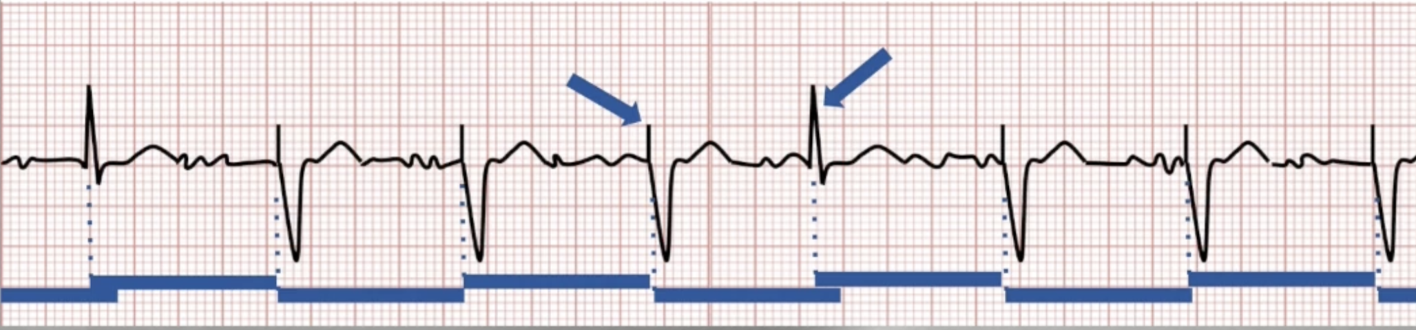
VVI Pacing Interval
Determined by LRL (60000/rate = interval)
Begins after every paced or sensed beat
Device paces when interval times out
Sensing will lead to inhibition and reset the pacing interval
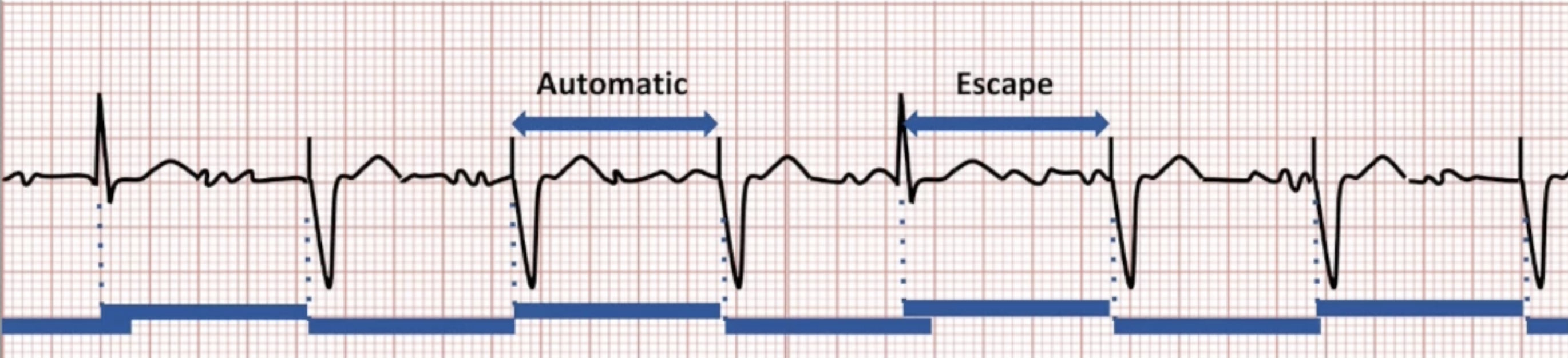
Automatic and Escape Interval
Automatic interval triggered by paced beat
Escape interval triggered by sensed beat
Can be programmed separately with rate hysteresis
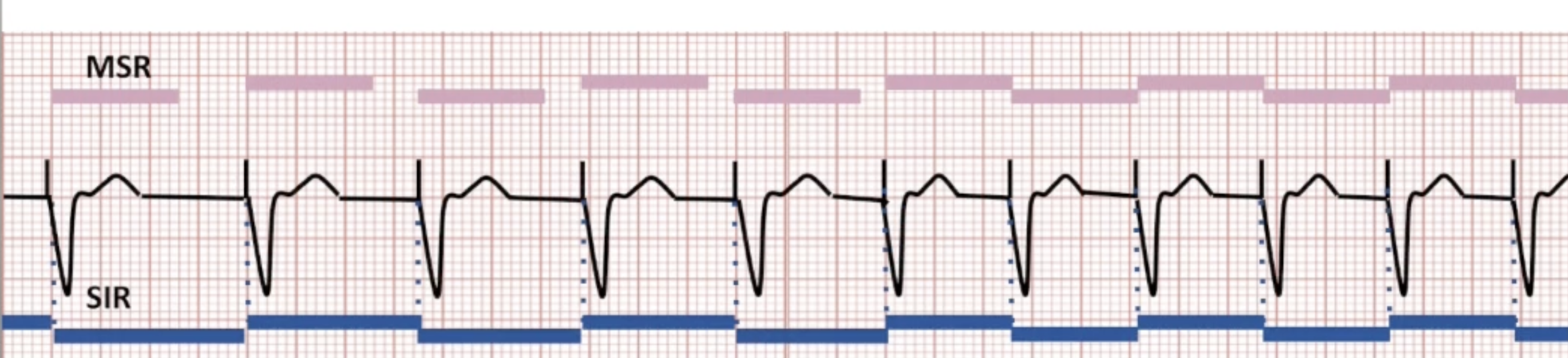
Rate Response
Sensor-indicated rate (SIR): rate controlled by rate response sensor
Max sensor rate (MSR): highest rate device will pace
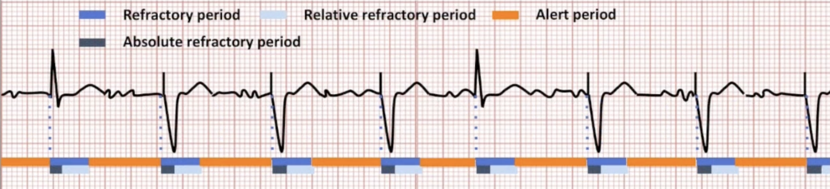
Refractory Period
Prevents sensing of T waves and non-physiologic signals
Divided into absolute and relative refractory periods
End of refractory period triggers alert period
Fixed or dynamic
DDD Pacemakers
More physiologic than VVI
Leads pace each chamber separately and communicate with each other to maintain AV synchrony
Sinus rate slows: pace atrium
AV conduction slows: pace ventricles
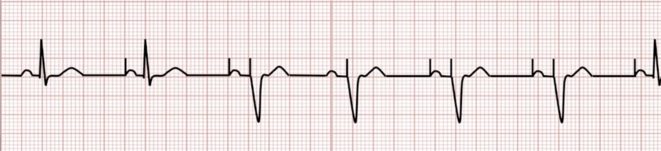
Ventricular-Based Timing
Atrial pacing driven by VA interval (AKA atrial escape interval)
VA interval = pacing interval - AV delay
Device adjusts other intervals to keep VA interval constant
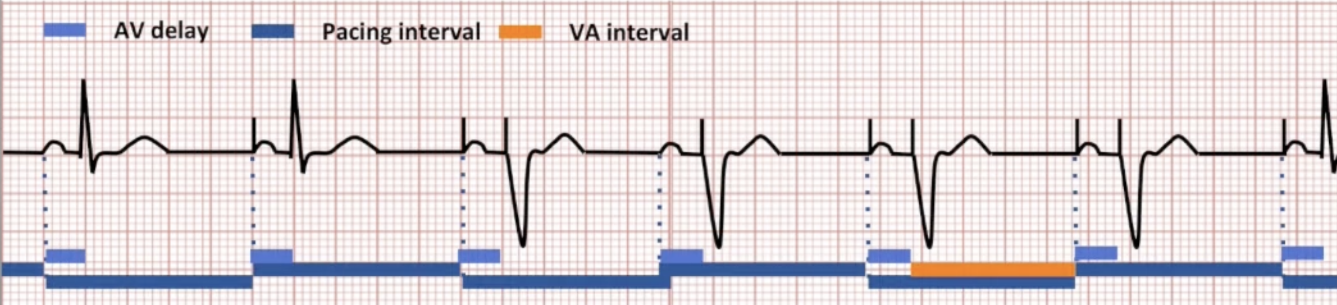
Atrial-Based Timing
Atrial pacing driven by A-A interval
Device adjusts other intervals to keep A-A interval constant
VA interval can be calculated, but is not used
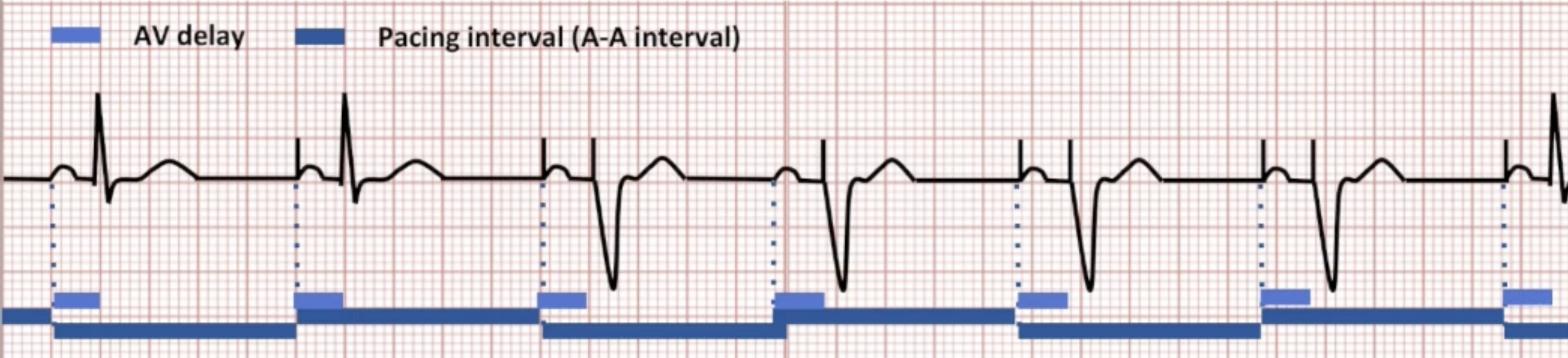
A Paced, V Paced
Ventricle-based vs Atrial-based AP/VP diagram
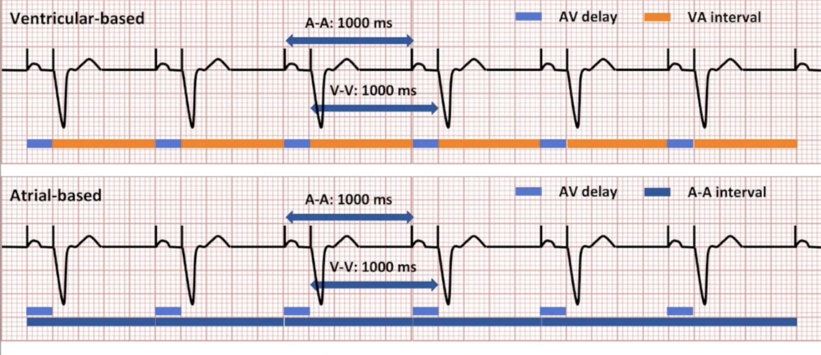
A Paced, V Sensed
Ventricle-based vs Atrial-based AP/VS diagram
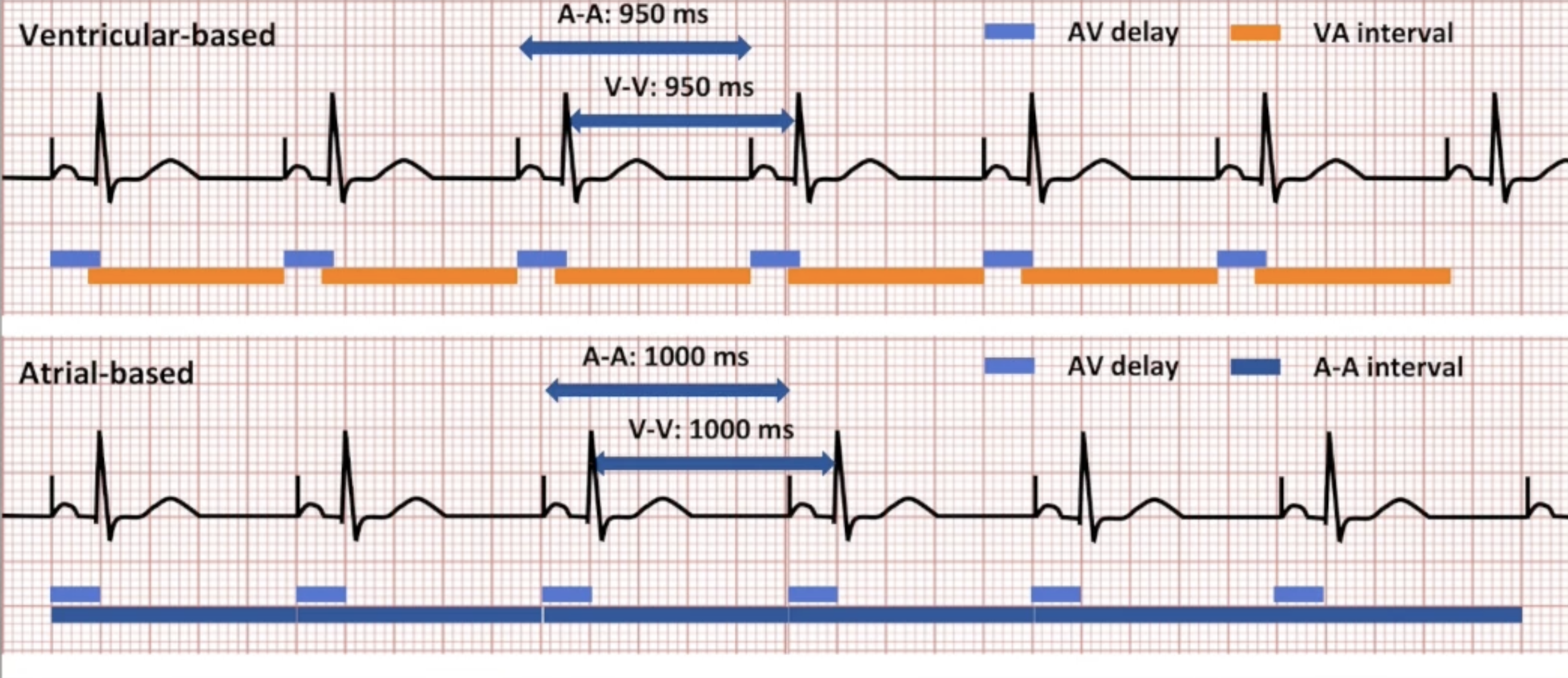
A Paced, V Paced to A Paced, V Sensed
Ventricle-based vs Atrial-based AP/VP to AP/VS diagram
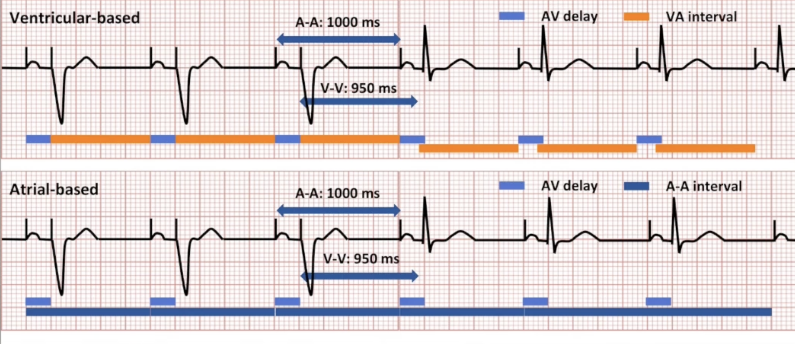
A Paced, V Sensed to A Paced, V Paced
Ventricle-based vs Atrial-based AP/VS to AP/VP diagram
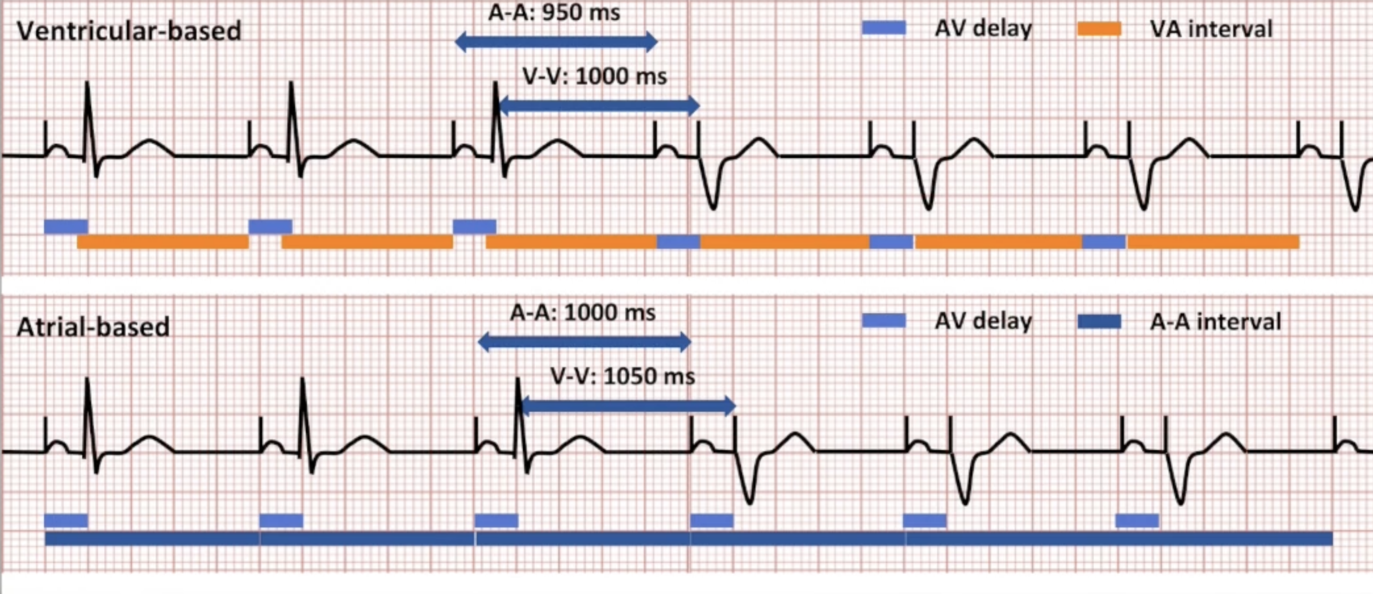
Importance of Timing Quirks
Changes in rate as patient transitions in and out of block
Telemetry unit: variations in rate can alarm nurses
Understand timing quirks, answer concerns
Know basic rules:
Ventricular-based: VA interval constant
Atrial-based: A-A interval constant
PVC Response
PVC defined by the device:
Two consecutive ventricular events
No intervening atrial events
Second event is VS
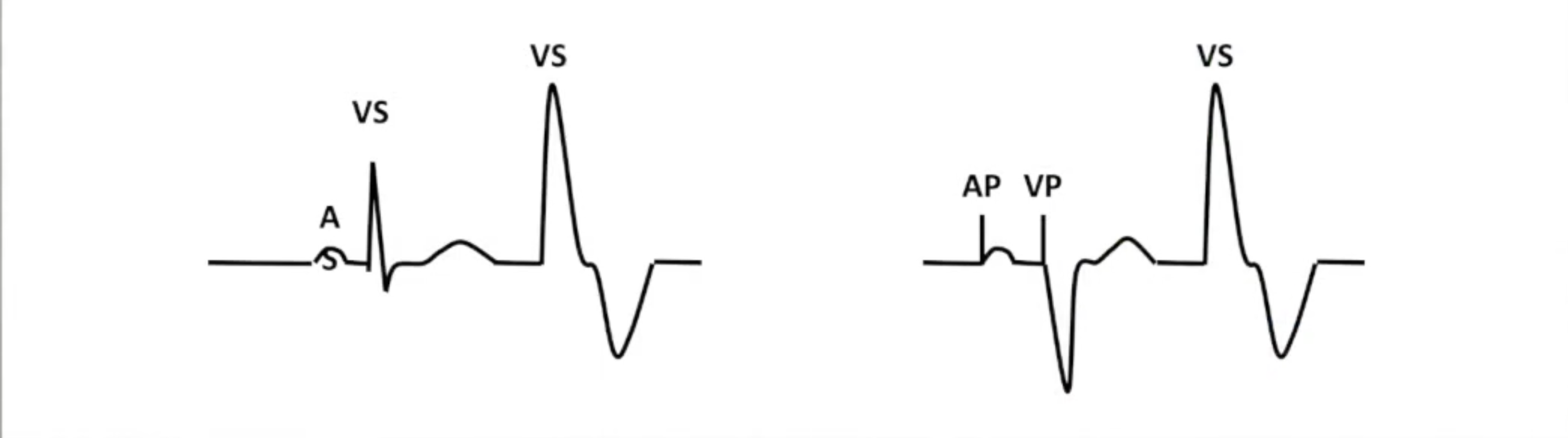
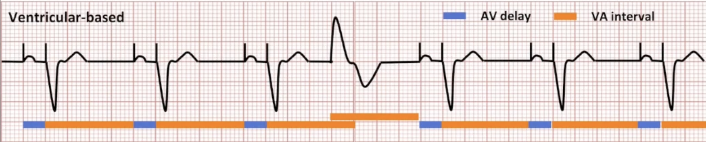
PVC Response: Ventricular-Based Timing
PVC defined by the device:
Two consecutive ventricular events
No intervening atrial events
Second event is VS
Ventricular-based timing: PVC triggers new VA interval
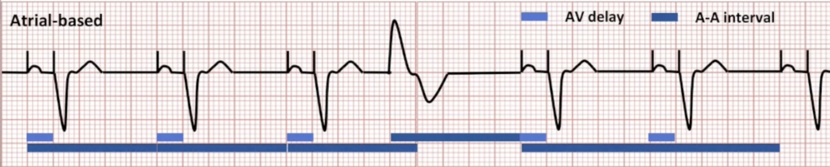
PVC Response: Atrial-Based Timing
PVC defined by the device:
Two consecutive ventricular events
No intervening atrial events
Second event is VS
Atrial-based timing: PVC triggers new A-A interval
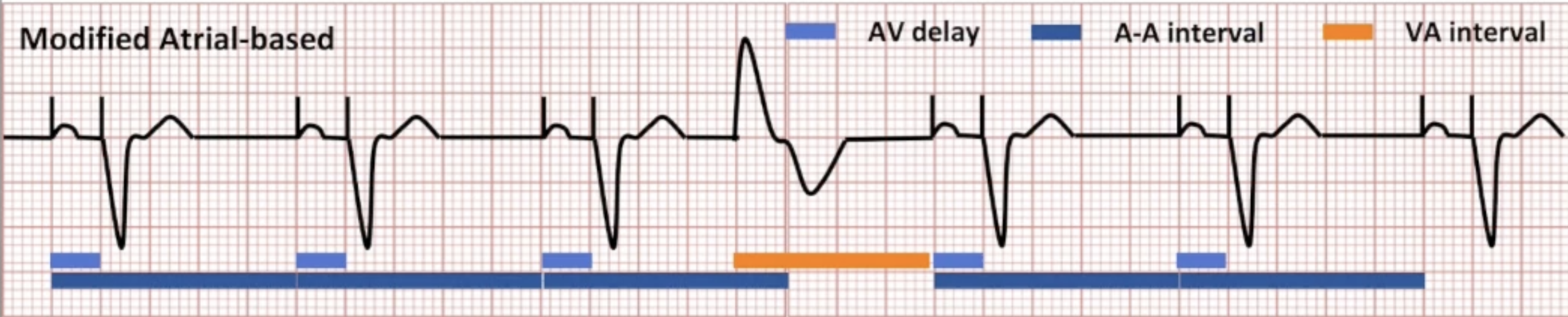
Modified Atrial-Based Timing
Normal beats: operates like atrial-based
PVCs: triggers VA interval instead of A-A interval
Impact of Atrial Fibrillation
Loss of atrial kick: 10-30% CO
Loss of AV synchrony: reduced ventricular filling, preload, CO
Rapid, irregular ventricular response: reduced diastole, reduced ventricular filling, reduced coronary perfusion, palpitations, decreased exercise capacity
Remodeling: worsening AF, tachycardia-induced cardiomyopathy
CIED Algorithms
How can CIEDs minimize these negative effects?
Prevent AF
Minimize frequency/duration
Manage/reduce symptoms
Terminate AF
Prevent recurrence
Type of Algorithms
Preemptive algorithms
Prevent/minimize AF
Pre-mode switch
Responsive algorithms
Manage/reduce symptoms
Terminate AF
Inside mode switch
Reactive algorithms
Reduce recurrence of AF
Post-mode switch
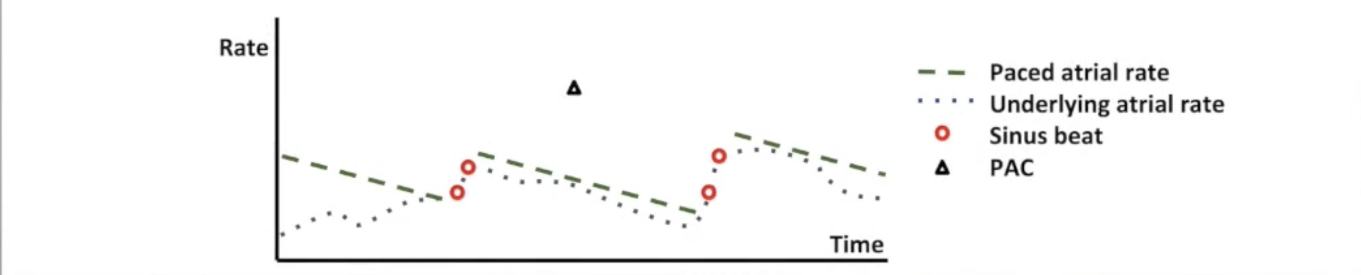
Pre-Mode Switch: Pace Conditioning
Adjusts atrial pacing rate above intrinsic rhythm
Pace ~95% of time
Keep atria refractory to suppress AF triggers
Sinus beats detected (usually 2+): raise pacing rate
Programmable maximum rate (seperate from MSR)
Rate slowly decreases towards base rate
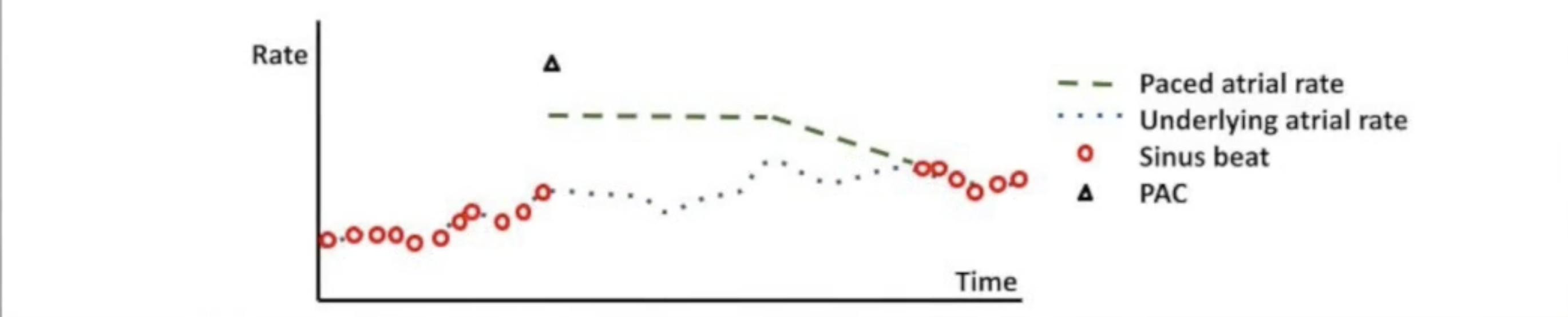
Pre-Mode Switch: PAC Response
PACs can trigger atrial tachyarrhythmias
PAC response activated by single rapid beat
Triggers atrial pacing at fast rate
Gradually decrements to base rate
Pace quickly to eliminate pause after PAC and prevent arrhythmias
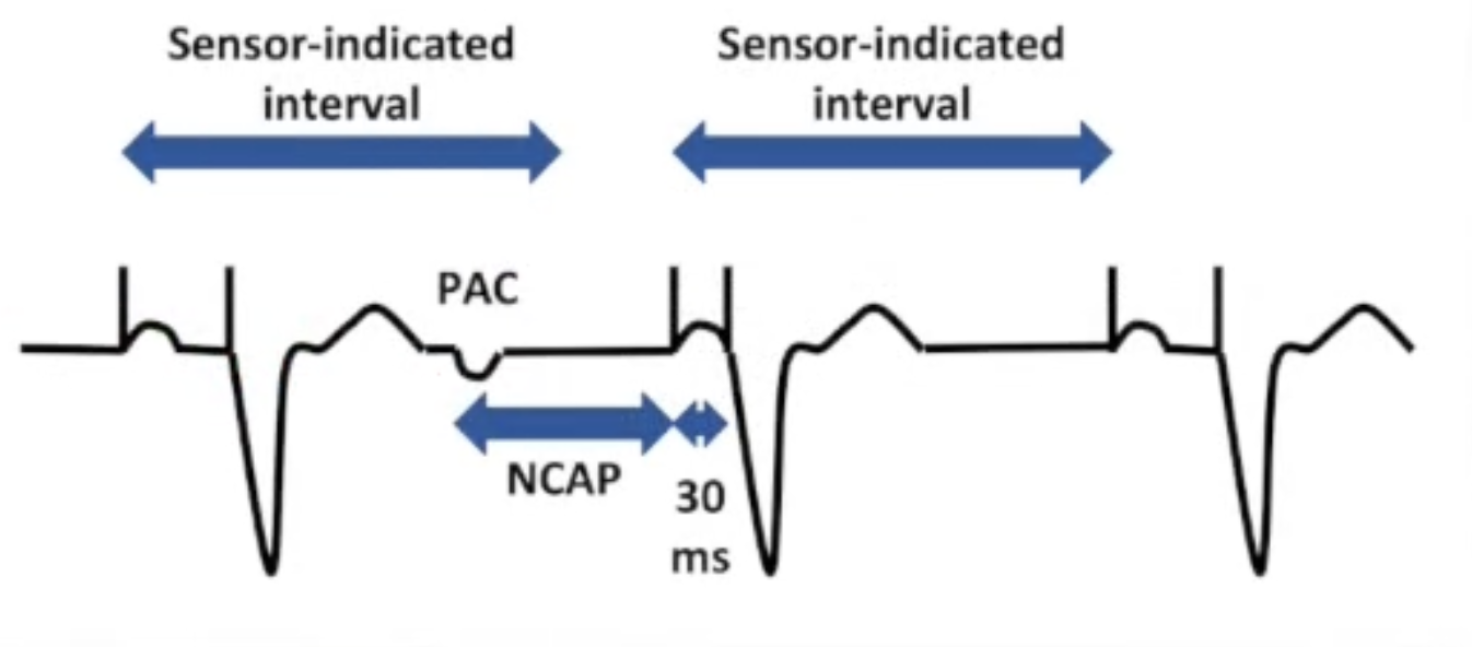
Pre-Mode Switch: NCAP
Non-competitive atrial pacing
Pacing in atrial vulnerable period can trigger atrial
tachyarrhythmiaNCAP delays time to next paced atrial event after PAC
Defines PAC as atrial signal inside PVARP
Extends NCAP window
Next AV interval shortened
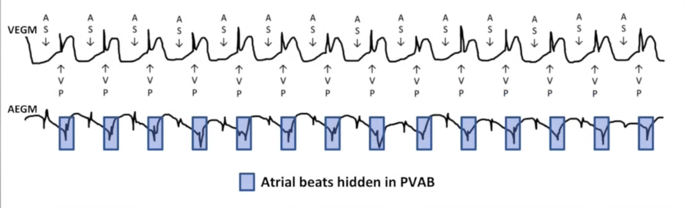
Functional Undersensing of Atrial Flutter
Atrial rate of 300 bpm
Every other beat in PVAB: no mode
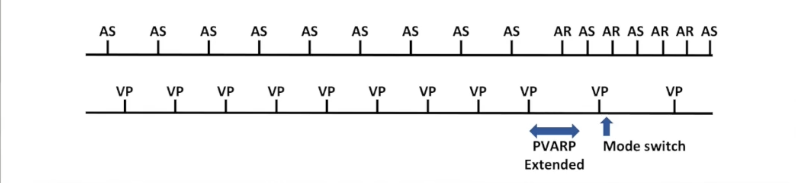
Blanked Flutter Search
Similar to PMT termination
Detects A sensed V paced beats at consistent, fast rate
Extends PVARP: next atrial beat will be refractory
Previously blanked flutter beat sensed: triggers mode switch
Atrial Flutter Response
Separately programmable atrial flutter response rate
Triggers flutter response window: atrial refractory period
Atrial event within window triggers new window
Continues until mode switch criteria met or rate slows
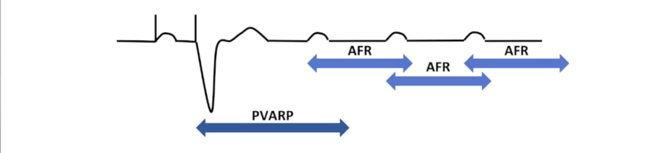
Pre-Mode Switch: Rate Smoothing
Reduces symptoms from rate changes
Beat-to-beat basis: maximum percentage change in interval
AT/AF starts: gradually increases ventricular rate
AT/AF stops: gradually decreases ventricular rate
Many mode switch algorithms have rate smoothing built in
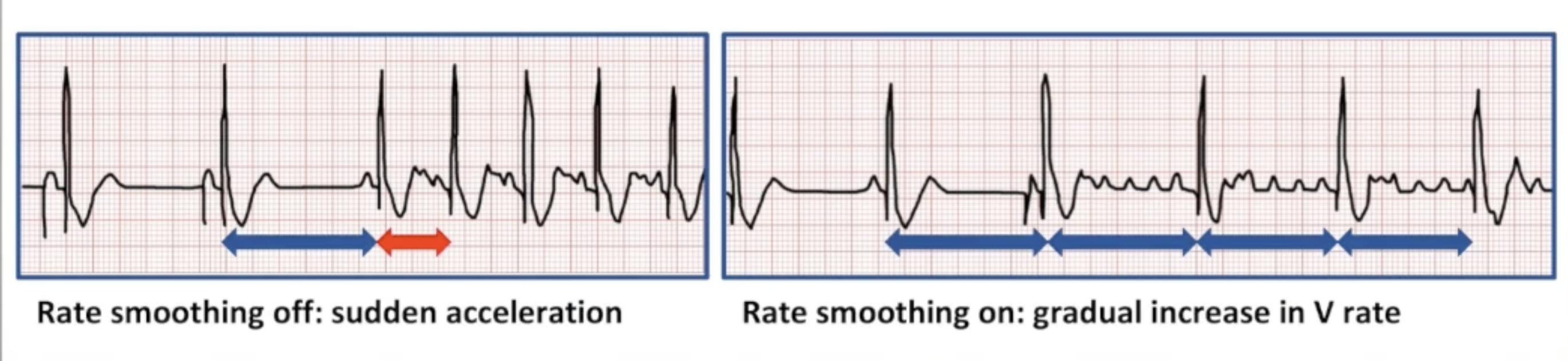
Inside Mode Switch: Regularization
Regularize rate of ventricular response to AF
Pace between intrinsic beats to prevent large variations in rate
Uses rolling window
Pace slightly faster than average rate
Increases % RV pacing: no longer used in non-CRT devices
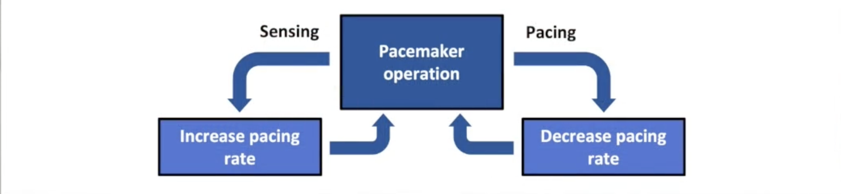
Inside Mode Switch: Termination
ATP in the atrium
Works best for regular, monomorphic, reentrant
Unlikely to work for AF
Risks: trigger VT, transient AV block, increase patient symptoms
More aggressive than normal burst
Ramp
Burst+
Reactive ATP
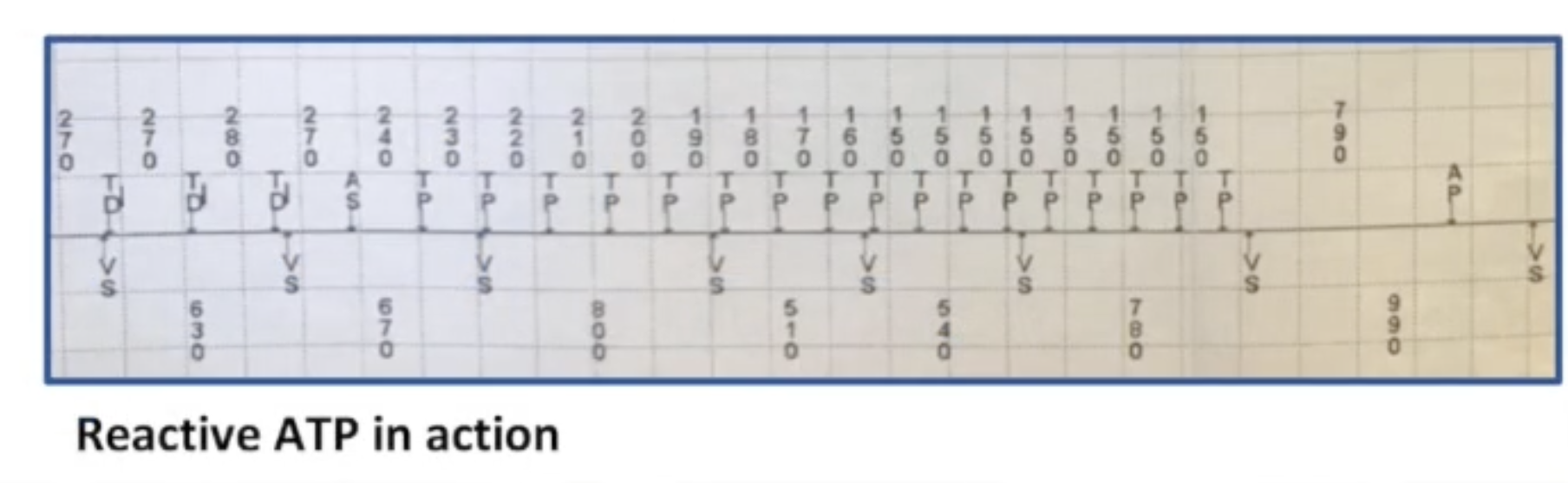
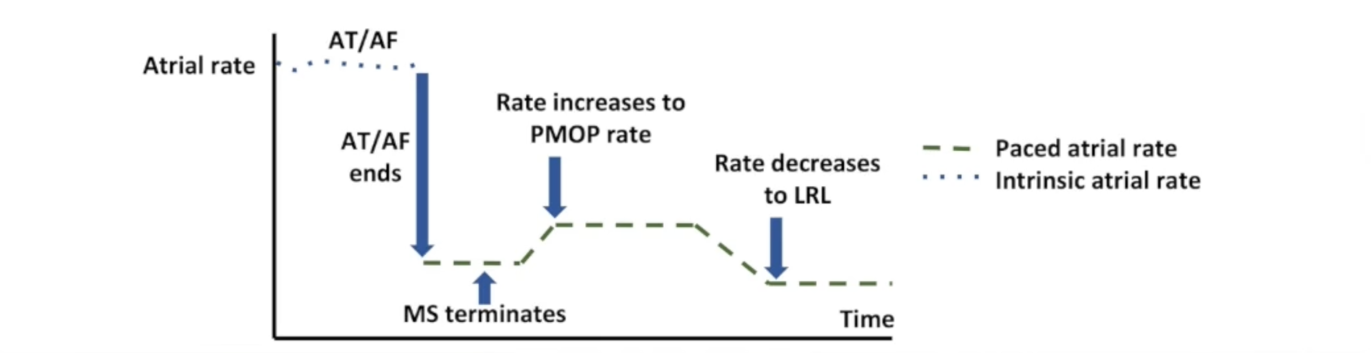
PMOP
Post-mode switch overdrive pacing
Pacing at elevated rate after termination of mode switch
Keeps atria refractory
Prevents early recurrence of atrial fibrillation (ERAF)
Minimizing RV Pacing
Increase AV delay
AAl mode
Algorithms that promote intrinsic conduction
AV search hysteresis
Algorithms that change modes
Alternative pacing site
Upgrade to CRT
Rate Smoothing
Prevent large fluctuations in rate
Used in either chamber
Separate percentages for increase/decrease
Useful scenarios:
AF, SVTs
Frequent PACs/PVCs
Brady-tachy syndrome
Sinus pauses
LQTS: Torsades initiated by long-short-long interval pattern
Turned off during certain algorithms
PMT termination
Search hysteresis
Programmed increase in LRL
Intrinsic rate > MTR
Mode switch with regularization algorithm
Increases RV pacing %
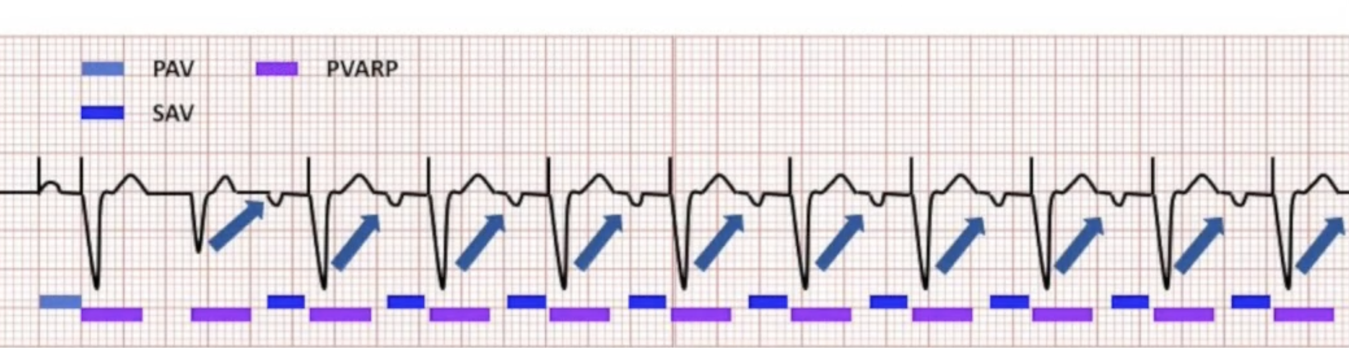
Pacemaker Mediated Tachycardia
Requirements for initiation:
Dual chamber tracking mode
Ability to conduct retrograde
Longer VA conduction time than PVARP
Triggering event causing loss of AV synchrony
VA conduction:
80% of patients with SSS
35% of patients with AV block
20% of patients with VA conduction have experienced PMT
PMT Triggers
PACS
PVCs
Loss of atrial capture
Atrial undersensing
Atrial oversensing
Long AV delays
AV search hysteresis
Magnet removal
PMT Termination
Self-termination: fatigue of conduction system, PAC/PVC
Active termination:
Vagal maneuvers (carotid sinus massage)
Medications (verapamil, beta blockers)
Magnet application: switch to asynchronous
PMT termination algorithms
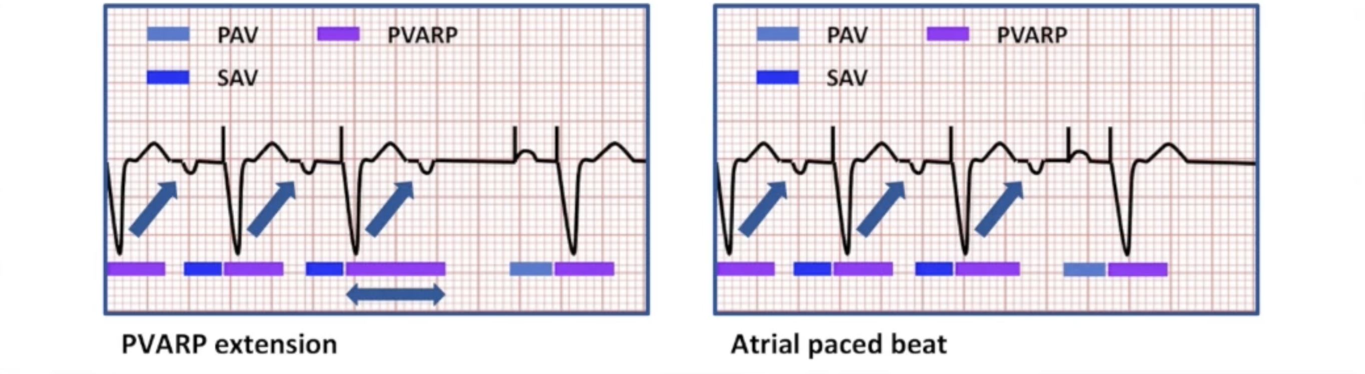
PMT Termination Algorithms
Look for specific number of P waves tracked at MTR
Extend PVARP for one interval
Alternative: deliver atrial paced beat
VA stability: alternative for PTM below MTR
Inappropriate PMT Termination
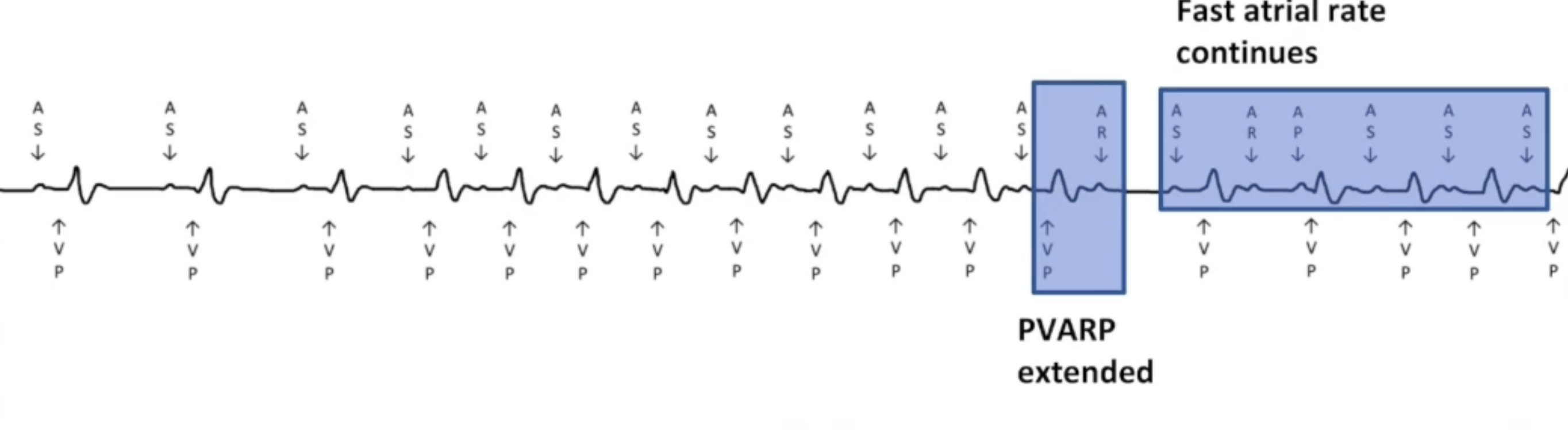
Sinus tachycardia: program MTR higher
Atrial flutter: blanked flutter search
PMT Prevention
Run VA conduction test, set PVARP 50 ms longer than RVAC
Not possible in all patients
RVAC varies over time
PVC response: extend PVARP or atrial pace in response to PVC
Correct underlying cause
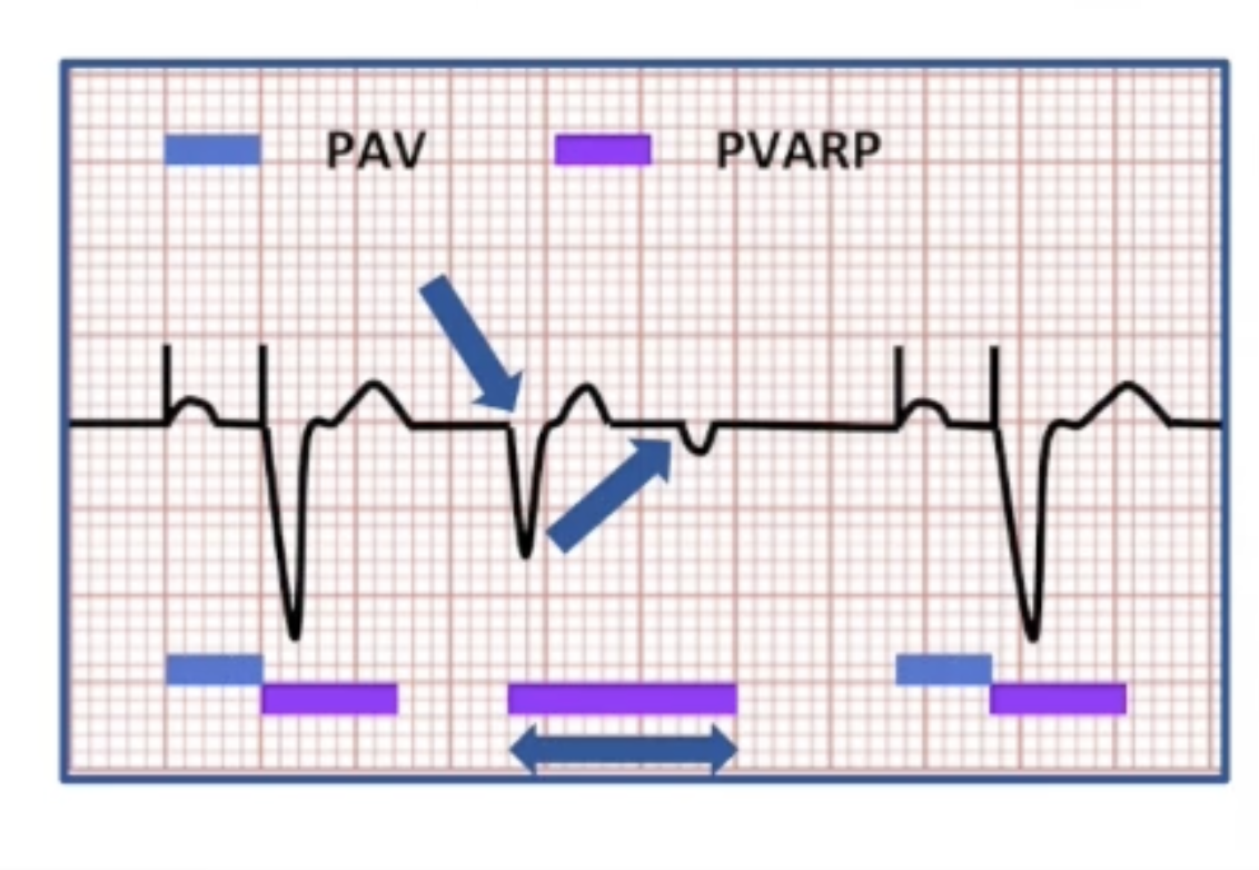
RNRVAS
Repetitive non-reentrant ventriculoatrial synchrony
Dual chamber pacemaker
Retrograde conduction
VA conduction time shorter than PVARP
Atrial pacing occurs within intrinsic atrial refractory period
Triggering event
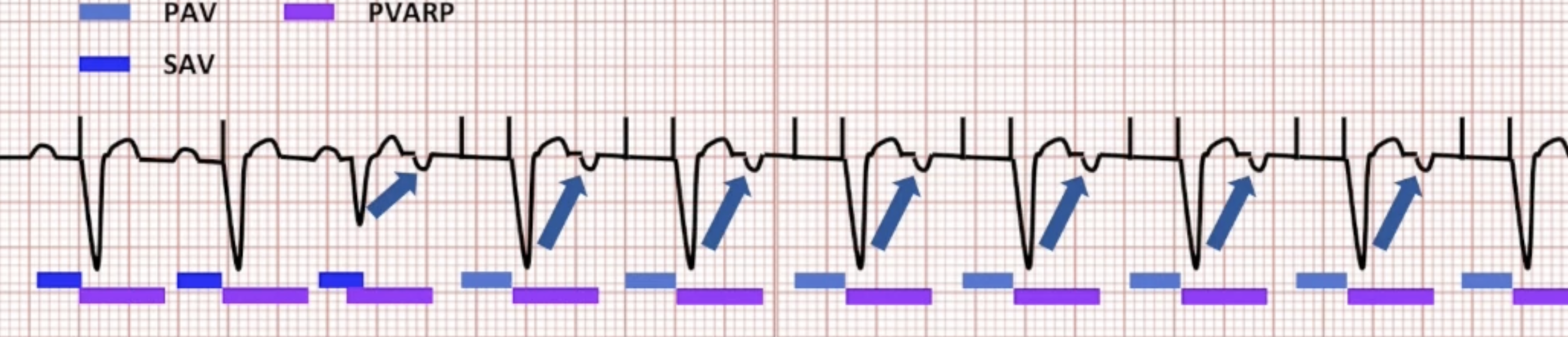
RNRVAS: FUNC & FUS Part 1
Triggering event allows retrograde conduction
Retrograde beat lands within PVARP: functional undersensing
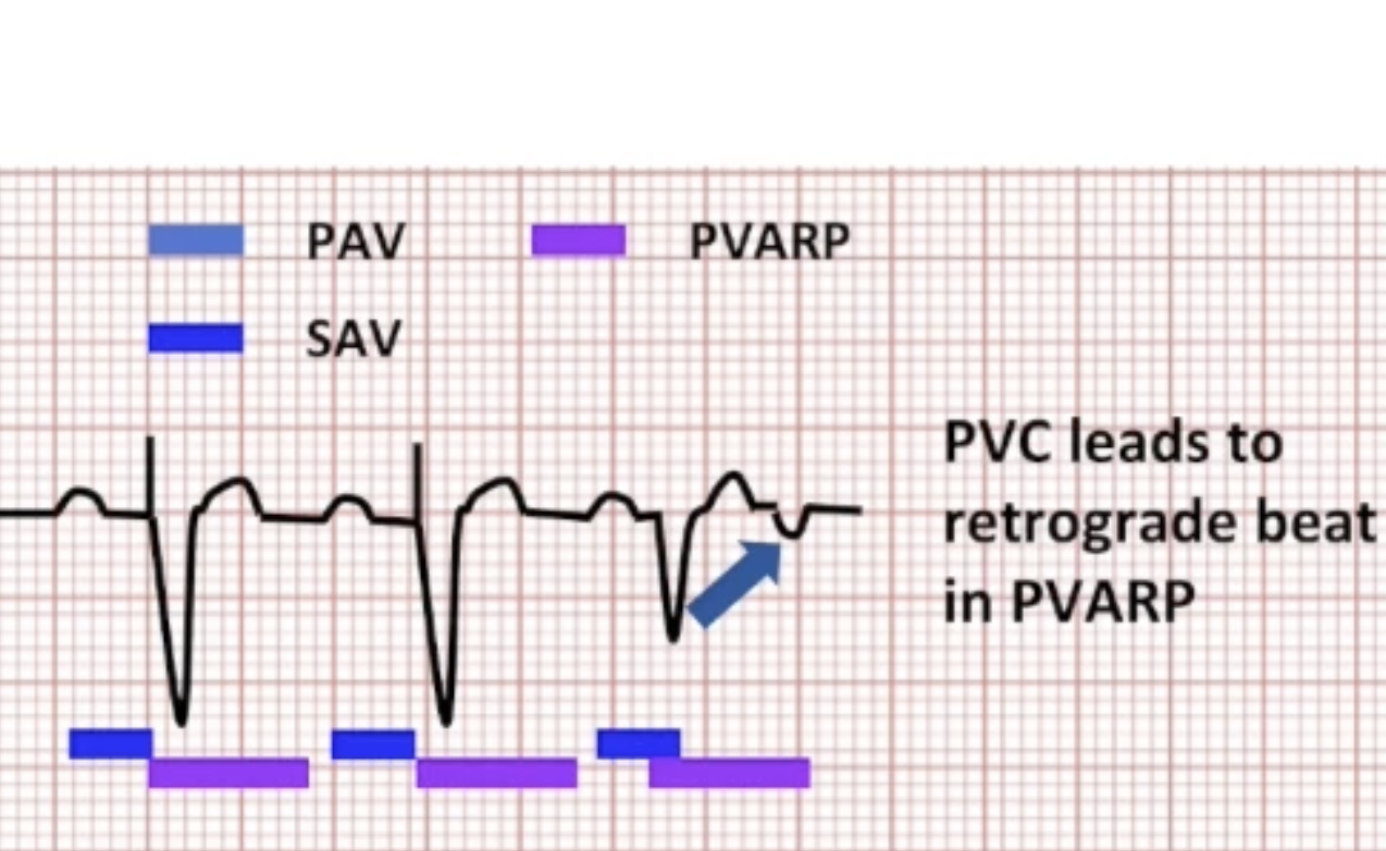
RNRVAS: FUNC & FUS Part 2
Fast atrial pacing rate causes atrial paced beat to land within intrinsic refractory period: functional non-capture
Sensor-indicated rate
High base rate
Atrial overdrive pacing
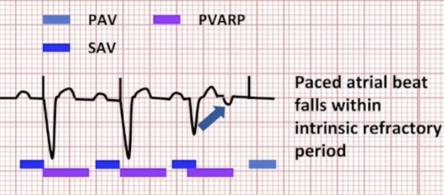
RNRVAS: FUNC & FUS Part 3
No intrinsic conduction: AV delay times out
V pacing causes retrograde conduction again: FUS
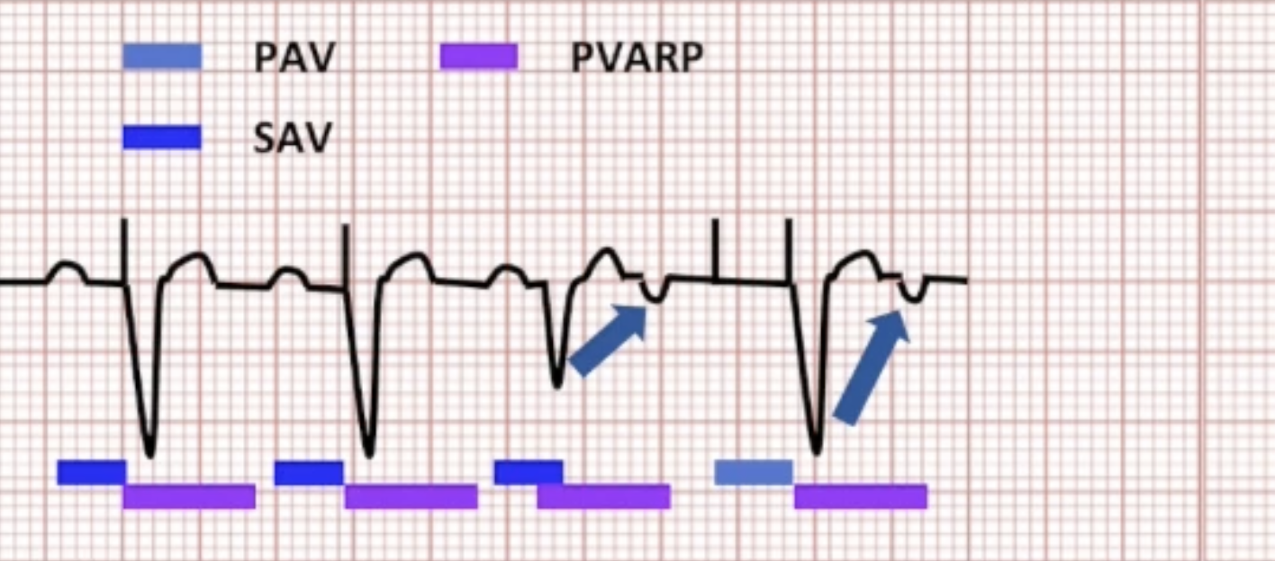
RNRVAS Pattern
Atrium paced again: FUNC
Pattern continues:
Loss of optimal AV delay
Unnecessary RV pacing
Possible triggering of arrhythmias/algorithms
RNRVAS Prevention
Extend PVARP after PVC: delays atrial pacing
Shorten PVARP
Dynamic PVARP
Shorten AV delay
Dynamic AV delay
Neurocardiogenic Syncope (NCS)
Vasovagal syncope, neurally-mediated syncope, carotid sinus syndrome, carotid sinus hypersensitivity
Inappropriate physiologic response to stress causing sudden drop in HR & BP
Decreased blood flow to brain: LOC
Many presyncopal symptoms: sit down to avoid falling
NCS Diagnosis
Rule out other possible causes:
Temperature: fever
Heart rate: tachyarrhythmia, ACS
Blood pressure: orthostatic hypotension
Blood tests
12 lead
Echocardiogram
Stress test
Tilt table test
NCS Treatment
Treatment often unnecessary
Avoid triggers:
Lifestyle changes
Increase salt in diet
Drink fluids
Avoid prolonged standing
Medications for hypotension
Sometimes goes away with age
Pacemaker as last resort
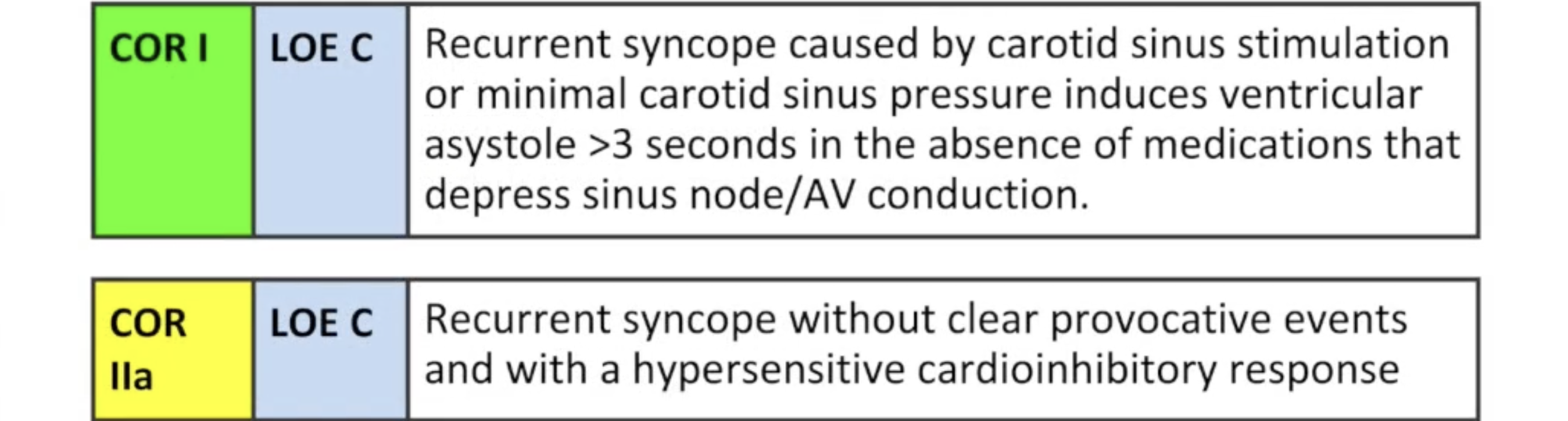
NCS Indications
NCS classes indication
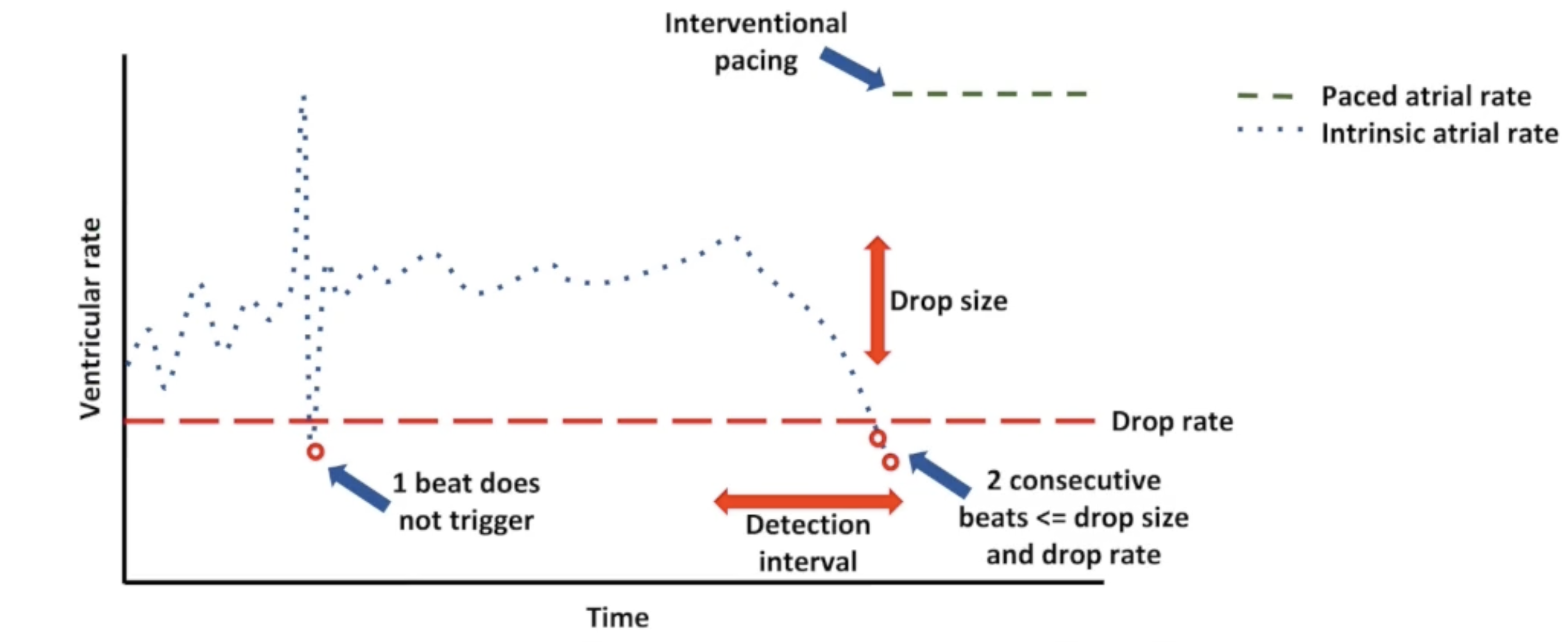
Rate Drop Response
Patient indicated for syncope turn this feature on
Unique Properties of the Cardiac Cell
Automaticity is the ability of cardiac cells to generate their own impulses spontaneously
Excitability is the ability of cardiac cells to respond to an electrical stimulus
Conductivity is the ability of cardiac cells to transfer or propagate an electrical stimulus
Contractility is the ability of cardiac cells to shorten and cause myocardial contraction in response to an electrical stimulus
Stimulation Threshold
Minimum output of electrical stimulation needed to consistently produce cardiac depolarization
Voltage @ pulse width
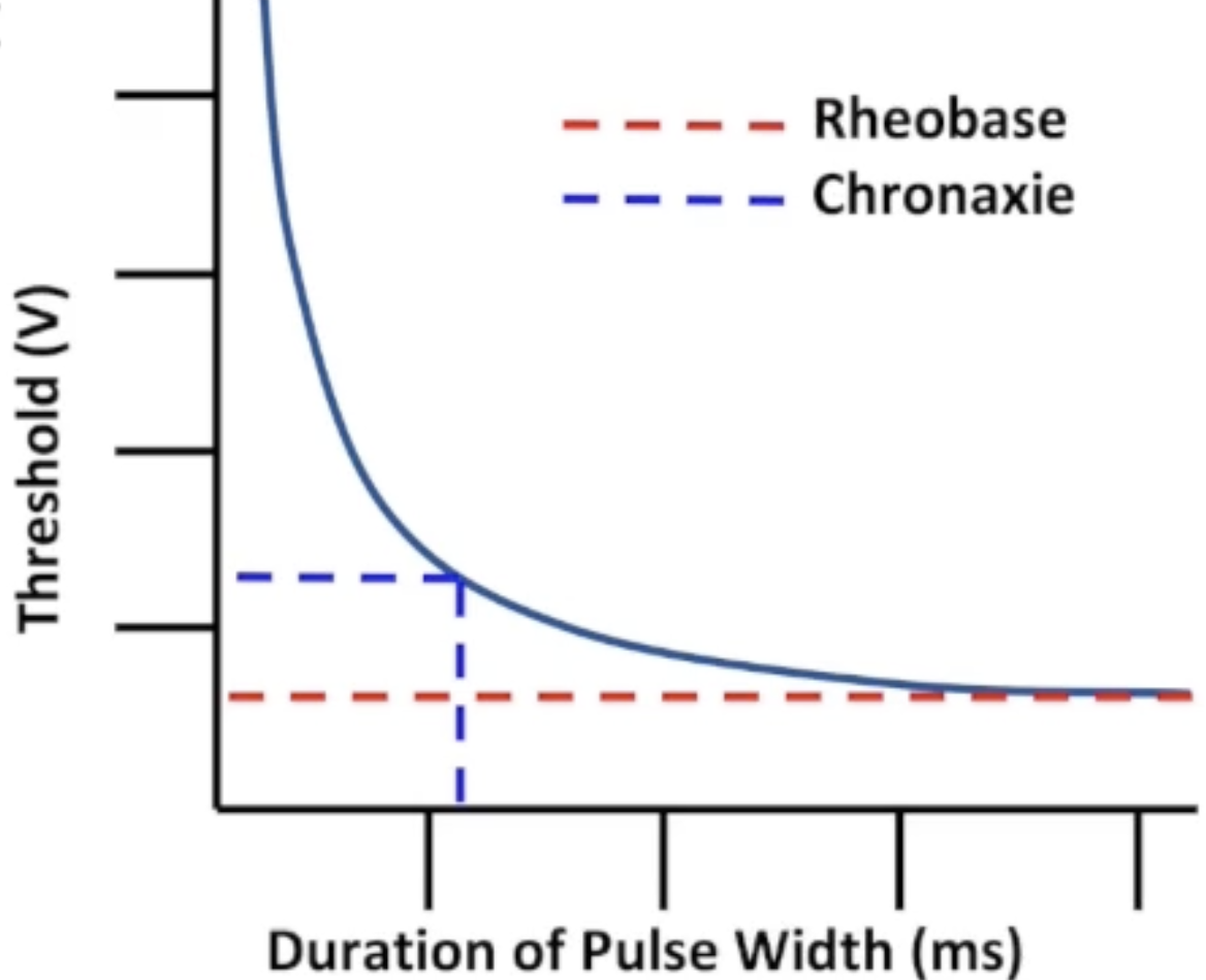
Map thresholds using strength-duration
Rheobase and Chronaxie
Rheobase: minimum voltage that captures at any pulse width
Chronaxie: shortest pulse width that captures at 2x rheobase voltage
Threshold Testing
Hold pulse width constant, decrement voltage
Threshold: voltage just before capture is lost
Set output 2x threshold voltage
Battery Longevity
Battery technology
Impedance (higher = longer)
Pacing outputs (lower = longer)
J = V^2xT/R
Raising voltage has greater effect on battery life
Battery Longevity: Cell Voltage
Lithium-iodide cell voltage: typically 2.8-3 V
Higher output requires voltage multiplier
Consider changing pulse width:
Decrement voltage at higher fixed pulse width
Decrement pulse width (3:1 safety margin)
Pulse width no higher than 1.0 ms
Battery Longevity: Other Factors
Percentage pacing (lower = longer)
Activity sensor
Remote monitoring
Interrogations: make sure you end the session
Threshold Changes
Safety margins set to accommodate threshold fluctuations
Loss of capture in 1-5% of patients
Dangerous for pacemaker-dependent patients
Renders device useless
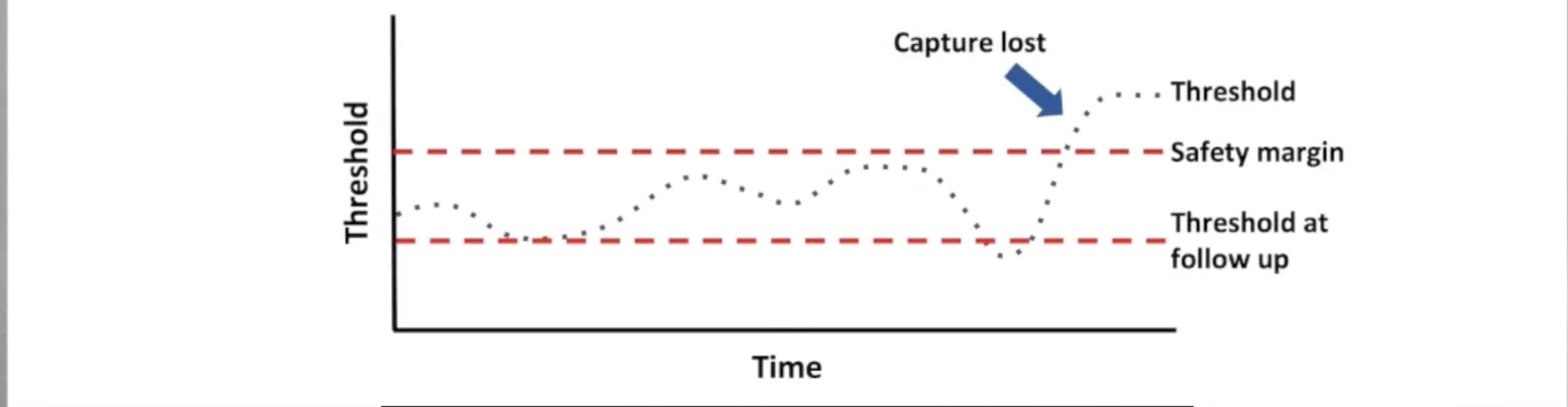
Threshold Changes: Lead Complications
Long term impacts on threshold
Lead fracture
Lead insulation break
Helix crystallization
Lead encapsulation
Threshold Changes: Autonomic Tone
Activity | Effect on autonomic tone | Effect on capture threshold |
Sleeping | Stimulates parasympathetic | Increased threshold |
Eating | Stimulates parasympathetic | Increased threshold |
Exercise | Stimulates sympathetic | Decreased threshold |
Threshold Changes: Other Abnormalities
Electrolyte imbalances (hyperkalemia)
Hyperglycemia
pH imbalance
Acidic (low pH)
Alkalotic (high pH)
Affected by many factors
Threshold Changes: Medications
Antiarrhythmics can raise thresholds
Spironolactone
Quinidine
Flecainide
Sotalol
Stimulants can lower thresholds
Epinephrine
Ephedrine
Steroids can lower thresholds
Glucocorticoids
Isoproterenol
Autocapture
Automatically tests thresholds and resets outputs
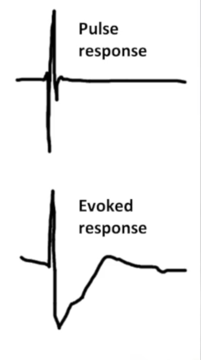
Pulse response (PR)
Traditional sensing amplifier
Used to sense intrinsic R waves
Evoked response (ER)
New sensing amplifier for autocapture
Electric potential recorded following pacing stimulus
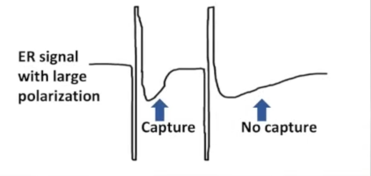
Morphology changes when capture is lost
Polarization
Residual charge (concentration of ions) that remains near electrode after pacing pulse
Consistent with or without capture
Autocapture will not function if polarization dominates ER
morphologyMust be minimized
Minimizing Polarization
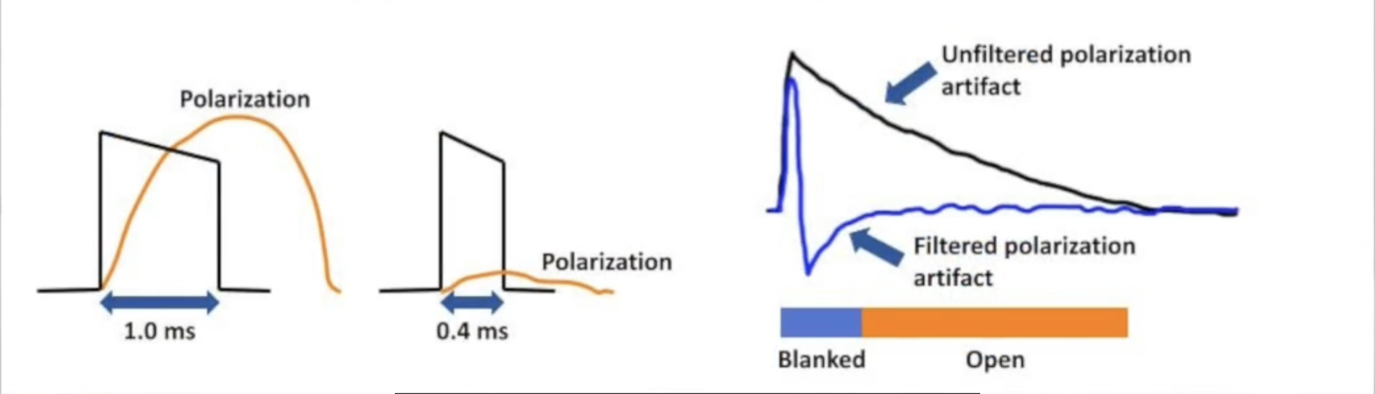
Smaller electrode size increases polarization
Increased current density at tip
Small electrode leads designed to increase impedance
Maximize surface area: fractal surface, coatings
Larger pulse width increases polarization
Autocapture uses 0.4 ms
Second order bandpass filter to distort polarization artifact
ER blanking period to block filtered polarization signal
Autocapture Sensing Test
Determine if:
Evoked response is large enough
Polarization is small enough
Large enough safety margin between sensitivity and ER
Large enough safety margin between ER and polarization
5-7% have inadequate signals
Automatic Threshold Test
Run periodically by device
Gradually decrements outputs to look for loss of capture
No 2:1 safety margin
Paces to override intrinsic activity
Delays test for high intrinsic rate, algorithms
Backup pulse at elevated outputs when capture is lost
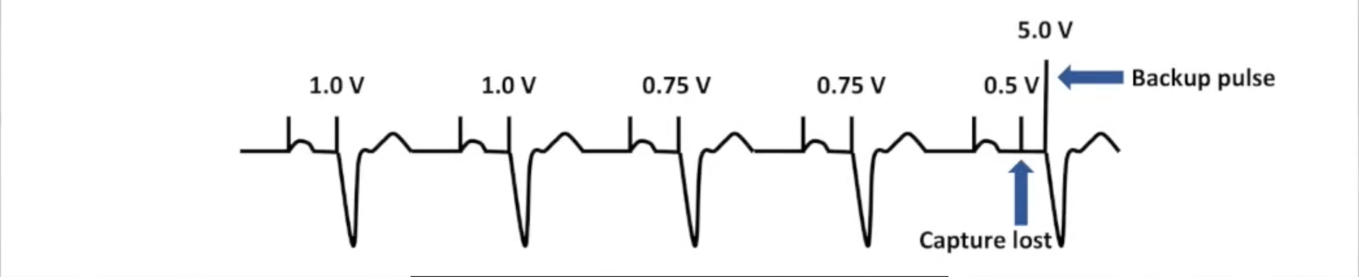
Automatic Threshold Test in Clinic
Can run an automatic test using programmer
Compare device's assessment with you own
Always use to test autocapture
Watch for loss of capture: device may get it wrong!
Capture Verification
Device constantly monitors evoked response
Beat to beat or rolling window
Capture lost: high output backup pulse
Runs another threshold test after certain # of beats
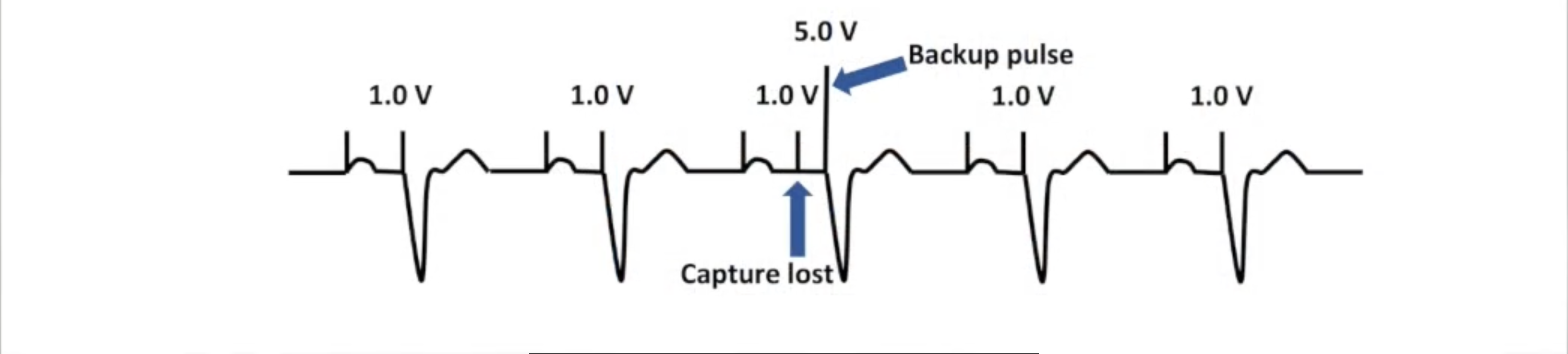
Atrial Autocapture
Atrial signals too small for ER
Algorithms test threshold periodically
Pace faster than intrinsic rate
Intrinsic or retrograde signals indicate loss of capture
No capture verification
Autocapture Benefits
Smaller safety margin: reduced battery consumption
Maintains capture during threshold fluctuations
Backup safety pacing
Sends alerts when thresholds rise
Runs threshold tests automatically: included in RM
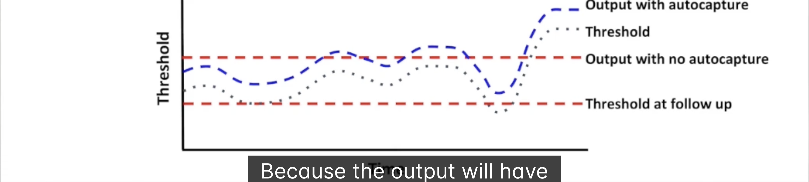
Autocapture Drawbacks
Algorithms not initially reliable
Backup pacing can be proarrhythmic
AF: fusion beats confuse algorithm
Questionable benefits in certain populations
Low pacing %
Low thresholds
Monitor mode if physician unsure
Heart Failure
Progressive disease
50% of HF patients die within 5 years of diagnosis
Begins with injury/stress to myocardium:
CAD
HTN
Diabetes
Obesity
Heart remodels: causes dyssynchrony
Electrical Dyssynchrony
Seen on ECG
Damaged myocardium alters electrophysiologic properties
Impairs velocity/direction of propagation
Abnormal ventricular depolarization: wide QRS
Systolic dysfunction
Mechanical Dyssynchrony
Interstitial fibrosis
Rearrangement of extracellular matrix and myocytes
More diffuse wavefront
Disorganized contraction
Causes further fibrosis/remodeling
Areas of Dyssynchrony
1 & 2: AV dyssynchrony:
Between RA and RV
Between LA and LV
Loss of atrial kick
Reduced cardiac output
Regurgitation
3: interventricular dyssynchrony:
Between RV and LV
Measured using doppler echo
Aortic vs. pulmonic outflow
Delay > 40 ms abnormal
4: intraventricular dyssynchrony:
Between walls of LV
Important abnormality in progression of heart failure
Apical Rocking
End diastole
Isovolumetric contraction
Ejection
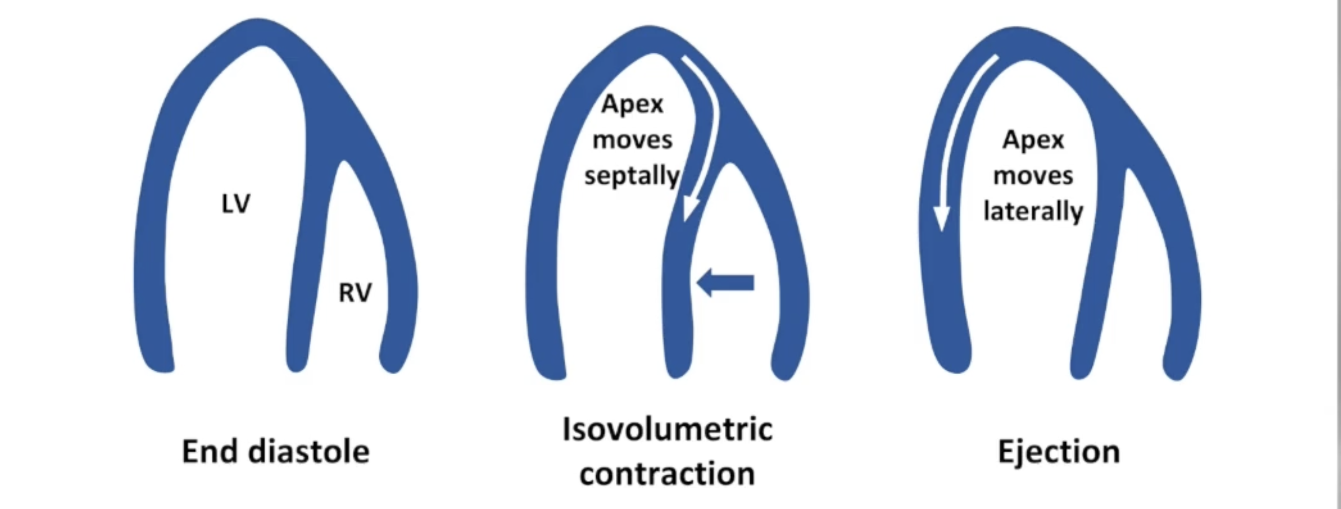
Cardiac Resynchronization Therapy
Biventricular device resynchronizes heart
Electrical depolarizations
Mechanical contractions
Goal of CRT: BIV pace 100% of the time in order to reverse remodel the heart
Reduce dyssynchrony
Improve CO/EF
Decrease EDV/ESV
Improve QOL
Categories of CRT Responses
Clinical measures assessment
LV reverse remodeling assessment
Outcome measures assessment
Clinical Measures
NYHA Class
Class I: asymptomatic with reduced EF
Class II: mildly symptomatic during ordinary exertion
Class III: moderately symptomatic during less than ordinary exertion
Class IV: symptomatic at rest
6 minute walk test
Exercise duration
Cardiopulmonary stress test
LV Reverse Remodeling
Acute measures:
Increased CO
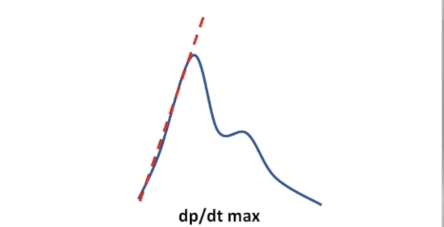
Increased LV dp/dt max: maximum rate of rise in LV pressure in early systole
Chronic measures:
Increased LVEF
Decrease in ESV/EDV
Reduced MR
Outcome Measures
Heart failure hospitalizations
Morbidity
All cause mortality
CRT Non-Responders
HF gets worse after they receive device
Increased remodeling, no improvement in functional class after 6 months
Initial improvement then worsening symptoms
Contributing factors:
Poor patient selection
Suboptimal LV lead positioning
Inappropriate device programming
CRT Class I Indication
CRT is recommended for patients with:
Sinus rhythm
LVEF <= 35%
LBBB
QRS >= 150 ms
NYHA Class II, III, ambulatory IV
On GDMT
Class IIa Indication for AF Patients
CRT is reasonable for patients with:
Atrial fibrillation
LVEF <= 35%
Symtoms on GDMT
If the patient requires ventricular pacing or otherwise meets CRT criteria
If AV nodal ablation or pharmacological rate control will allow near 100% BIV pacing
CRT Patient Selection: ECG
Best results with LBBB
Results improved with wider QRS
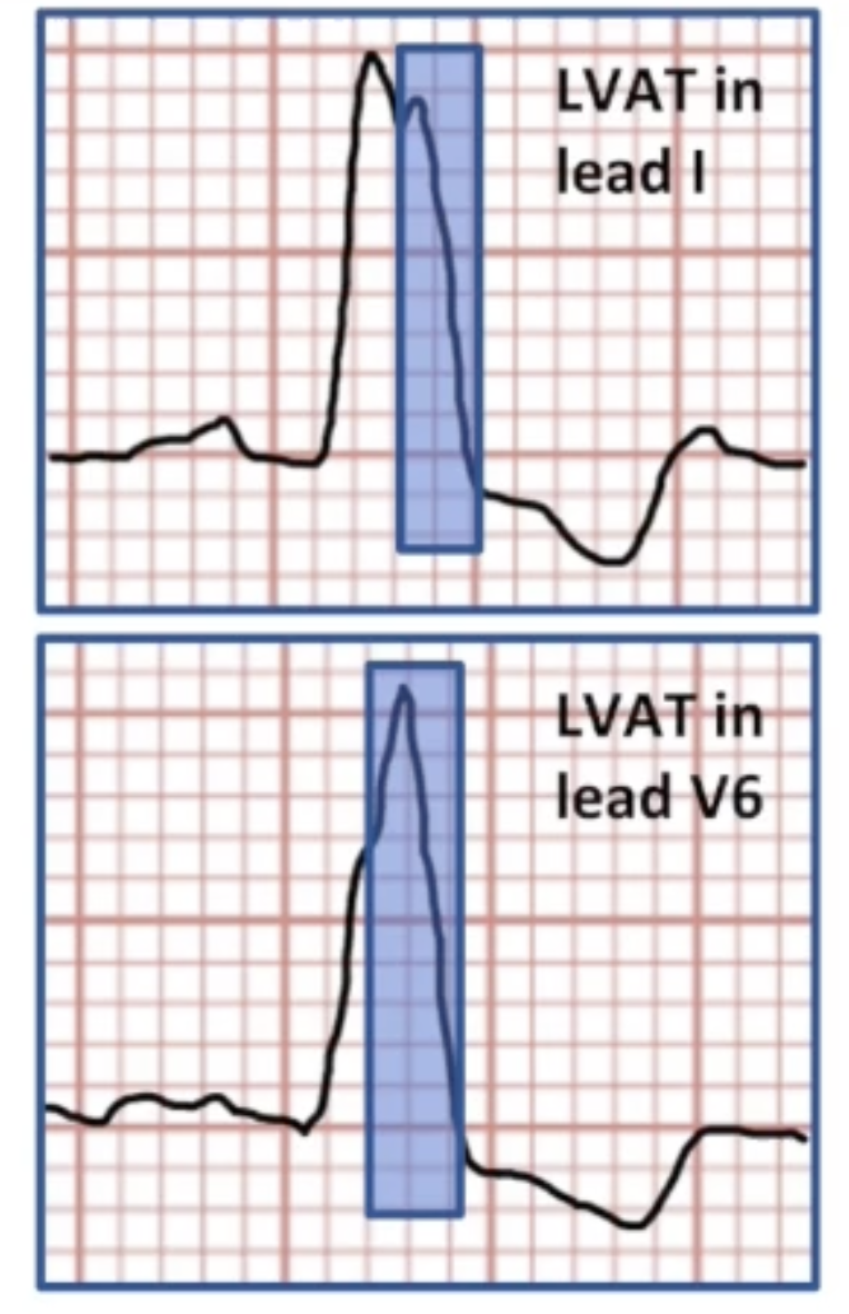
Longer LV activation time
Time difference between first notch after 40 ms of QRS and end of QRS
Factors in CRT Response
~30% of patients with reduced LV systolic function and narrow QRS have mechanical dysfunction
Research into patients with wide QRS, no mechanical dyssynchrony
Data suggests patients with both electrical and mechanical dyssynchrony respond best
Other factors:
Female sex
Non-ischemic cardiomyopathy
Genetic factors
Other HF Management Strategies
Pharmacological therapy
Surgical procedure
Heart transplant
Other devices:
Mechanical circulatory support to pump blood
ICD to prevent SCD
Barostimulation to address compensatory mechanisms
During CRT Implant
Achieve optimal stimulation configuration
LV lead position
Pacing vector
Choose optimal device settings
Reprogram post-implant if needed
May need lead revision
After the Implant
Patient education and continued care
Remote monitoring
Continue OPT
Communication between EP and HF specialist
Manage underlying disorders
Anemia
Myocardial ischemia
Mitral regurgitation
Treat arrhythmias
Antiarrhythmics
Ablation
Physiologic Pacing
More physiologic as it goes down
Asynchronous
VVI mode
RV apical pacing
Demand
DDD mode
Rate response
Dynamic intervals
His bundle pacing
Biventricular pacing
Resynchronization
AV dyssynchrony
AV sequential pacing
Atrial tracking
Interventricular dyssynchrony
Pacing RV and LV in unison
Pacing with calculated delay between RV and LV
Intraventricular dyssynchrony
Pacing lateral wall of LV early
AV Synchrony
Maximizes ventricular preload
Increases contractility
Increases stroke volume
Allows AV valves time to close
Prevents regurgitation
Maintains low atrial pressures
Easier venous return
Prevents congestion
Regulates autonomic & neurohormonal reflexes
Dual Chamber CIED
Degree of Heart Block | Appropriate Algorithm |
None | Fixed extended AV delay, intrinsic promotion algorithm |
1st Degree | Fixed extended AV delay, intrinsic promotion algorithm |
Intermittent 2nd Degree | Intrinsic promotion algorithm, AV search hysteresis |
Consistent 2nd Degree | AV search hysteresis |
3rd Degree | Fixed or dynamic AV delay |
CRT-P or CRT-D
100% BIV pacing required
Program AV delay shorter than intrinsic conduction while keeping it as physiologic as possible
Suboptimal AV timing: #1 reason for poor CRT response
Mitral Velocity Doppler Echo
Flow of blood through mitral valve
Assess mechanical dyssynchrony between atria and ventricles
E (early) wave: passive ventricular filling
A (atrial) wave: atrial systole
E Waves & A Waves Part 1
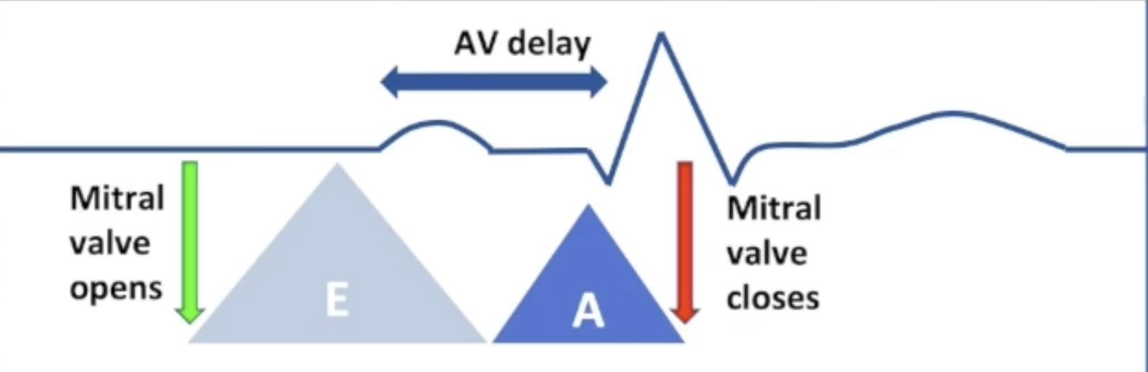
AV delay optimal:
E wave begins after isovolumetric relaxation
A wave begins at end of E wave
Isovolumetric contraction begins at end of A wave
E Waves & A Waves Part 2
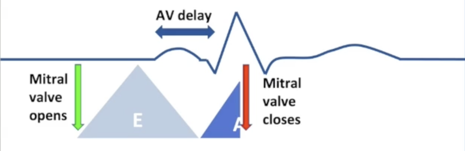
AV delay too short:
Ventricular contraction begins during atrial systole
A wave is truncated by closing of mitral valve
Less time for filling
Systolic MR
E Waves & A Waves Part 3
AV delay too long:
Ventricular contraction delayed
Ventricular relaxation delayed: late E wave
Fused E/A wave: A wave begins part way through E wave
Diastolic MR
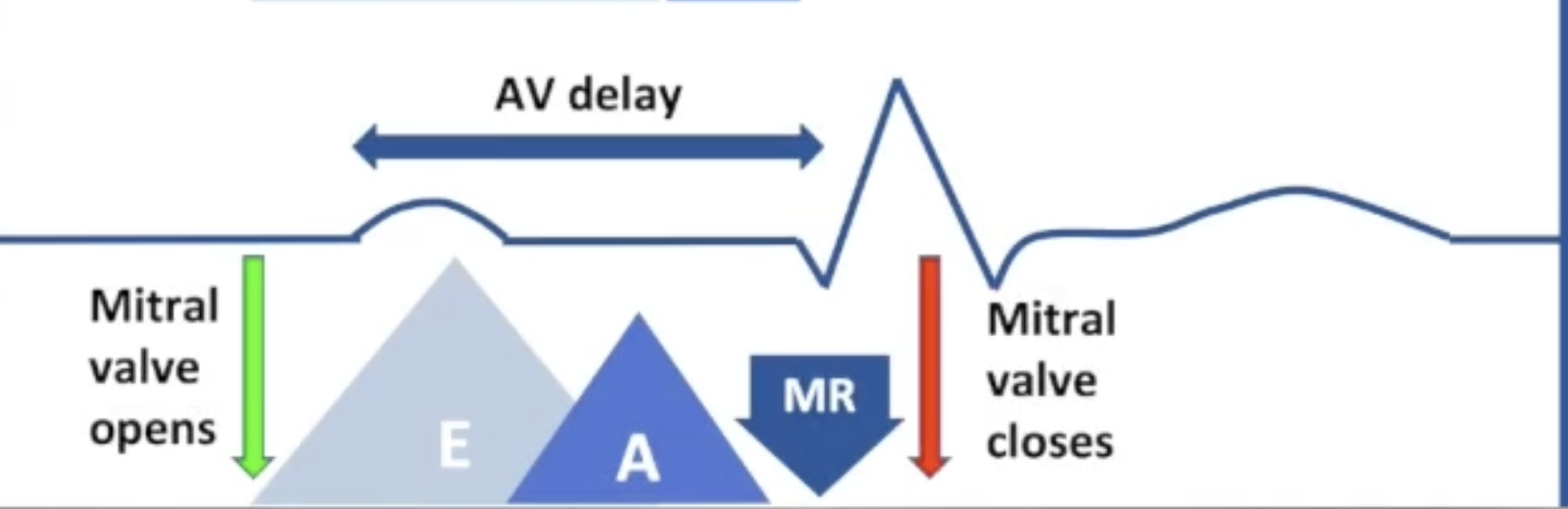
Optimizing AV Delays
Goal: ventricular contraction directly after A wave
Too early: A wave truncation
Too late: E/A wave fusion
Check percentage A paced
0% A Paced: SAV optimization only
Permanent AF: no AV optimization
Otherwise: SAV and PAV optimization
Ritter Method
Program SAV/PAV to 75% intrinsic PR interval
Shorten delay in 20 ms increments while recording on echo
Continue to shorten until A wave first begins to truncate
Lengthen delay in 10 ms increments until truncation disappears
Program this delay as the permanent SAV/PAV
V-V Optimization
LBBB: LV depolarized before RV
Most have mechanical interventricular dyssynchrony as well
BIV pacing:
Pace both ventricles at same time
Offset pace LV before RV
Offset pace RV before LV