Chapter 11 Cranium
1/213
There's no tags or description
Looks like no tags are added yet.
Name | Mastery | Learn | Test | Matching | Spaced | Call with Kai |
|---|
No analytics yet
Send a link to your students to track their progress
214 Terms
The small horizontal plate of the ethmoid, seen on Fig. 11.6, is called the
Cribiform plate
The vertical plate of the ethmoid bone forming the upper portion of the bony nasal septum is the
Perpendicular plate
A structure found in the middle of the sphenoid bone that surrounds the pituitary gland is the
sella turcica
The posterior aspect of the sella turcica is called the
Dorsum Salle
Which structure of the sphenoid bone allows for the passage of the optic nerve and is the actual opening into the orbit?
Optic foramen
Which structures of the sphenoid bone help to form part of the lateral walls of the nasal cavities?
medial and lateral pterygoid processes
Which radiographic projection best demonstrates the sella turcica and dorsum sellae?
lateral projection
Which aspect of the frontal bone forms the superior aspect of the orbit?
orbital or horizontal portion
name all the sutures of the skull
Coronal, sagittal, lambdoid, squamous, sphenosquamous, sphenofrontal, sphenoparietal.
name all the asterions of the skull
bregam, lamdoidal, pterion, asterion
Name all the fontanels in the fetal skull
Posterior Fontanel
Anterior Fontanel
Mastoid
Sphenoid
Cranial sutures are classified as being _____ joints.
fibrous or synarthrodial
Small, irregular bones that sometimes develop in adult skull sutures are called __________________________ or __________________________ bones and are most frequently found in the __________________________ suture.
sutural or wormian, lambdoidal
Which term describes the superior rim of the orbit? (Include the abbreviation also.) __________________________
supraorbital margin (SOM)
What is the name of the notch that separates the orbital plates from each other? __________________________
ethmoidal notch
Which cranial bones form the upper lateral walls of the calvarium?
right and left parietals
Which cranial bone contains the foramen magnum?
occipital bone
A small prominence located on the squamous portion of the occipital bone is called the
External occipital protuberance, or inion
What is the name of the oval processes found on the occipital bone that helps form the atlanto-occipital joint?
Occipital condyles, or lateral condylar portions
List the three aspects of the temporal bones.
A. Squamous
B. Mastoid
C. Petrous
True/False: The mastoid portion of the temporal bone is the densest of the three aspects of the temporal bone.
False (petrous portion)
Which external landmark corresponds with the level of the petrous ridge? __________________________
Top of the ear attachment (TEA)
Which opening in the temporal bone serves as a passageway for nerves of hearing and equilibrium?
Internal acoustic meatus
List the three aspects of the temporal bones
A. Squamous
B. Mastoid
C. Petrous
Which aspect of the temporal bone is considered the densest?
Petrous portion
Which structure makes up the cartilaginous external ear?
Auricle or pinna
How long is the average external acoustic meatus (EAM)?
1 inch (or 2.5 cm)
Which small membrane marks the beginning of the middle ear?
Tympanic membrane (eardrum)
Which of the following bones is not a facial bone?
A. Middle nasal conchae
B. Vomer
C. Lacrimal bone
D. Mandible
A. Middle nasal conchae
What is the largest immovable bone of the face?
Maxilla
List the four processes of the maxilla.
A. Frontal process
B. Zygomatic process
C. Alveolar process
D. Palatine process
Which of the processes mentioned in question number 3 is considered most superior?
Frontal process
Which soft tissue landmark is found at the base of the anterior nasal spine?
Acanthion
Which facial bones form the posterior aspect of the hard palate?
Horizontal portion of the palatine bones
Which two cranial bones articulate with the maxilla?
Frontal and ethmoid
Which facial bones are sometimes called the "cheek bones"?
Zygomatic or malar bones
Which of the following bones does not articulate with the zygomatic bone?
B. Mandible
Which facial bone is associated with the tear ducts?
Lacrimal bones
The purpose of the __________________________, or __________________________, is to divide the nasal cavity into compartments and to circulate air coming into the nasal cavities. (Include both terms for these bones.)
Conchae, turbinates
True/False: The right and left nasal bones form the largest part of the nose.
False (Most of the nose is composed of cartilage.)
A deviated nasal septum is most likely to occur at the junction between __________________________ and __________________________.
Septal cartilage, vomer (pushed laterally to one side)
Match each of the following mandibular terms to the correct definition or description. (Use each choice only once.)
A. Gonion
B. Mandibular notch
C. Body
D. Condyloid process
E. Coronoid process
F. Ramus
G. Mentum
H. Symphysis menti
1. Vertical portion of mandible
2. Chin
3. Mandibular angle
4. Point of union between both halves of the mandible
5. Bony process located anterior to mandibular notch
6. Horizontal portion of mandible
7. Posterior process of the upper ramus
8. U-shaped notch
A. 3
B. 8
C. 6
D. 7
E. 5
F. 1
G. 2
H. 4
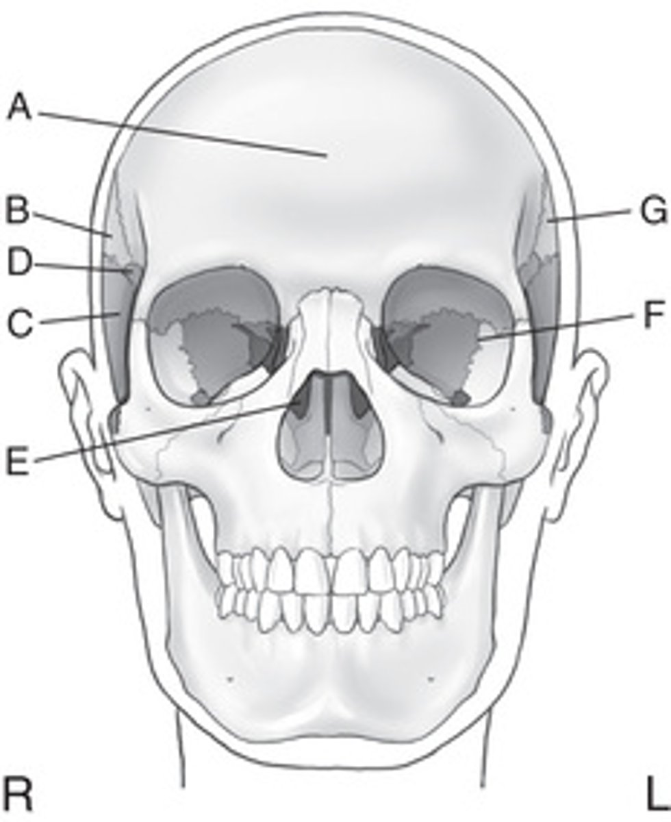
Identify the cranial bones labeled in Figs. 11.1 and 11.2. (Note: All eight cranial bones, including each paired bone, are visible in at least one of the following drawings.)
A. Frontal
B. Right parietal
C. Right temporal
D. Sphenoid
E. Ethmoid
F. Left temporal
G. Left parietal
H. Occipital
Fill in the total number of bones.
A. Cranium ___________
B. Facial bones ___________
A. 8
B. 14
List the four cranial bones that form the calvaria (skull cap).
A. Frontal
B. Right parietal
C. Left parietal
D. Occipital
List the four cranial bones that form the floor of the cranium
A. Right temporal
B. Left temporal
C. Sphenoid
D. Ethmoid
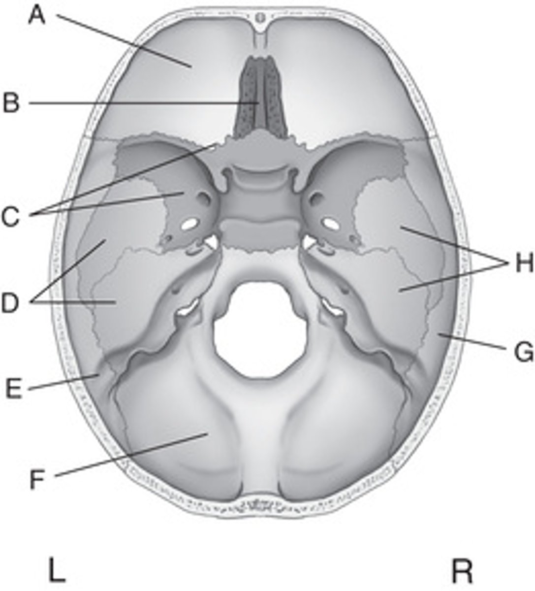
Identify all eight cranial bones on the two superior-view drawings (Figs. 11.3 and 11.4)
A. Frontal
B. Ethmoid
C. Sphenoid
D. Left temporal
E. Left parietal
F. Occipital
G. Right parietal
H. Right temporal
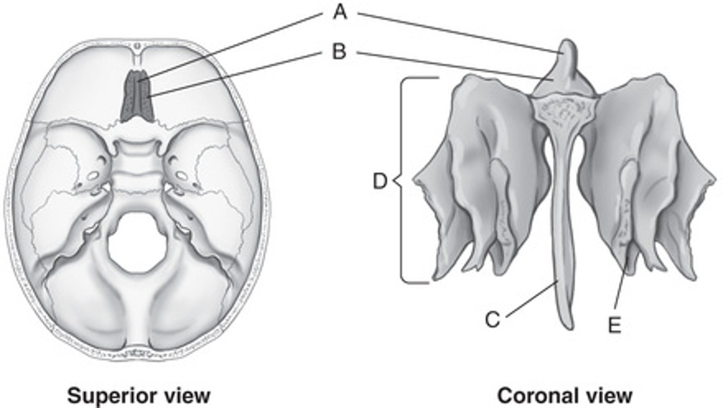
Identify the labeled parts on the three views of the ethmoid bone (Figs. 11.5 and 11.6).
A. Crista galli
B. Cribriform plate
C. Perpendicular plate
D. Lateral labyrinth (mass)
E. Middle nasal conchae (turbinate)
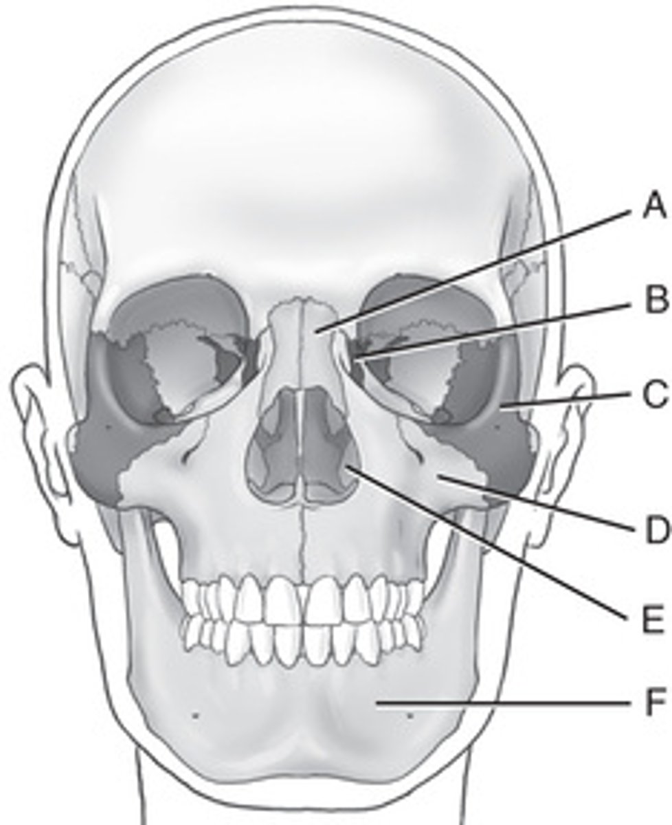
Identify the labeled facial bones visible in Figs. 11.18
Paired Bones
A. ___________
B. ___________
C. ___________
D. ___________
E. ___________
Single Bone
F. ___________
A. Nasal bones
B. Lacrimal bones
C. Zygomatic bones
D. Maxillary bones
E. Inferior nasal conchae
F. Mandible
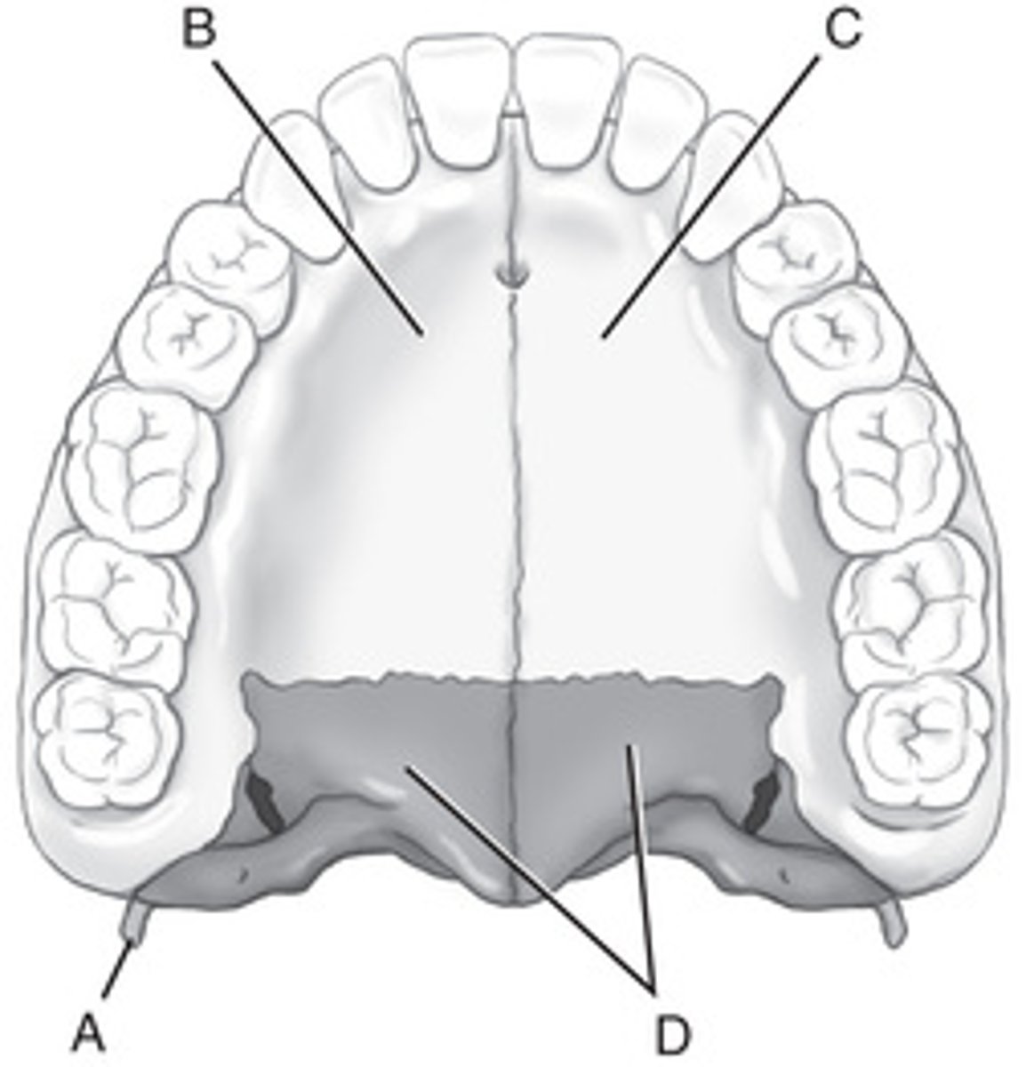
The single facial bone and the one pair of facial bones not visible from the exterior and not demonstrated in Figs. 11.18 and 11.19 are the __________________________ and the __________________________, respectively. (These are demonstrated in special-view drawings in the following questions.)Identify the labeled structures (and the facial bones of which they are a part) on this inferior surface view of the maxillae (Fig. 11.20).
Vomer and the palatine bones
Structure / Bone(s)
A. ___________
(___________)
B. ___________
(___________)
C. ___________
(___________)
D. ___________
(___________)
A. Pterygoid hamulus, sphenoid
B. Right palatine process, right maxilla
C. Left palatine process, left maxilla
D. Horizontal portions, right and left palatine bones
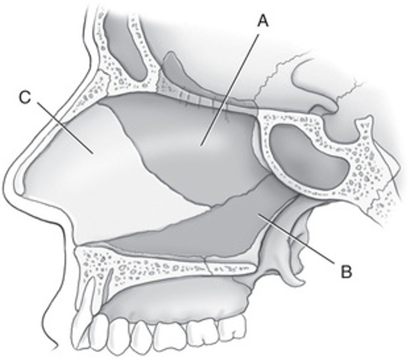
List the three structures that form the nasal septum as shown in Fig. 11.21.
A. Perpendicular plate of ethmoid
B. Vomer
C. Septal cartilage
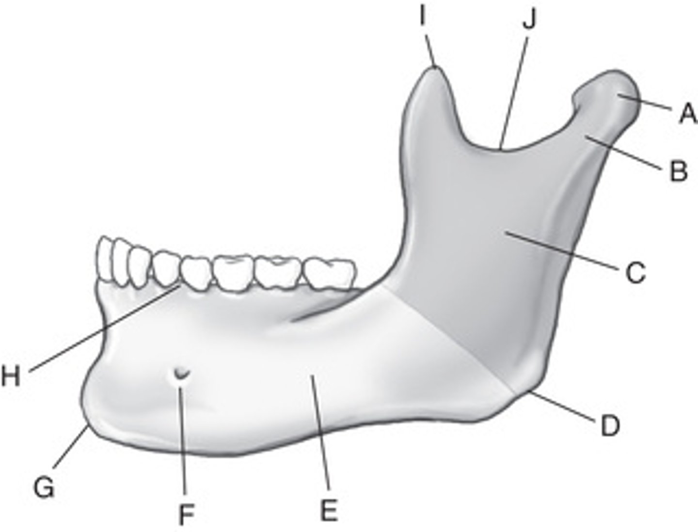
Identify the parts of the mandible and skull as labeled in Figs. 11.22 and 11.23.
A. ___________
B. ___________
C. ___________
D. ___________
E. ___________
F. ___________
G. ___________
H. ___________
I. ___________
J. ___________
K. (Cranial bone) ___________
L. (Joint) ___________
M. (Key landmark) ___________
N. (Landmark) ___________
A. Condyle
B. Neck
C. Ramus
D. Gonion or mandibular angle
E. Body
F. Mental foramen
G. Mentum or mental protuberance
H. Alveolar process
I. Coronoid process
J. Mandibular notch
K. Temporal bone
L. Temporomandibular joint (TMJ)
M. External auditory meatus (EAM)
N. Mastoid process
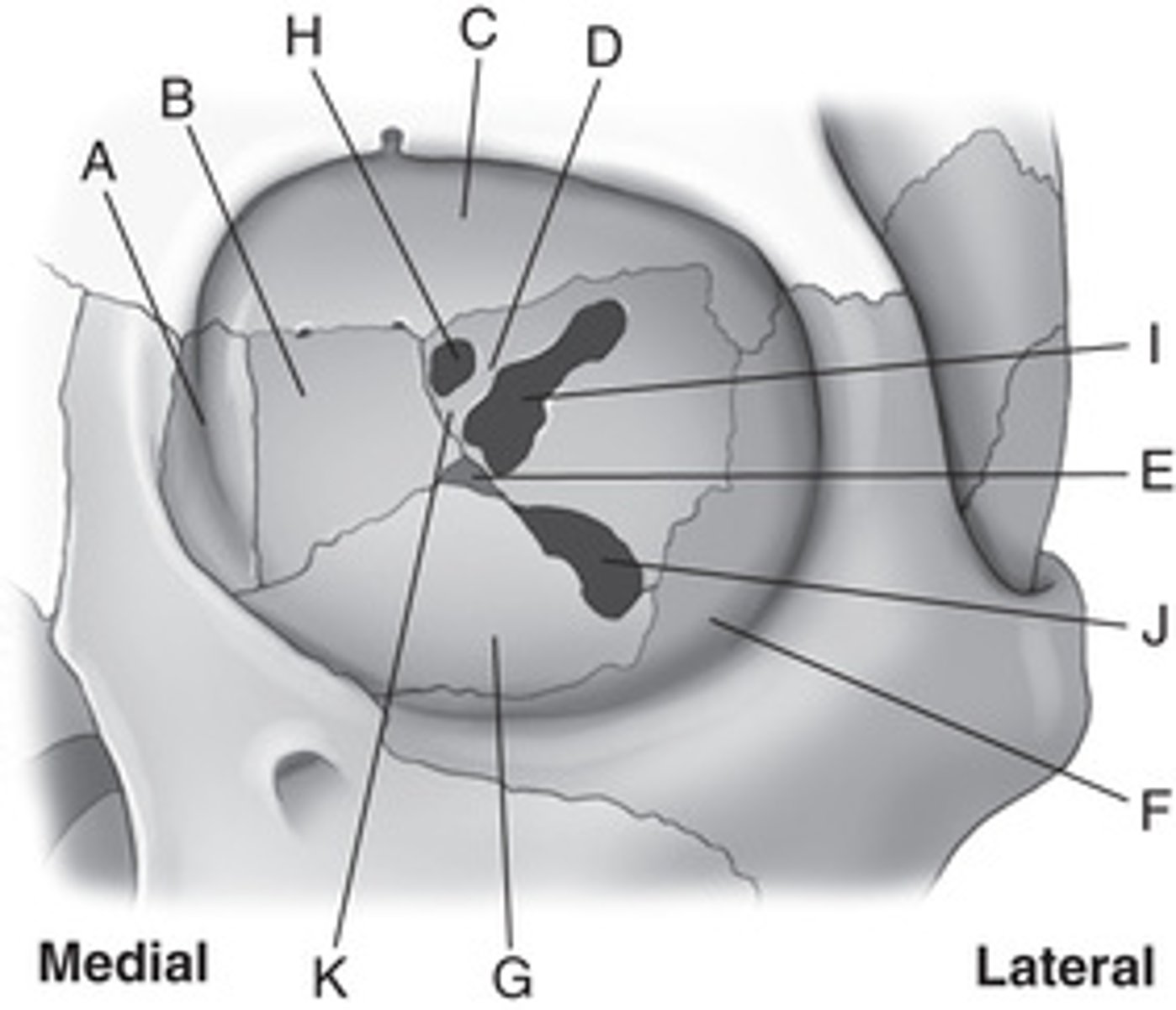
16. Identify the seven bones that form the orbit and indicate whether they are cranial or facial bones (Fig. 11.24). (See pp. 398-399.)
BoneCranial or Facial?
A. ___________
(___________)
B. ___________
(___________)
C. ___________
(___________)
D. ___________
(___________)
E. ___________
(___________)
F. ___________
(___________)
G. ___________
(___________)
A. Lacrimal (facial)
B. Ethmoid (cranial)
C. Frontal (cranial)
D. Sphenoid (cranial)
E. Palatine (facial)
F. Zygomatic (facial)
G. Maxilla (facial)

Identify the three foramina found within the orbits as labeled in Fig. 11.24.
H. ___________
I. ___________
J. ___________
Small section of bone:
K. ___________
H. Optic foramen
I. Superior orbital fissure
J. Inferior orbital fissure
K. Sphenoid strut
From anterior to posterior, the cone-shaped orbits project upward at an angle of __________________________ degrees and toward the midsagittal plane at an angle of __________________________ degrees.
30 degrees, 37 degrees
Which facial bone opening has the maxillary branch of the fifth cranial nerve passing through it?
Inferior orbital fissure
Which of the facial bone openings is formed by a cleft between the greater and lesser wings of the sphenoid bone?
A. Superior orbital fissure
B. Optic foramen
C. Inferior orbital fissure
D. Optic canal
A. Superior orbital fissure
What is another term for the second cranial nerve?
B. Optic nerve
What is the older term for the maxillary sinuses?
Antrum of Highmore
An infection of the teeth may travel upward and involve the __________________________ sinus.
Maxillary
Specifically, where are the frontal sinuses located?
Between the inner and outer tables of the skull, posterior to the glabella
The frontal sinuses rarely become aerated before the age of
6 years
Which specific aspect of the ethmoid bone contains the ethmoid sinuses?
Lateral masses or labyrinths
The drainage pathway for the paranasal sinuses is called the:\
A. Uncinate process
B. Ostiomeatal complex
C. Paranasal meatus
D. Lateral masses
B. Ostiomeatal complex
Which sinus is projected through the open mouth with a PA axial transoral projection?
Sphenoid sinus
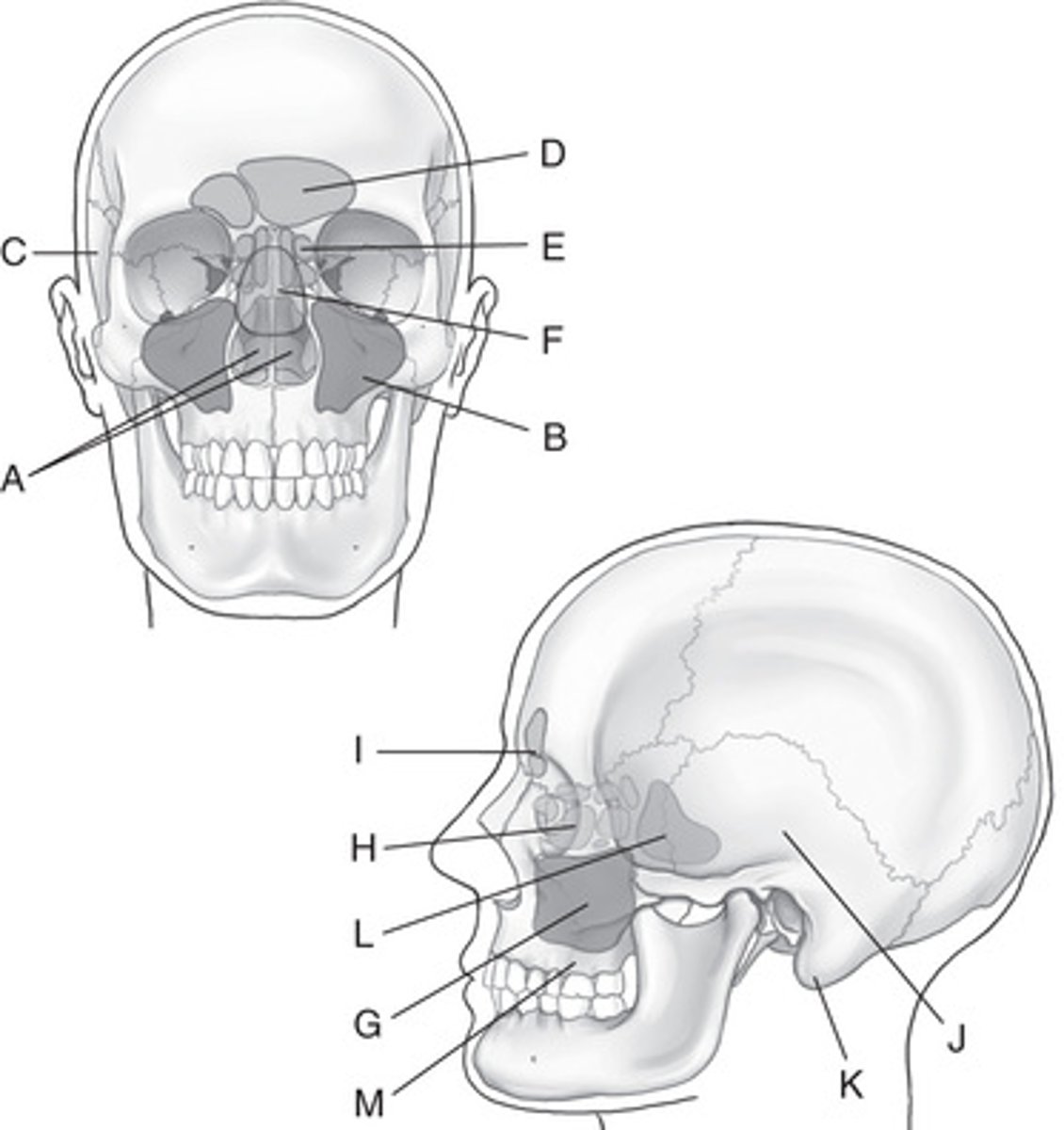
Identify the sinuses, structures, or bones labeled in Fig. 11.25.
A. ___________
B. ___________
C. ___________
D. ___________
E. ___________
F. ___________
G. ___________
H. ___________
I. ___________
J. ___________
K. ___________
L. ___________
M. ___________
A. Nasal cavity (fossae)
B. Maxillary sinuses
C. Right temporal bone (squamous portion)
D. Frontal sinuses
E. Ethmoid sinuses
F. Sphenoid sinuses
G. Maxillary sinuses
H. Ethmoid sinuses
I. Frontal sinuses
J. Squamous portion of left temporal bone
K. Mastoid portion of left temporal bone
L. Sphenoid sinus
M. Roots of upper teeth (alveolar process)
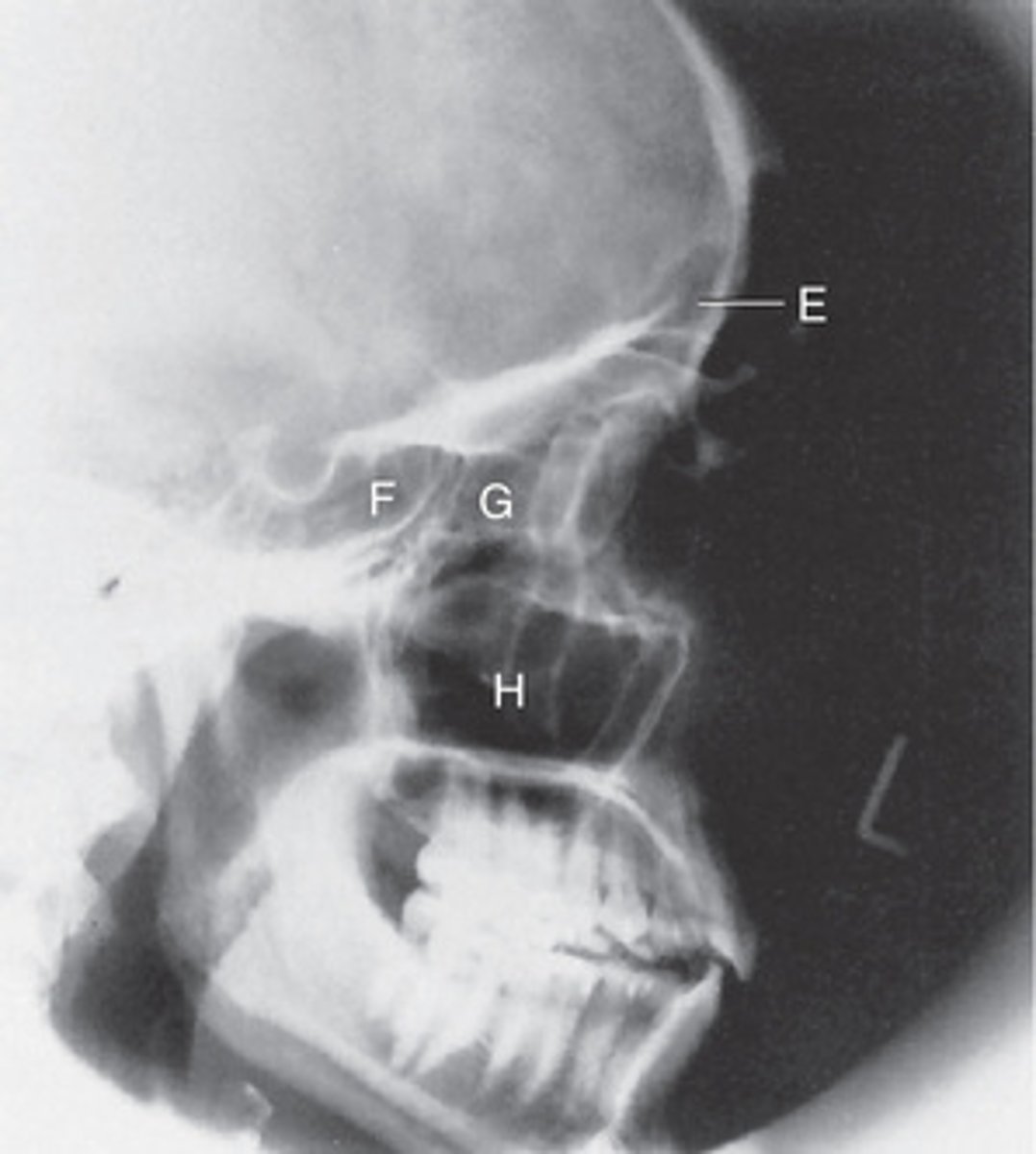
Identify the following paranasal sinuses labeled in Figs. 11.27.
Fig. 11.27
E. ___________
F. ___________
G. ___________
H. ___________
E. Frontal sinuses
F. Sphenoid sinus
G. Ethmoid sinuses
H. Maxillary sinuses
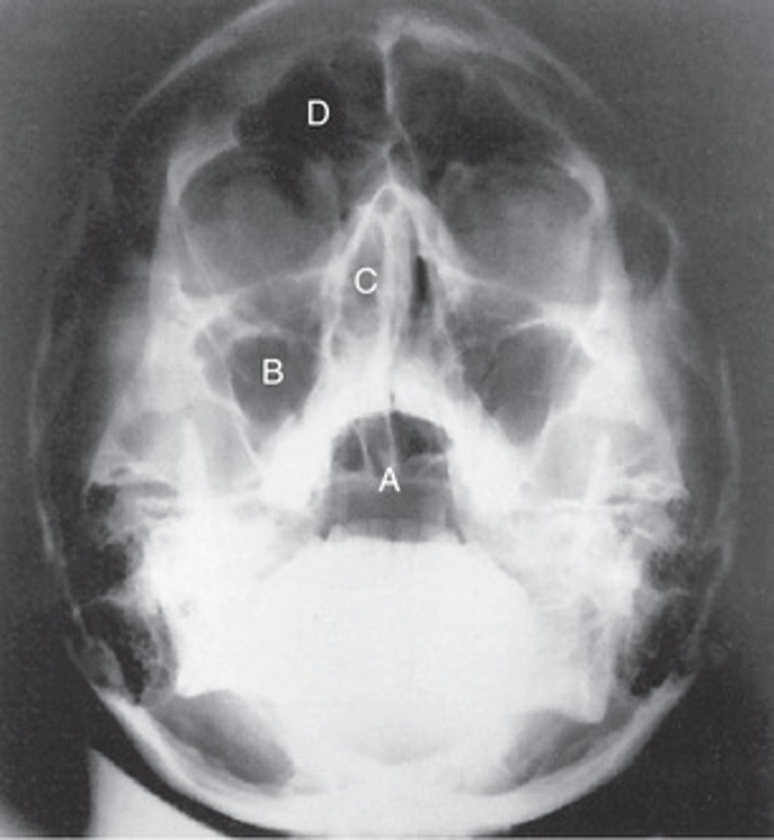
Identify the following paranasal sinuses labeled in Figs. 11.26.
Fig. 11.26
A. ___________
B. ___________
C. ___________
D. ___________
A. Sphenoid sinus
B. Maxillary sinuses
C. Ethmoid sinuses
D. Frontal sinus
What is the name of the passageway between the maxillary sinuses and the middle nasal meatus?
Infundibulum
True/False: Most CT studies of the sinuses do not require the use of contrast media.
True
Which position is most often used when performing a CT study of the sinuses?
B. Prone
1. List the three classifications of the skull; then match them with the correct shape description listed on the right.
A. Mesocephalic - Width between 75% and 80% of length
B. Brachycephalic - Width ≥80% of length
C. Dolichocephalic - Width <75% of length
Central ray angles and degree of rotation stated for basic skull positions are based on the __________________________ (average) skull, which has an approximate angle of __________________________ between the midsagittal plane and the long axis of the petrous bone.
Mesocephalic, 47 degrees
The long, narrow-shaped skull has an angle of approximately __________________________ degrees between the midsagittal plane and the long axis of the petrous bone.
±40
True/False: Two older terms for the orbitomeatal line (OML) are Reid's base line and the anthropologic base line.
False (These are other terms for the infraorbitomeatal line.)
There is a __________________________-degree difference between the orbitomeatal and infraorbitomeatal lines, and __________________________ degrees between the orbitomeatal and glabellomeatal lines.
7- to 8-degree; 7 to 8 degrees (same degrees of difference)
Match each of the following cranial landmarks and positioning lines with the correct definition. (Use each choice only once.)
______ 1. Lateral junction of the eyelid
______ 2. Posterior angle of the jaw
______ 3. A line between the infraorbital margin and the EAM
______ 4. Corresponds to the highest "nuchal" line of the occipital bone
______ 5. A line between the glabella and alveolar process of the maxilla
______ 6. A line between the mental point and the EAM
______ 7. Located at the junction of the two nasal bones and the frontal bone
______ 8. The small cartilaginous flap covering the ear opening
______ 9. Corresponds to the highest level of the facial bone mass
______ 10. A line between the midlateral orbital margin and the EAM
______ 11. The center point of the EAM
______ 12. A positioning line that is primarily used for the modified Waters projection
______ 13. A line used in positioning to ensure that the skull is in a true lateral position
______ 14. Corresponds to the level of the petrous ridge
______ 15. A smooth, slightly depressed area between the eyebrows
A. TEA
B. Supraorbital groove
C. Interpupillary line
D. Nasion
E. Gonion
F. Tragus
G. Outer canthus
H. Glabelloalveolar line
I. OML
J. Infraorbitomeatal line (IOML)
K. Mentomeatal line
L. Lips-meatal line
M. Glabella
N. Inion
O. Auricular point
1. G
2. E
3. J
4. N
5. H
6. K
7. D
8. F
9. B
10. I
11. O
12. L
13. C
14. A
15. M
What is the average kV range for analog skull radiography? kV range for digital imaging?
75 to 85 kV (analog) and 80 to 90 kV (digital systems)
List the five most common errors made during skull radiography.
A. Rotation
B. Tilt
C. Excessive neck flexion
D. Excessive neck extension
E. Incorrect central ray angulation
Of the five causes listed in the previous question, which two are the most common?
A. Rotation
B. Tilt
Bilateral horizontal fractures of the maxillae describe a __________________________ fracture.
A. Le Fort
B. Blowout
C. Tripod
D. Contrecoup
A. Le Fort
Which of the following imaging modalities is the most common neuroimaging procedure performed for the cranium?
A. Computed tomography (CT)
B. Ultrasound
C. Magnetic resonance imaging (MRI)
D. Nuclear medicine
A. Computed tomography (CT)
Which of the following imaging modalities is commonly performed on neonates with a possible intracranial hemorrhage?
A. CT
B. Ultrasound
C. MRI
D. Nuclear medicine
B. Ultrasound
Which of the following imaging modalities is most commonly performed to evaluate patients for Alzheimer disease?
A. CT
B. Ultrasound
C. MRI
D. Nuclear medicine
D. Nuclear medicine
Match each of the following clinical indications to the correct definition or statement. (Use each choice only once.)
A. Fracture that may produce an air-fluid level in the sphenoid sinus
B. Destructive lesion with irregular margins
C. Also called a "ping-pong" fracture
D. Proliferative bony lesion of increased density
E. A tumor that may produce erosion of the sella turcica
F. Also known as osteitis deformans
G. A bone tumor that originates in the bone marrow
1. Osteoblastic neoplasm
2. Pituitary adenoma
3. Basal skull fracture
4. Paget's disease
5. Osteolytic neoplasm
6. Depressed skull fracture
7. Multiple myeloma
A. 3
B. 5
C. 6
D. 1
E. 2
F. 4
G. 7
Which of the following clinical indications may require an increase in manual exposure factors?
A. Advanced Paget's disease
B. Metastatic neoplasm
C. Multiple myeloma
D. Basal skull fracture
A. Advanced Paget's disease
Which cranial bone is best demonstrated with an AP axial (Towne method) projection of the skull?
Occipital
When using a 30-degree caudad angle for the AP axial (Towne method) projection of the skull, which positioning line should be perpendicular to the image receptor?
A. OML
B. IOML
C. GAL
D. AML
A. OML
A properly positioned AP axial (Towne method) projection should place the dorsum sellae into the middle aspect of the:
A. Orbits
B. Clivus
C. Foramen magnum
D. Anterior arch of C1
C. Foramen magnum
A lack of symmetry of the petrous ridges indicates which of the following problems with a radiograph of an AP axial projection?
A. Tilt
B. Central ray angle
C. Flexion or extension
D. Rotation
D. Rotation
If the patient cannot flex the head adequately for the AP axial (Towne method) projection, the technologist could place the __________________________ perpendicular to the image receptor and angle the central ray __________________________ degrees caudad.
IOML; 37
What evidence on an AP axial (Towne method) radiograph indicates whether the correct central ray angle and correct head flexion were used?
Dorsum sellae and posterior clinoids should be projected into the foramen magnum.
What central ray angle should be used for the PA axial (Haas method) projection for the cranium?
25 degrees cephalad
Where is the central ray centered for a lateral projection of the skull?
2 inches (5 cm) above the EAM
Which specific positioning error is present if the mandibular rami are not superimposed on a lateral skull radiograph?
A. Tilt
B. Rotation
C. Overflexion of head and neck
D. Incorrect central ray angle
B. Rotation
Where will the petrous ridges be projected with a 15-degree PA axial (Caldwell) projection of the cranium?
In the lower 1/3 of the orbits
Which specific positioning error is present if the petrous ridges are projected higher in the orbits than expected for a 15-degree PA axial projection?
Excessive flexion or insufficient central ray angle
Which projection of the cranium produces an image of the frontal bone with little or no distortion?
0 degrees posteroanterior (PA)
For a patient with possible trauma, what must be determined before performing the submentovertical (SMV) projection of the skull?
Rule out any possible cervical fractures or subluxation.