Topic 8: Grey Matter
0.0(0)
Card Sorting
1/185
Earn XP
Study Analytics
Name | Mastery | Learn | Test | Matching | Spaced |
|---|
No study sessions yet.
186 Terms
1
New cards
central nervous system (CNS)
brain and spinal cord.
2
New cards
peripheral nervous system
sensory nerves and motor nerves → somatic nervous system and autonomic nervous system (divided into sympathetic and parasympathetic).
3
New cards
sensory nerves
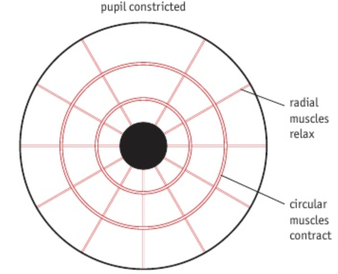
carry sensory informations from the receptor to the CNS.
4
New cards
motor nerves
carry the motor commands from the CNS to the effectors.
5
New cards
somatic nervous system
voluntary → stimulates skeletal muscle.
6
New cards
autonomic nervous system
involuntary → stimulates smooth muscle, cardiac muscle, and glands. subdivided into the sympathetic and parasympathetic nervous systems.
7
New cards
sympathetic nervous system
prepares body for ‘fight or flight’ responses (e.g. accelerates heartbeat, stimulates glucose production and release, secretes adrenaline).
8
New cards
parasympathetic nervous system
prepares body for ‘rest and digest’ (e.g. slows heartbeat, stimulates bile release, stimulates peristalsis).
9
New cards
neurone
single nerve cell. consists of dendrites, the axon, and a cell body, containing the nucleus and organelles.
10
New cards
nerve
bundle of the axons of neurones surrounded by a protective covering (myelin sheath).
11
New cards
dendrites
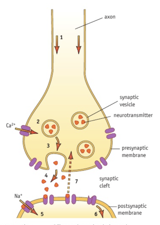
very fine extensions from the cell body that conduct impulses towards the cell body. dendritic branches can form.
12
New cards
axon
single long process that transmits impulses away from the cell body.
13
New cards
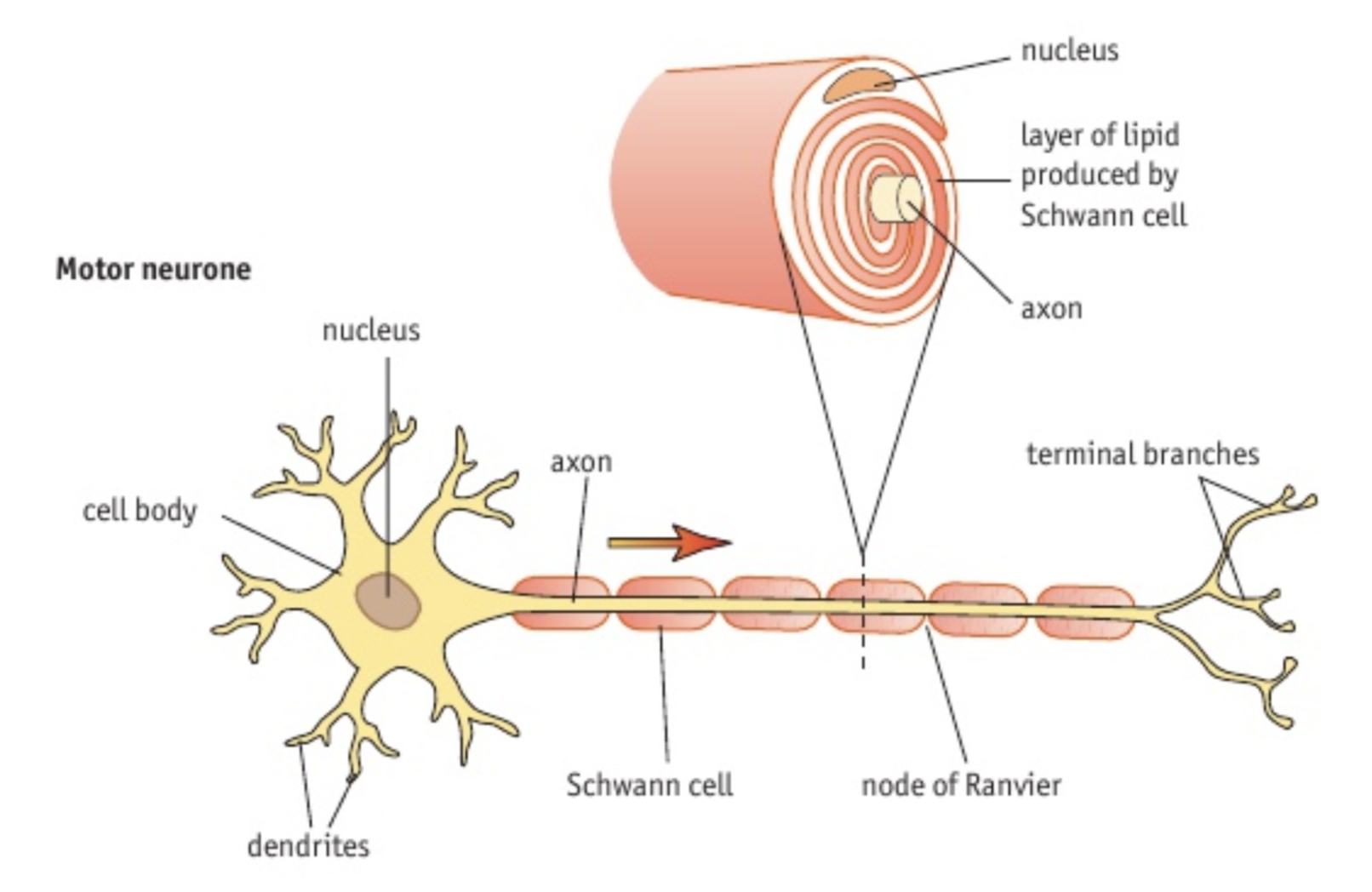
motor neurones
the cell body always situated within the CNS and the axon (of varying length) extends out, conducting impulses from the CNS to the effectors (muscles or glands).
14
New cards
sensory neurones
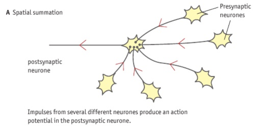
these carry impulses from the sensory cells to the CNS.
15
New cards
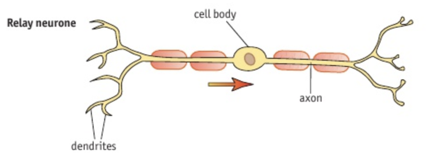
relay neurones
found mostly within the CNS. they have a large number of connections with other nerve cells. also known as connector neurones or interneurones.
16
New cards
schwann cell
type of glial cell that keeps neurones alive and sometimes cover them with a myelin sheath. they are the major glial cell type in the peripheral nervous system and play essential roles in the development, maintenance, function, and regeneration of peripheral nerves.
17
New cards
node of ranvier
myelin sheath gaps that allow the generation of a fast electrical impulse along the axon (saltatory conduction), as the myelin sheath is composed of insulating, fatty substance.
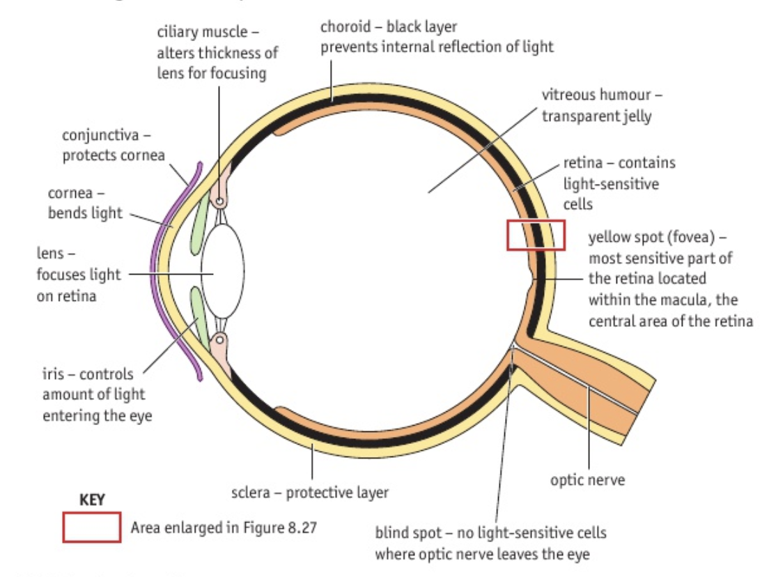
18
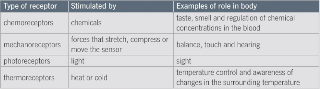
New cards
terminal branches
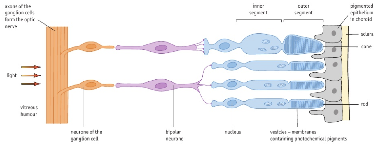
change electrical impulses or action potentials within a neuron into chemical messages in the form of neurotransmitters. these neurotransmitters are released into synapses to relay messages to other neurons or other types of cells like muscle cells.
19
New cards
reflex arc
neural pathway controlling a reflex (e.g. knee-jerk reflex or pupil reflex).
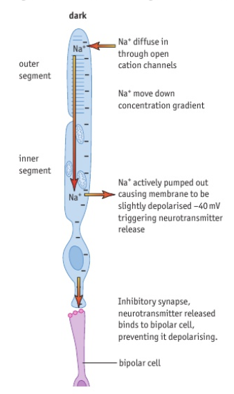
20
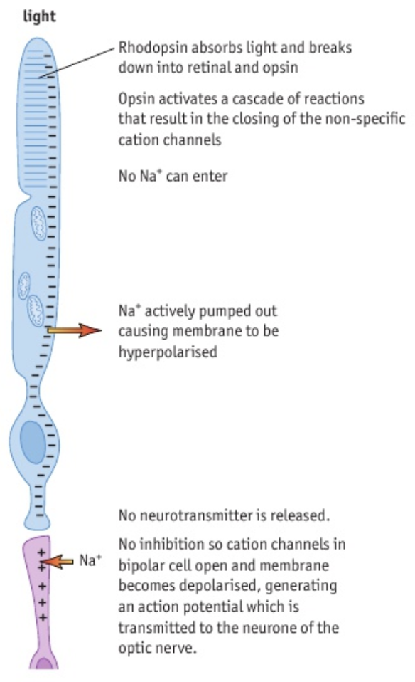
New cards
reflex
rapid, involuntary response to stimuli.
21
New cards
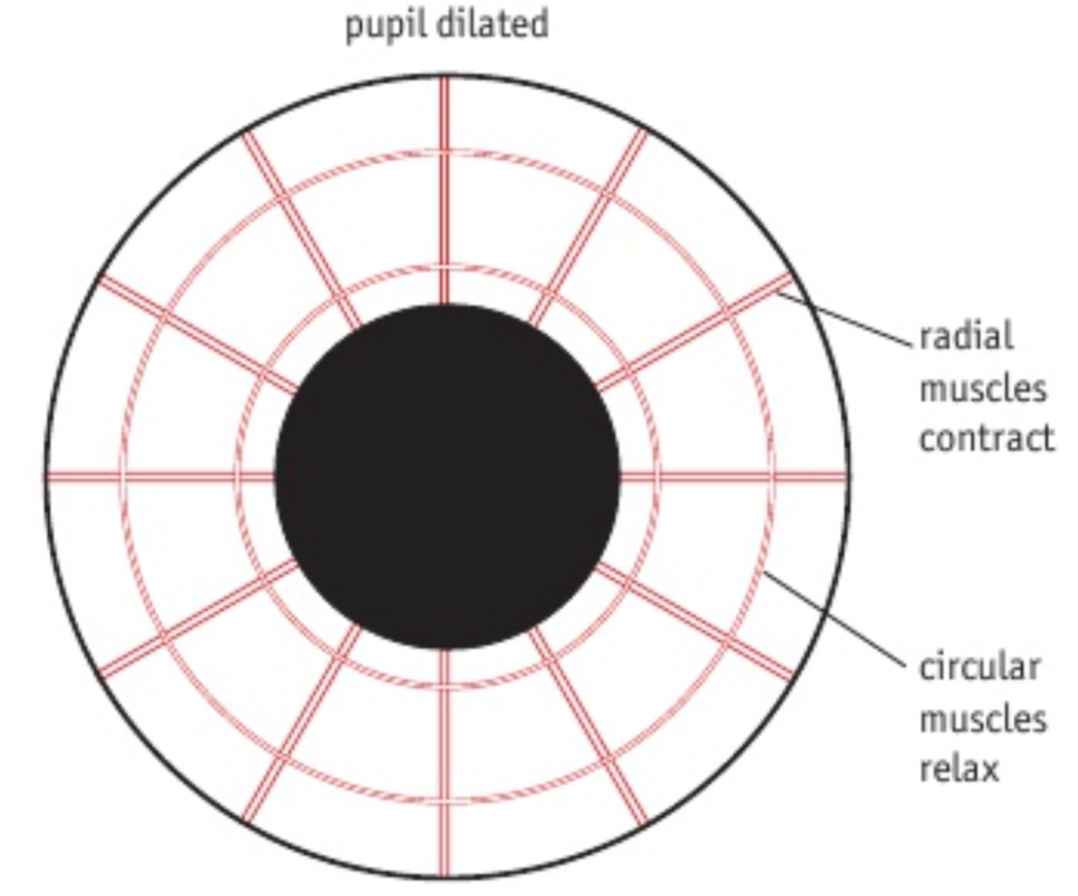
pupil reflex
reflex arc that causes a change in diameter of the pupils by contracting different muscles in the iris.
22
New cards
iris muscles
antagonistic muscles controlling the size of the pupil → radial and circular muscles. controlled by the autonomic nervous system.
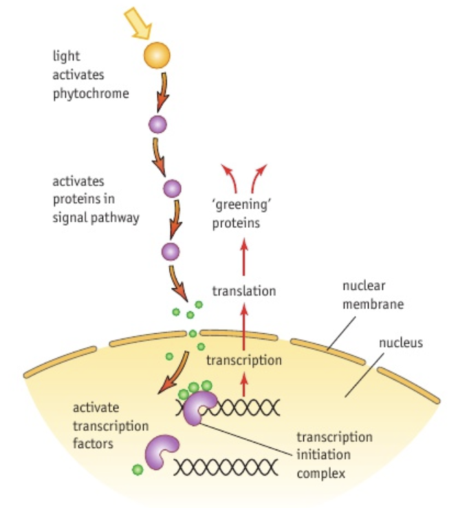
23
New cards
radial muscles
controlled by a sympathetic reflex → contracts to dilate pupils.
24
New cards
circular muscles
controlled by a parasympathetic reflex → contracts to constrict the pupils.
25
New cards
pupil dilation
radial muscles contract, circular muscles relax.
26
New cards
pupil constriction
radial muscles relax, circular muscles contract.
27
New cards
pupil constriction neural pathway
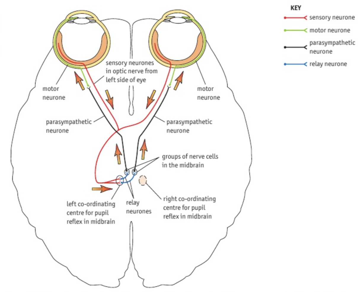
high light levels strike the photoreceptors in the retina causing nerve impulses to pass along the optic nerve to a number of different sites within the CNS, including a group of coordinating cells in the midbrain. impulses from these cells are sent along parasympathetic motor neurones to the circular muscles of the iris causing them to contract. at the same time, the radial muscles relax.
28
New cards
why is there a potential difference across the membrane of an axon?
the distribution of ions extracellularly and intracellularly are uneven.
29
New cards
how does the axon ion distribution change?
sodium-potassium pumps in the membrane carry Na+ out of the cell and K+ into the cell. they act against the concentration gradient and use energy from the hydrolysis of ATP. organic anions (e.g. negatively charged amino acids) are large and stay in the cell, so Cl- ions move out of the cell to help balance the charge across the membrane.
30
New cards
resting potential
voltage across the membrane of a non-signaling neuron. its value is -70mV.
31
New cards
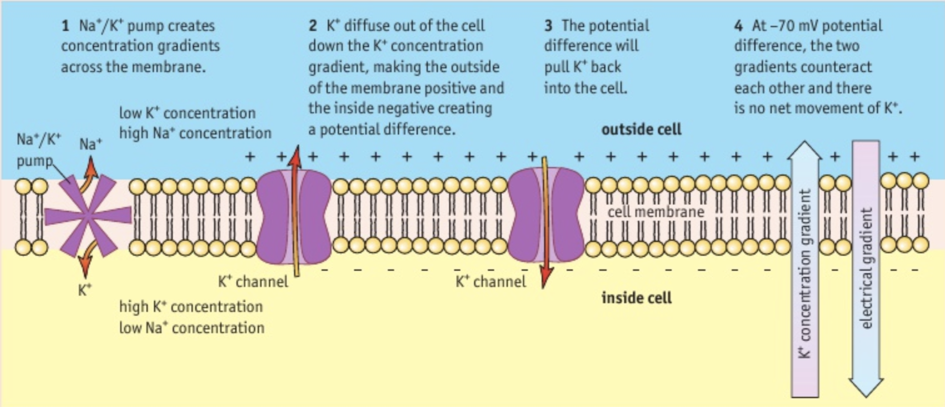
how is the resting potential established?
once the concentration gradients are established by the sodium-potassium pumps and there is no difference in charge between the inside and outside of the cell membrane, K+ ions diffuse out of the membrane down the potassium concentration gradient. the K+ ions pass through potassium channels, making the outside of the cell surface membrane positive. the membrane is permeable to K+ ions but is virtually impermeable to Na+ ions. there is some leakage of Na+ into the neurone down the concentration gradient, but it does not balance the difference in charge from the movement of K+. the difference in charge causes a potential difference across the membrane - the resting potential.
32
New cards
why is the resting potential -70mV?
when the potential difference across the membrane reaches -70mV, the electrical gradient exactly balances the chemical gradient, resulting in no net movement of K+. an electrochemical equilibrium is established and the membrane is polarised.
33
New cards
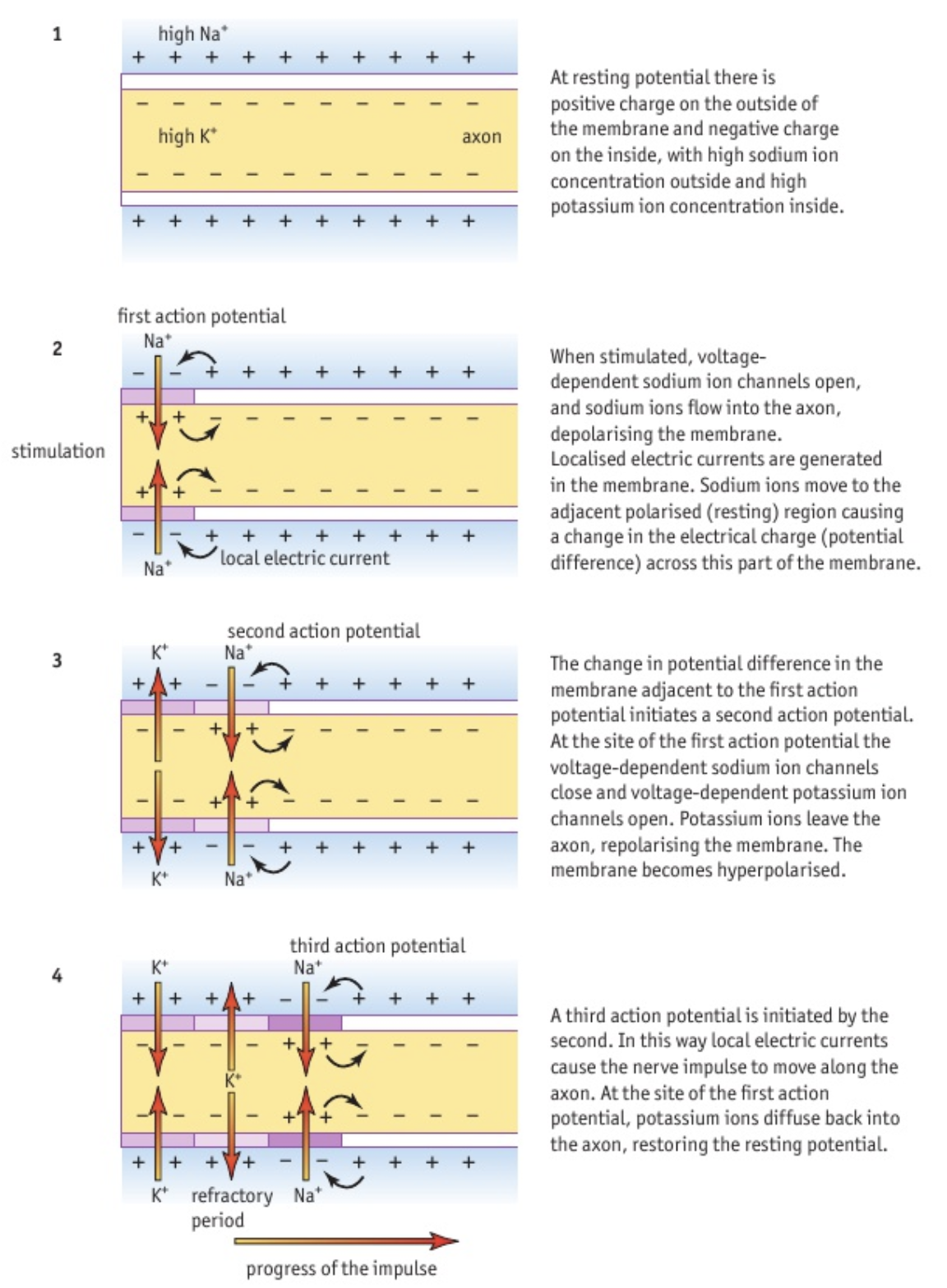
action potential
rapid change in voltage across the membrane that allows nerves to transmit information.
34
New cards
what causes an action potential?
changes in the permeability of the cell surface membrane to Na+ and K+ upon threshold stimulation, due to the opening and closing of voltage-dependent Na+ and K+ channels. at the resting potential, these channels are blocked by gates preventing the flow of ions through them. there are three different stages: depolarisation, repolarisation, and restoring the resting potential.
35
New cards
depolarisation
when a neurone is stimulated (e.g. an electric current above the threshold voltage), it causes a massive change in potential difference. the potential difference is locally reversed, making the inside of the axon positive and the outside negative.
36
New cards
how does depolarisation occur?
the change in potential difference across the membrane causes a change in shape of the Na+ gate, opening some of the voltage-dependent sodium ion channels. as the sodium ions flow in down the concentration gradient, depolarisation increases, triggering more gates to open once a certain potential difference threshold has been reached. the opening of more gates increases depolarisation further → positive feedback. the polarity of the membrane is reversed.
37
New cards
all-or-nothing depolarisation
as depolarisation is a positive feedback loop, there is no way of controlling the degree of depolarisation - there is either an action potential or not.
38
New cards
repolarisation
a quick attempt to return to resting potential so that the neuron can conduct more impulses. the potential difference ends up being more negative than the resting potential (hyperpolarisation).
39
New cards
how does repolarisation occur?
after about 0.5ms, the voltage-gated Na+ channels spontaneously close and Na+ permeability of the membrane returns to its usual very low level. voltage-dependent K+ channels open due to the depolarisation of the membrane and K+ ions move out of the axon, down the electrochemical gradient. as potassium ions move out of the cell, a negative potential difference is established, due to the inside of the axon being negative.
40
New cards
why does hyperpolarisation occur?
the membrane is highly permeable to K+ ions due to the open voltage-dependent K+ channels. more ions move out of the membrane than occurs at resting potential, resulting in a more negative potential difference.
41
New cards
restoring the resting potential
voltage-dependent K+ channels are closed and K+ ions diffuse into the axon until resting potential is re-established.
42
New cards
sodium-potassium pumps in action potential
only used if a cell is transmitting many action potentials, to maintain the original ion concentrations when Na+ concentration gets too high intracellularly. pumps Na+ ions back out of the cell if there is leakage at rest.
43
New cards
how are impulses passed along an axon?
neurone stimulation triggers a series of action potentials along the axon. the site of the action potential causes depolarisation of that part of the membrane, and the charged sodium ions move from that area to the adjacent resting region, causing a local electrical current. the depolarisation will spread to the adjacent resting region and open Na+ gates, triggering another action potential. as this is repeated, a wave of depolarisation is passed along the membrane. this is a nerve impulse.
44
New cards
refractory period
after a nerve impulse, a new action potential cannot be generated in the same section of membrane for around 5ms. this is because all the voltage-dependent sodium and potassium channels must return to their resting state (closed) and the resting potential has to be restored. this ensures the impulses only travel in one direction.
45
New cards
how is the intensity of a stimulus communicated?
action potentials are always the same size - they are always generated if the stimulus is above the threshold level. the size of stimulus affects the frequency of impulses and the number of neurones in a nerve that are conducting impulses.
46
New cards
what controls the speed of conduction?
the diameter of the axon. the wider the axon, the faster the impulse.
47
New cards
myelin sheath in conduction
acts as an electrical insulator along most of the axon, preventing any flow of ions across the membrane.
48
New cards
nodes of ranvier in conduction
gaps in the myelin sheath where depolarisation can occur, as ions can flow across the membrane.
49
New cards
saltatory conduction
ions are allowed to flow across the membrane at one node, creating a circuit that reduces the potential difference of the membrane at the next node, triggering an action potential. the impulse jumps from one node to the next, which is much faster than a wave of depolarisation across the whole membrane and means that a myelinated axon has a higher impulse velocity than an unmyelinated axon.
50
New cards
propagation of an impulse along an axon
51
New cards
how does a nervous impulse pass between cells?
the gap between cells: synaptic cleft.
52
New cards
synapse
where two neurones meet.
53
New cards
structure of a synpase
synaptic cleft separates the presynaptic membrane of the stimulating neurone from the postsynaptic membrane of the other cell. the gap is around 20-50nm and a nerve impulse cannot jump across it, meaning neurotransmitters are needed.
54
New cards
neurotransmitter
chemical messengers in the nervous system that are contained in synaptic vesicles in the cytoplasm at the end of the presynaptic neurone.
55
New cards
how does the synapse transmit an impulse?
an action potential arrives at the presynaptic membrane, causing the release of a neurotransmitter into the synaptic cleft. the neurotransmitter diffuses across the gap, resulting in events that cause the depolarisation of the postsynaptic membrane and hence the propagation of the impulse along the next cell. the presynaptic cell expends a lot of energy to produce the neurotransmitter and package it into vesicles ready for transport out of the cell.
56
New cards
neurotransmitter release
the action potential causes the depolarisation of the presynaptic membrane, opening calcium ion channels that allow these ions to diffuse into the cytoplasm down the concentration gradient. the increased Ca2+ concentration intracellularly causes synaptic vesicles containing the neurotransmitter to fuse with the presynaptic membrane and release their contents into the synaptic cleft by exocytosis.
57
New cards
stimulation of the postsynaptic membrane
specific receptor proteins embedded in the postsynaptic membrane will have a complementary binding site for the neurotransmitter. when the neurotransmitter reaches the postsynaptic membrane, it will bind to the protein, causing a conformational change of the protein that opens cation channels, making the membrane more permeable to sodium ions. sodium ions flow across the postsynaptic membrane, causing depolarisation that, if crossing the threshold level, will produce an action potential that will be propagated along the postsynaptic neurone.
58
New cards
how is the extent of depolarisation determined?
the number of functioning receptors in the postsynaptic membrane. also, the amount of neurotransmitter reaching the postsynaptic membrane, which will depend partly on the frequency of impulses reaching the presynaptic membrane. several impulses are usually required to generate enough neurotransmitter to depolarise the postsynaptic membrane.
59
New cards
inactivation of the neurotransmitter
some neurotransmitters are actively taken up by the presynaptic membrane to be used again. others rapidly diffuse away from the synaptic cleft or are taken up by other cells of the nervous system. some are broken down by enzymes.
60
New cards
inactivation of acetylcholine
a specific enzyme at the postsynaptic membrane, acetylcholinesterase, breaks down the acetylcholine so that it can no longer bind to receptors. some of the breakdown products are then reabsorbed by the presynaptic membrane and reused.
61
New cards
role of synapses in nerve pathways
control of nerve pathways, allowing flexibility of response, and the integration of information from different neurones, allowing a coordinated response.
62
New cards
what determines whether or not the postsynaptic membrane will generate an action potential?
the overall effect of all of the synapses the postsynaptic cell is receiving inputs from. two main factors affect the likelihood that the postsynaptic membrane will depolarise: type of synapse, and the number of impulses received.
63
New cards
types of synapses
excitatory synapses help stimulate an action potential, whereas inhibitory synapses and make it less likely that the postsynaptic membrane will depolarise. a postsynaptic cell can have many inhibitory and excitatory synapses, so the generation of an action potential depends on the balance of excitatory and inhibitory synapses acting.
64
New cards
excitatory synapses
make the postsynaptic membrane more permeable to sodium ions. a single one will not depolarise the membrane enough, but several impulses will produce sufficient depolarisation to produce an action potential (summation).
65
New cards
types of summation
spatial summation and temporal summation.
66
New cards
spatial summation
impulses come from different synapses, usually from different neurones. the number of different sensory cells stimulated can be reflected in the control of the response.
67
New cards
temporal summation
several impulses arrive at a synapse having travelled along a single neurone one after the other. their combined release of neurotransmitter generates an action potential in the postsynaptic membrane.
68
New cards
inhibitory synapses
these lower the likelihood of an action potential being generated in the postsynaptic cell. the neurotransmitter from these synapses open channels for chloride and potassium ions in the postsynaptic membrane, which will travel down their concentration gradients. negative Cl- ions will move into the cell and positive K+ ions will leave the cell, causing a more negative potential difference. as the cell becomes hyperpolarised, subsequent depolarisation becomes less likely unless more excitatory synapses transmit an impulse.
69
New cards
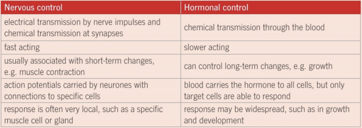
differences between nervous control and hormonal control
\
70
New cards
phototropism
bending of plants toward a light source.
71
New cards
mechanism of phototropism
as it was found that plants with the tip cut off did not display phototropism, the purpose of the tip was explored. it was shown that a chemical called auxin (indoleacetic acid - IAA) that was made in the tip passed down and stimulated growth by cell elongation.
72
New cards
how does the plant curve towards the light?
increased concentration of auxin on the shaded side of the plant increases cell elongation; reduced concentration on the unshaded side of the plant inhibits cell elongation.
73
New cards
effects of auxin on transcription
they are transported long distances in the phloem and shorter distances between cells, via specific carrier proteins in the cell membrane, from their place of synthesis (actively growing root and shoot tips, and in developing leaves, seeds, and fruits). the auxins bind to protein receptors in the target cells, activating intracellular second messenger signal molecules, which activate transcription factors. these control the transcription of auxin-regulated genes, and the proteins produced bring about metabolic changes that result in a range of responses through changes in cell expansion, division, and differentiation.
74
New cards
how does cell expansion occur?
the cell wall must be loosened. it is thought that auxin does this through stimulating the activity of proton pumps that move H+ ions out of the cytoplasm and into the cell wall, causing acidification. the low pH will activate proteins called expansins, which disrupt the bonds that hold the cellulose microfibrils and hemicelluloses together. there is slippage of the polysaccharides relative to each other, causing bonds to reform in new locations, allowing the expansion of the cell. the acidification of the cell wall also increases the potential difference across the membrane, enhancing the uptake of ions into the cell, causing water uptake by osmosis, causing the cell to swell resulting in cell elongation.
75
New cards
plant growth and differentiation
in the meristem, cells are actively dividing, and most of these cells then enlarge, forming a region of elongation adjacent to the meristem. these cells go on to mature and differentiate.
76
New cards
receptor cells
these cells detect stimuli and send electrical impulses to the central nervous system. many are spread throughout the body, but some types of receptor cells are grouped together into sense organs.
77
New cards
sense organs
organs, such as the eyes, that help protect the receptor cells and improve their efficiency. structures within the sense organ ensure that the receptor cells are able to receive the appropriate stimulus.
78
New cards
the eye as a sense organ
the receptor cells that detect light are found in the retina of the eye. the lens and cornea refract the light so that it is focused on the retina.
79
New cards
types of receptors
receptors can either be cells that synapse with a sensory neurone, or can themselves be part of a specialised sensory neurone, like skin thermoreceptors.
80
New cards
structure of the eye
81
New cards
photoreceptors in the retina
rod cells and cone cells
82
New cards
cone cells
allow colour vision in bright light.
83
New cards
rod cells
only give black and white vision but work in both dim light and bright light.
84
New cards
proportion of rods and cones in the retina
there is a small area in the centre of the retina where there are only cones, allowing us to pinpoint accurately the source and detail of what we are looking at. over the remainder of the retina, rods outnumber cones by a factor of about 20:1.
85
New cards
how does light stimulate photoreceptor cells?
a photochemical pigment in both rods and cones absorbs the light resulting in a chemical change. rods have a purplish pigment called rhodopsin, which is located in the membranes of the layers of flattened vesicles in the outer segment.
86
New cards
receptors in the dark
sodium ions flow into the outer segment through non-specific cation channels, moving down the concentration gradient into the inner segment where pumps continuously transport them back out of the cell. the influx of Na+ produces a slight depolarisation of the cell, changing the potential difference to about -40mV. the slight depolarisation triggers the release of a neurotransmitter, thought to be glutamate, from the rod cells. in the dark, rods release this neurotransmitter continuously. the neurotransmitter binds to the bipolar cell, stopping it depolarising.
87
New cards
receptors in the light
rhodopsin breaks down into retinal and opsin, non-protein and protein components. the opsin activates a series of membrane-bound reactions, ending in hydrolysis of a cyclic nucleotide molecule attached to the cation channel in the outer segment. the breakdown of this molecule results in the closing of the cation channels. the influx of Na+ into the rod decreases, while the inner segment continues to pump Na+ out. this makes the inside of the cell more negative, and it becomes hyperpolarised, stopping the release of glutamate. the bipolar neurone which the rod synapses with can thus depolarise, causing depolarisation in the ganglion neurones producing an action potential.
88
New cards
dark adaptation
once rhodopsin has been broken down, it is essential that it be rapidly converted back to its original form so that subsequent stimuli can be perceived. each individual rhodopsin molecule takes a few minutes to do this. the higher the light intensity, the more rhodopsin molecules are broken down and the longer it takes for all the rhodopsin to reform. the quick reformation of rhodopsin is what allows rod cells to work in dim light and adapt to darkness.
89
New cards
phytochromes
plant photoreceptors that consist of a protein component bonded to a non-protein light-absorbing pigment molecule. there are five different ones that have been identified which each differ in their protein component.
90
New cards
non-protein component of phytochromes
exists in two isomeric forms: Pr - phytochrome red, absorbs red light (660nm); Pfr - phytochrome far-red; absorbs far-red light (730nm).
91
New cards
photoreversibility
Pr and Pfr convert into each other. absorption of red light converts Pr into Pfr. absorbtion of far red light converts Pfr into Pr. in sunlight, both Pr is converted into Pfr and Pfr into Pr, however the former reaction dominates considering more red than far red light is absorbed. this means that Pfr accumulates in the light. in the dark, any Pfr present is slowly converted to Pr.

92
New cards
phytochromes triggering germination
it has been observed that seeds require light to germinate. experiments with lettuce indicate that a flash of red light will trigger germination, but a second flash of far-red light inhibits germination, suggesting their reversible effects. the final flash of light determines whether or not germination occurs. Pfr is the contributory form of the phytochrome that triggers germination.
93
New cards
photoperiod
the relative length of day or night which acts as the environmental cue that determines the time of flowering. it is determined by the ratio of Pr to Pfr in a plant.
94
New cards
effects of the photoperiod on Pr and Pfr
long winter nights give ample time for Pfr to convert back into Pr, so that by sunrise all phytochrome will be Pr. summer nights may not be long enough, so there may still be some Pfr present in the morning.
95
New cards
long-day plants
only flower when day length exceeds a critical value. they flower when the period of uninterrupted darkness is typically less than 12 hours; they need Pfr to stimulate flowering.
96
New cards
short-day plants
flower when the period of uninterrupted darkness is greater than 12 hours. long hours of darkness are needed in order to convert all Pfr present back to Pr. in these plants, Pfr inhibits flowering. a flash of red light in the middle of the dark period will negate the effects of the dark period in these plants.
97
New cards
greening
the changes in its form and biochemistry that a plant undergoes once a shoot has broken through soil into sunlight. once in the light, phytochromes promote the development of primary leaves, leaf unrolling, and the production of pigments. they can also inhibit certain processes, such as elongation of internodes.
98
New cards
how do phytochromes switch processes on or off?
exposure to light causes phytochrome molecules to change from one form to another, bringing about a change in shape. it is thought that each activated phytochrome then interacts with other proteins. the phytochromes may bind to the protein or disrupt the binding of a protein complex. these signal proteins may act as transcription factors or activate transcription factors that bind to DNA to allow transcription of light-regulated genes. the transcription and translation of proteins result in the plant’s response to light. for example, in seedlings, synthesis of the enzymes that control chlorophyll production will result in greening of the shoot.
99
New cards
gravity as an environmental cue
this stimulus ensures that developing shoots reach the light while roots grow in the soil, as light cannot be used as a cue more than a short distance under the soil surface.
100
New cards
touch and mechanical stress as an environmental cue
rubbing plant stems can result in shorter stems than in controls. it is thought that mechanical stimuli activate signal molecules whose end result is the activation of genes that control growth. in mimosa pudica plants, the leaf folds rapidly and collapses upon being touched. the specialised cells lose potassium ions, and water follows by osmosis, causing the cells to become flaccid, and so no longer support the leaf and keep it upright. this is an adaptation that helps to prevent grazing by herbivores.