ECG Ch2
 Call Kai
 Learn
Learn Practice Test
 Spaced Repetition
 Match
Match Flashcards
 Knowt Play
1/11

There's no tags or description
Looks like no tags are added yet.
Name | Mastery | Learn | Test | Matching | Spaced | Call with Kai | Chat |
|---|
No analytics yet
Send a link to your students to track their progress
12 Terms
What are the basic measurements and representations on EKG paper?-
Smallest division =
Between heavy lines =
Horizontal axis =
Vertical axis =
10 small squares of height =
Upward deflection =
Downward deflection =
5 large squares (25 small squares) of width =
1 small square =
1 large square =
Smallest division = 1 mm square
Between heavy lines = 5 small squares
Horizontal axis = time, read left to right
Vertical axis = voltage
10 small squares of height = 1 mV
Upward deflection = positive (+)
Downward deflection = negative (−)
5 large squares (25 small squares) of width = 1 second
1 small square = 0.04 seconds
1 large square = 0.20 seconds
What leads make up the standard 12-lead EKG?
The standard EKG is composed of 12 separate leads:
6 limb leads, using electrodes placed on the right arm, left arm, and left leg
6 chest leads: V1–V6
What are bipolar limb leads?
Bipolar limb leads are the foundational electrocardiogram (ECG) configurations. Selecting a different pair of electrodes for each lead creates 3 separate bipolar limb leads: I, II, and III
Recorded using 2 electrodes
One is (+)
One is (−)
Designated with Roman numerals
Moving each lead to the center forms three intersecting lines of reference
The angles remain the same relative to each other.
What are the augmented limb leads?
Sometimes called “unipolar” leads
Each limb, in turn, is designated as (+), while the remaining limbs are designated as negative
AVF = Augmented Voltage Foot
AVR = Augmented Voltage Right
AVL = Augmented Voltage Left
Each augmented lead is oriented as a “cross” between two bipolar leads
Example: AVF is a cross between bipolar leads II and III.
How are the six limb leads oriented?
AVR, AVL, and AVF intersect at different angles than those produced by the bipolar leads. Together, the 3 bipolar + 3 augmented limb leads form 6 intersecting leads that lie in a flat FRONTAL PLANE on the patient’s chest.
Why do the six limb leads produce different-looking waves, and which are lateral versus inferior leads?
The positive electrode of each lead records the same cardiac activity, but the waves look different because the heart’s electrical activity is recorded at a different angle in each lead.
Lateral leads: AVL and I → (+) electrode on left arm
Inferior leads: AVF, II, and III → (+) electrode on left foot
This allows us to determine whether depolarization is moving toward or away from the left side or left foot.
What are the six chest leads?
Each electrode position is considered (+)
Chest leads cover the heart in its normal anatomical position
They are numbered progressively from right to left anatomically as V1–V6.
How are the chest leads oriented and how does the QRS complex change from V1 to V6?
Chest leads are oriented through the AV node and project through the patient’s back in the HORIZONTAL PLANE
Waves show progressive changes from V1 → V6
QRS in V1 should be mainly negative
QRS in V5 and V6 should be mainly positive
A depolarization wave moving toward a (+) chest electrode causes an upward deflection on the EKG tracing.
Which chest leads correspond to the right heart, left heart, and interventricular septum?
V1 and V2: “RIGHT chest leads” → oriented over the right side of the heart
V5 and V6: “LEFT chest leads” → oriented over the left side of the heart
V3 and V4: oriented over the interventricular septum.
What are the five general areas of proper routine EKG interpretation?
Rate
Rhythm
Axis
Hypertrophy
Infarction
They are all equally important.
Review
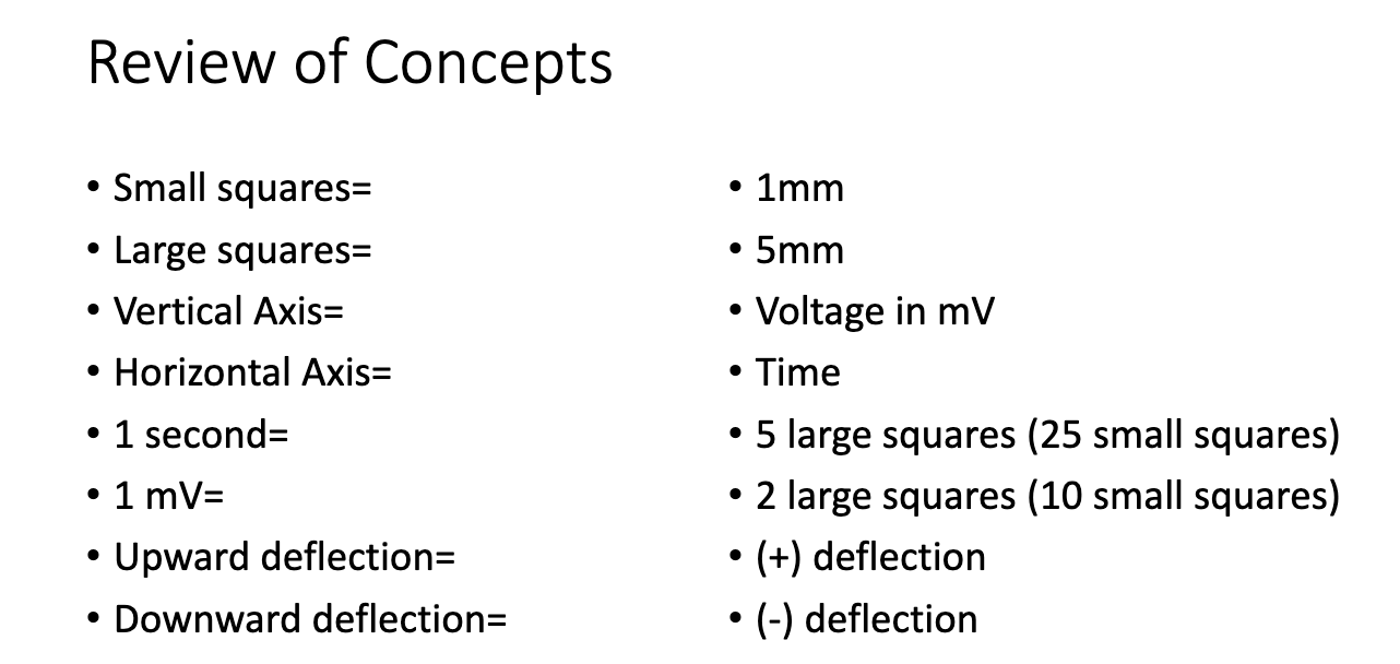
• Small squares=
• Large squares=
• Vertical Axis=
• Horizontal Axis=
• 1 second=
• 1 mV=
• Upward deflection=
• Downward deflection=
Review of Concepts
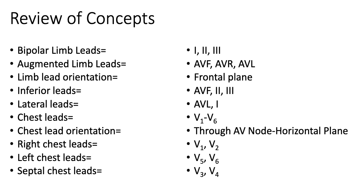
• Bipolar Limb Leads=
• Augmented Limb Leads=
• Limb lead orientation=
• Inferior leads=
• Lateral leads=
• Chest leads=
• Chest lead orientation=
• Right chest leads=
• Left chest leads=
• Septal chest leads=