Clinical Imaging - Exam 1: Get your Magnifying Glasses (Musculoskeletal)
 Call Kai
 Learn
Learn Practice Test
 Spaced Repetition
 Match
Match Flashcards
 Knowt Play
1/147

There's no tags or description
Looks like no tags are added yet.
Name | Mastery | Learn | Test | Matching | Spaced | Call with Kai | Chat |
|---|
No analytics yet
Send a link to your students to track their progress
148 Terms
What two etiologies make a bone lesion aggressive?
Neoplasia and infection
What are the three characteristics of an aggressive lesion?
Cortical destruction, irregular periosteal reaction, and wide transition zone
How many aggressive characteristics need to be present to classify a lesion as aggressive?
One
What are the five types of periosteal reactions in order of aggression?
Smooth
Lamellar
Columnar
Spiculated
Amorphous
(Some Lads Can't Spell August)

What type of periosteal reaction is this?
Spiculated (spine like)

What type of periosteal reaction is this?
Lamellar (layered, onion like)

What type of periosteal reaction is this?
Smooth

What type of periosteal reaction is this?
Amorphous (no form)

What type of periosteal reaction is this?
Columnar (columns)
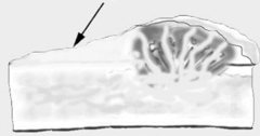
What is the smooth periosteal reaction at the edge of an aggressive lesion?
Codman's triangle
What are the three types of bone loss in order of aggression?
Geographic
Moth eaten
Permeative

What type of bone loss is this?
Permeative (pin points)

What type of bone loss is this?
Moth eaten

What type of bone loss is this?
Geographic
Primary bone tumors tend to be in what parts of the legs?
Away from the elbow and toward the knee


You see several bones with "punched out" lesions. What do you suspect?
Multiple myeloma
What are the five radiographic signs of DJD?
Osteophytes
Enthesiophytes
Sclerosis
Swelling
Narrowing of the joint

Enthesiopathy of the attachment of vertebral discs to vertebral body is called what?
Spondylosis Deformans
Narrowing the joint space can only be evaluated when?
During weight bearing
What sign is an indication of cranial cruciate ligament instability?
Cranial drawer sign (tibia moves cranially)

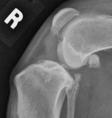
What is your diagnosis?
DJD

Failure of cartilage to turn to bone is called what?
Osteochondrosis (OC)

Detachment of mineralized cartilage is called what?
Osteochondrosis Dessicans (OCD)
What is the most common site of OC(D) in dogs?
Caudal humeral head

What is your diagnosis?
Nothing, it's normal
T/F: OC(D) is often bilateral
True

What is your diagnosis?
OC

What is your diagnosis?
DJD
What is the first radiographic indication of hip dysplasia?
Joint laxity
We want __% or greater coverage of the femoral head by the acetabular rim
50

Hip dysplasia?
No
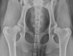
Hip dysplasia?
Yes

Bone proliferation on the acetabular rim due to increased friction from hip dysplasia is called what?
Morgan's line

ID
Osteophytes

Besides laxity and morgan's line, what is another sign of hip dysplasia?
Femoral neck thickening
You want a quantitative review of your patient's hip dysplasia. Who do you send the rads to, how many do you need to send, and what age must your patient be?
PennHIP; 3 views; 16 weeks
You realize that PennHiP is too expensive, so you decide to send it to OFA for qualitative review. How many rads do you need to send, and how old must your patient be?
1 view; 2 years

Osteophytes on the anconeal process is the first radiographic sign of what?
Elbow dysplasia

Diagnosis?
Hip dysplasia

Diagnosis?
None, it's normal
Panosteitis increases the opacity of what part of the bone?
Diaphysis

Diagnosis?
Normal

Diagnosis?
OC
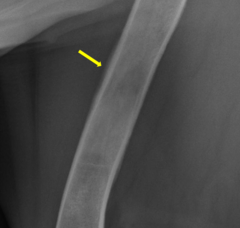
What type of periosteal reaction is this?
Smooth
What part of the bone is affected by hypertrophic osteodystrophy (HOD)?
Metaphysis
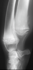
Diagnosis?
HOD

What sign characteristic of HOD is seen here?
Double physis
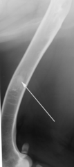
Diagnosis?
Panosteitis

Diagnosis?
Normal

Diagnosis?
HOD

Avascular necrosis of the femoral head is what?
Legg-Calve-Perthes
Unlike panosteitis and HOD, which affect (big/little) dogs, Legg-Calve-Perthes (LCP) and patellar luxation affect (big/little) dogs
Big; little

What is a key sign of LCP?
Widened joint space

Failure of the femoral physes to close, leading to fracture is what?
Physeal dysplasia
What is the poster child for physeal dysplasia?
Obese male cats
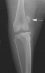
Diagnosis?
Patellar luxation
What view is useful to diagnose physeal fractures?
Frog leg

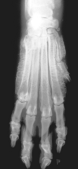
Periosteal proliferation of the limbs secondary to thoracic tumors in dogs is what?
Hypertrophic osteopathy (HO)
T/F: HO will affect all four limbs, including the joints
False, won't include joints

Metastasis of a lung tumor to the feet in a cat is what?
Feline Lung-Digit Syndrome

Diagnosis?
Hypertrophic Osteopathy

Diagnosis?
IVDD

Incomplete rupture of the annulus fibrosis, leading to protrusion into the spinal canal is what type of IVDD?
Hansen type II
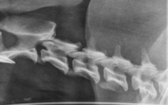
Which space has IVDD?
None, it's normal
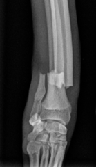
Describe the fracture's orientation
Transverse (90 degree angle)

Describe the fracture's orientation
Long oblique (

Describe the fracture's orientation
Spiral
All fractures are assumed to be what five adjectives unless stated otherwise?
Closed, simple, complete, traumatic, and acute
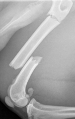
Comminuted or segmental?
Segmental, because the two fractures lines do not communicate

What fracture occurs in young patients due to their bendy bones?
Greenstick fractures

What is the angulation of this fracture?
Caudal (direction Pacman faces)

Cranial or caudal displacement?
Caudal (compare distal to proximal segment)

Incomplete fractures extending from the fracture margins are called what?
Fissures
A type 1 Salter-Harris fracture involves what parts of the bone?
Growth plate (1P)
A type 2 Salter-Harris fracture involves what parts of the bone?
Growth plate and metaphysis (2P)
A type 3 Salter-Harris fracture involves what parts of the bone?
Growth plate and epiphysis (3P)
A type 4 Salter-Harris fracture involves what parts of the bone?
Growth plate, metaphysis, and epiphysis (4P)
A type 5 Salter-Harris fracture involves what parts of the bone?
Comppppression of the growth plate (5P)
What is the most common Salter-Harris fracture?
Type 2
What is the most common site for a Salter-Harris type 5 fracture?
Distal ulnar physis

Diagnosis?
Avulsion fracture (excessive force at tendon)
Rigid fixation of the bone occurs in what type of healing?
Direct (primary)
T/F: Direct healing will not result in an observable callus
True

Diagnosis?
Chip fracture

What are two radiographic features of pedal osteitis?
Decreased bone density and increased vascular channels
Infectious osteitis often develops secondary to what?
Abscess
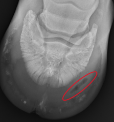
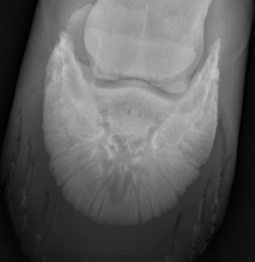
Diagnosis?
Normal
The most visible radiographic feature of infectious osteitis is what?
Focal marginal bone loss


Diagnosis?
Pedal osteitis
What is the main radiographic sign of keratomas?
Focal radiolucent defect at the solar margin

How can you distinguish keratoma from infectious osteitis?
Keratomas generally have smoother lesions

Ossification of the collateral cartilage of the foot is known as what?
Side bones

Diagnosis?
Side bones
What are the six radiographic signs of DJD?
Osteophytes
Enthesiophytes
Swelling
Joint narrowing
Subchondral sclerosis
Subchondral erosions

Which of these is DJD?
The right (osteophytes)
ID the osteophytes


T/F: You have studied the fractures of the coffin joint
Sure . . .

Fracture type?
Type 4

Fracture type?
Type 2

Fracture type?
Type 3