Health and Illness 3: Exam 1
 Call Kai
 Learn
Learn Practice Test
 Spaced Repetition
 Match
Match Flashcards
 Knowt Play
1/45

Earn XP
Description and Tags
September 14
Name | Mastery | Learn | Test | Matching | Spaced | Call with Kai | Chat |
|---|
No analytics yet
Send a link to your students to track their progress
46 Terms
Formula for Cardiac Output
HR (BPM) x Stroke Volume = CO (L/min)
Normal range 5-6 L/min
Diastole vs Systole
Diastole: All chambers of heart relax, blood flows in
Systole: Heart contraction - pushing blood out
Ischemia vs Infarction
Ischemia: Blood supply available but decreased
NSTEMI, unstable angina
Infarction: Death of tissue r/t lack of blood supply (ie M.I)
STEMI
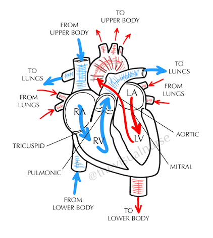
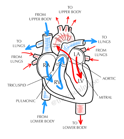
Flow of Blood Through the Heart
Deoxygenated blood enters
Vena Cava
Right Atrium
Right AV valve (tricuspid)
Right Ventricle
Pulmonary valve/arteries
Lungs
Oxygenated blood enters via
Veins
Left Atrium
Left AV valve (mitral/bicuspid)
Left Ventricle
Aortic valve/aorta
Body
Patient Lifestyle Management for Hypertension
Reduce weight by 5% (1 kg = 1 mmHg down)
DASH eating plan
<1500 mg/day of sodium (if substituting salt, watch for potassium if on a potassium sparing diuretic (spironolactone))
No alcohol
Increased exercise (150 min/week)
Stage 1 (130/80): Diet/lifestyle mods (try for 3-6 months before adding med if low risk) + one medication
Stage 2 (140/90): Diet/lifestyle mods + two medications
Medications for Hypertension (1st Line)
Thiazide diuretics (hydrochlorothiazide)
ACE inhibitor (lisinopril)
CCBs (amlodipine)
ARBs (losartan) - can cause renal problems if used alongside ACEs
Hypertensive Crisis
>180/120
Urgency
May require hospitalization
Emergency
Evidence of target organ damage
Hospitalization
S/S
Severe HA
Blurry vision
Dizziness
Epistaxis
Vomiting
Often caused by not taking HTN medications, or abrupt stopping of meds
Hypertensive Emergency Treatment
Vasodilators - Sodium Nitroprusside IV
Nicardipine IV (CCB)
Labetalol (BETA blocker)
Bring down BP by 20-40% in first hour - not less than 140/90
BP monitoring q 5-15 min (ART line?)
Neuro assessment (pupils, LOC) ( can cause hypertensive encephalopathy, intracranial hemorrhage)
Cardiac status assessment (can cause MI, aortic aneurysm, HF)
Measure UOP
High Creatinine, High BUN - RENAL FAILURE
MAP Calculation
SBP + 2 (DBP) / 3 = MAP
What Causes Hypertension
Increased CO (from increased fluid/SV, from increased cardiac activity/HR)
Increased resistance/SVR, PVR (from vasoconstriction, RAAS system activation)
Hypertension Medications that Reduce SV (and in turn reducing CO)
If cause of increased CO is from high fluid/SV in body
Diuretics to get fluid out
Furosemide - strongest, loop, K wasting (watch for digoxin tox!) - not first line
Hydrochlorothiazide - thiazide - watch for hypokalemia
Spironolactone - potassium sparing - blocks aldosterone instead of providing direct diuresis - watch for hyperkalemia
Hypertension Meds that reduce HR (and in turn reduce CO)
If cause of increased CO is from increased cardiac activity (HR)
Decrease HR/cardiac activity
Metoprolol - cardioselective (beta - 1 only- only affects heart)
Propranolol - non- cardioselective (affects heart and lungs beta 1 and 2) - caution with asthma
Diltiazem - CCB - decreases HR and contraction strength - use for HTN, NOT HF! - No grapefruit
Clonidine - CCB - stops SNS system - mostly used to decrease SVR but can also decrease HR - DONT stop abruptly - rebound htn
Hypertension Meds that Decrease Resistance (SVR, PVR) - Inhibitors of RAAS System
If cause of increased resistance is due to RAAS system activation
Lisinopril - ACE inhibitor - stops angiotensin 1-2 conversion which leads to vasoconstriction - Stops RAAS system - Monitor creatinine and BUN for kidney function
Losartan - ARB - Blocks angiotensin receptors preventing vasoconstriction - Stops RAAS system
Hypertension Meds that Decrease Resistance (SVR, PVR) - General Vasodilators
If cause of increased resistance is general vasoconstriction
Amlodipine, Nicardipine - CCBs - IV used for HTN crisis - Prevents Ca, which is needed for muscle contraction from entering into heart cells
Diltiazem - CCB
Hydralazine - Direct vasodilator - causes arterial vasodilation -may be sudden and quick drop
Nitroglycerin - Direct vasodilator- venous dilation in small doses, arterial vasodilation in larger doses
Sodium Nitroprusside - Direct vasodilator - used for HTN crisis
Coronary Artery Disease
Caused (usually) by artherscleorotic plauques that narrow the coronary arteries and reduce blood flow to the heart muscle, leading to ischemia and potential heart attacks.
Angiogenesis may occur - new BV pathways to heart if arteries are blocked - compensatory blood flow occurs - may not be adequate in events such as strenuous exercise which causes angina
Diagnostics for CAD
ECG
Coronary CT angiogram/Cardiac Cath - Gold standard for dx
Asses for contrast dye/iodine allergy and for kidney function (BUN, Creatinine) (taking metformin? Hold for 48h)
Keep extremity (usually femoral, radial or brachial) where catheter was inserted straight and flat after
Keep pressure dressing on for 24h
Brace insertion site when coughing, monitor site for bleeding
Troponin
CRP/ESR (general inflammation)
Echocardiogram - Have pt lay on L side
Increased LDL lab
Decreased HDL lab
Cardiac Stress Test - Goal to reach 85% of predicted HR for age - If pt has angina, the test is stopped and dx of CAD is made - if ECG shows ST elevation, depression or arrythmias test is also stopped
Chronic Stable Angina vs Unstable Angina
Chronic Stable Angina:
Decreased blood flow to heart from CAD
Often occurs at predictable times (ie during exercise)
Position pt upright, encourage rest
Apply O2
Nitroglycerin (can pre-treat)
As plaque increases and blood flow decreases over time, unstable angina may occur
Unstable Angina:
Partial rupture of artery - no permanent damage - a type of acute coronary syndrome
Often new onset
Less predictable
Occurs at rest
More prolonged and severe >10 min
Cardiac markers not elevated - distinguishes from NSTEMI and STEMI
ST segment depression, T wave inversion
STEMI vs NSTEMI
STEMI:
Classic heart attack - extensive muscle damage
Occur abruptly from stopped blood flow - usually from disrupted arthroscelertoic plauqe that becomes a thrombus and occludes blood flow
Cardiac markers are elevated
ST segment elevation. - appears as tombstones - T wave inversion
NSTEMI:
Intermediate form of ACS - less extensive heart damage
Cardiac markers are elevated
ST segment depression, T wave inversion (also seen in unstable angina)

Proper Nitroglycerin Storage
Keep away from light, heat
Once opened, replace in 6 mo
If long acting (ointment) ensure 10-14 hours nitrate free periods
Keep in dark bottle
Cardiac Markers
CKMB - enzyme specific to heart muscle - seen within 4 hours
Troponin - seen within 4 hours, eleated for days after
Myoglobin - Heme protein in cardiac and skeletal muscle seen within 1h - most immediate cardiac marker
BNP - from stretching of heart muscle, eval of HF severity and risk of death
>400 = HF
S/S of Acute Coronary Syndrome (NSTEMI, STEMI)
S/S
Severe chest pain not relieved by nitro or rest
Feeling of doom
Positive levine’s sign (clutching chest)
Rapid respirations
Dyspnea
Increased HR and BP at first, then decreased (uh oh)
Crackles
Abnormal heart sounds (S3, S4)
JVD
Liver enlargement
Peripheral edema
Decreased UOP
Pale, cool, clammy
N/V r/t blood shunting away from GI tract
Treatment for ACS
THROMBINS
Thienopyridines (clopidogrel)
Heparin/enoxaparin
RAAS system blockers (ACE inhibitors)
Oxygen
Morphine (pain and vasodilation)
Beta Blocker (decrease O2 demand of heart)
Intervention (PCI, CABG)
Nitroglycerin (SL) if B/P is okay
Stain/Salicylate (high dose)
Time Frames for STEMI and NSTEMI Treatment
STEMI:
PCI within <90 min
If no PCI available, thrombolytics in <30 min (TPA)
Thrombolytics cause bleeding EVERYWHERE - Contraindicated in hx of bleeding disorders, ischemic stroke, uncontrolled HTN, ect.
- Re-occlusion is a big concern - Heparin drip!
If PCI does not work - CABG
NSTEMI:
If manifestations are controlled, schedule heart cath
NO thrombolytics
Percutaneous Coronary Intervention (PCI)
Catheter threaded into artery - blockage visualized with contrast - balloon catheter into narrowed vessel and inflates to open the artery - stent placement
If radial artery used - no lifting >5 lbs for 48 hours
No submersion of cath site
If bleeding occurs, pressure for 10 min
Report new or worsening bruising and pain
Similar management to coronary angiogram insertion site
Pre-Procedure
Labs/ECG
Allergies (contrast?)
Prior meds (metformin??)
Kidney function
IV access - conscious sedation (fentanyl and midazolam) (heparin and direct thrombin inhibitors)
NPO
Clean skin/shave
Post-Procedure
EKG- watch for reperfusion arrhythmias! Very common
UOP, fluid volume status/labs
Monitor for bleeding at puncture sites q15 min for first hour
IV fluids
IS, OOB, ambulation ASAP
Dual antiplatelet therapy (clopidogrel, ticagrelor) for at least 1 year s/p procedure
ASA for life (prevents stent clotting)
Assess neurovascular status of involved extremity q15 min for 1st hour
CABG
Usage of transplanted mammary or radial artery, saphenous vein in leg to bypass blockage
Takes 3-6 hours
Requires median sternotomy and bypass to circulate blood while heart is stopped during surgery
NPO after midnight
Anticoagulant discontinuation
Post-Op Care for CABG
Continuous ECG - reperfusion arrhythmias (A-fib most common)
Arterial Blood Pressure via Arterial Line
Pulmonary Artery Pressure (PAP) and cardiac output via swan-ganz catheter
Central venous pressure (CVP) aka right atrial pressure RAP via central venous catheter
Mechanical ventilator weaning
Chest tubes, pacing wires for emergency
UOP monitoring
Diuretics/titration of IV meds (vasopressors and vasodilators)
Ambulation, IS, TCDB with splinting incision
Aspirin forever
Clopidogrel for 1 year
Beta Blockers to decrease O2 demand and prevent reperfusion arrhythmias
Statins to lower cholesterol, long term maintenance
Potential Complications:
Dysrhythmias
Bleeding
Cardiac tamponade (if chest tube clogged) Muffled heart sounds, JVD, hypotension (dx with echocardiogram)
Hypothermia r/t bypass
Stroke
Cardiogenic shock
Infection (sepsis)
Heart failure
Renal problems
Repeat MI :(
Medications for CAD
Decrease cholesterol
Statins - atorvastatin (lipitor) - liver damage/rhabdomyolysis (rare)
Niacin/vitamin B3 - flushing, purritis
Gemfibrozil - GI issues (cholesterol is part of GI tract, so decreasing it can cause GI issues, applicable for all meds)
Keep blockages from forming
Aspirin
Plavix (clopidogrel) - antiplatelet - longer half life
Warfarin - narrow therapeutic range
Preload
Degree of stretching of the ventricular muscle at the end of diastole (volume of blood is highest in the ventricle at end of diastole - ie stretching of the muscle is the greatest)
Can be estimated by measuring pulmonary capillary wedge pressure (PCW)
Preload affects stroke volume (SV) and ultimately CO
Afterload
Resistance/pressure the ventricle must overcome to eject the blood into circulation during ventricular contraction
Smaller BVs increase afterload as seen in hypertension
Directly affects arterial BP
Also affects SV and CO
Contractility
Force the myocardium generates during contraction
Affected by SNS
Positive inotropic meds increase, and negative inotropic meds decrease
Also affects SV and CO
Increased contractility = INCREASED O2 DEMAND
Heart Failure Pathophysiology and Symptoms
Insufficient blood supply/oxygen to tissues and organs
Can be associated with
HTN
CAD + MI
Valve defects
Heart rhythm disorders
DM
Kidney disease
Stimulant drugs
S/S:
SOB (how many pillows do you sleep on?)
Edema (specifically peripheral)
Fatigue
Chest congestion
Left Sided HF
HF where the left ventricle cannot effectively pump blood to the body, leading to fluid buildup in the lungs.
LEFT is for LUNGS
Crackles
Nocturnal dyspnea
Cough w/ frothy, pink sputum (pulmonary congestion)
Tachypnea
Cyanosis
Displaced apical pulse r/t hypertrophy (LV becomes dilated and hypertrophied over time - remodeling causes dysrhythmias)
S3 heart sound (gallop)
Can have reduced or preserved ejection fraction (% of blood in LV pumped out with each contraction)
Normal EF = 55-65%
Right Sided HF
Right ventricle not pumping into lungs effectively - fluid backs up into the body
Most common cause is LHF, second most common cause is cor pulmonale (r/t COPD, pulmonary fibrosis)
JVD
Edema (dependant peripheral)
Ascites
Weight gain
Polyuria
Nursing Management for HF
Oxygen therapy + high fowler’s position
Structured exercise program (associated with better outcomes)
Renal monitoring (EKG, UOP)
Avoid aspirin, NSAIDs - may cause increased sodium retention which can worsen HF
Low sodium diet
DASH diet
Fluid restriction (maybe) <2L/day
Daily weights at same time, same clothes (report wt gain of 3lb in 2 days or 3-5lb in 1 week)
Cardiogenic Shock
Heart cannot pump enough blood to vital organs - catastrophically low CO
Mainly caused by
MI
Severe HF
S/S
Pulmonary congestion
Pallor, cool, clammy skin (r/t blood shunting)
Decreased UOP r/t decreased renal perfusion
Dyspnea
Tachycardia
Hypotension
Treatment:
Vasopressors
Positive Inotropes (ie digoxin) to increase CO
Diuretics low and slow, after BP is OK
Nitrates if BP is OK
Medications for HF
Reduce afterload (lowers heart workload)
Via vasodilation
ACEs/ARBs - Stop RAAS system - Monitor creatinine and BUN for kidney function
Nitrates
Hydralazine
Valsartan
CCBs (not diltiazem)
Reduce preload (lowers heart workload)
Diuretics
Inhibit SNS
BETA blockers
Improve Contractility (Makes heart beat BETTER, not faster) (+ Inotropes)
Dobutamine (increases contraction strength and vasodilates)
Milrinone (increases contraction strength and vasodilates)
Digoxin (watch potassium lab!) (if on furosemide or HCTZ especially) (low K = digoxin toxicity)
Norepinephrine and epinephrine - weaker but used to vasoconstrict during shock
Cellulitis
Bacterial infection (usually of subcutaneous tissue) r/t a break in the skin (wounds, radiation)
Usually staphylococci or streptococci
S/S:
Heat
Redness
Tenderness
Fever, chills, malaise
Edema
Can lead to systemic/septic infection if not treated (blood, joints, bone, heart, thrombophlebitis, necrotizing fasciitis, death)
Dx:
Ultrasound to rule out DVT
CT if deep infection suspected
May be chronic or acute, mark borders with sharpie - spreading quickly can be indicative of necrotizing fasciitis
Tx:
Moist heat
Elevation, immobilization
Antibiotics (PO) - amoxicillin, cephalexin, clindamycin
IV Abx: Penicillin-G, Nafcillin, cefazolin, ceftriaxone
Severe IV Abx Tx: Vancomycin + piperacillin/tazobactam
May need surgical irrigation and debridement
Urinary Tract Infection (UTI)
Can be lower (bladder, urethra), upper (renal and pelvic), or cause urosepsis (systemic and life threatening)
Uncomplicated
Complicated - occurs with underlying disease (obstruction, stones, catheters, DM, abx resistance, pregnancy)
S/S
Dysuria
Frequency
Spasms, abd pain
Flank pain, chills, fever (upper UTI)
Confusion and cognitive impairment in older adults
Non- localized abd pain in older adults
Prevention for CAUTI:
Position foley bag below bladder with no kinks or “dependant loops”
Empty if over 400 mL
Dx:
Dipstick UA (+ Leukocytes)
Urine C/S
CBC w/ Diff
Ultrasound, CT urogram, pyelogram
Tx:
Abx r/t C/S
Nitrofurantoin, fosfomycin (shorter course, uncomplicated)
Longer tx for complicated (7-14 days)
Phenazopyridine (AZO)
Increased fluid intake
Pathophysiology of Shock
Massive vasodilation in the presence of endotoxins and other mediators
Inflammatory cytokines are released in the presence of sepsis
Cytokines damage internal layers of blood vessels
Clotting mechanisms are triggered
Other proinflammatory mediators are released such as histamines, serotonin, and bradykinin
This negative feedback loop initially increases CO
Increase in CO causes blood flow to bypass capillaries, which decreases perfusion to organs
CO decreases, hypotension occurs
Stages of Shock
Initial Stage
Homeostasis is altered (at a cellular level) Bodies cells change from aerobic to anaerobic metabolism r/t less avaliable O2
SIRS response is activated, but not yet affecting VS
Compensatory Stage
SNS is activated - body tries to re-perfuse vital organs - tachycardia, tachypnea, RAAS activation, oliguria, dim bowel sounds, cool skin (r/t blood shunting)
Progressive Stage
Compensatory actions begin to fail. CO is low and decreasing. Ischemia, lactic acid release (causes acid base imbalances like metabolic acidosis) hypotension, dysrhythmias, liver dysfunction, renal dysfunction, anuria, absent bowel sounds, low GCS
Refractory Stage
Irreversible cellular damage from hypoperfusion and lack of oxygen - multiple organs fail - ARDS, DIC, MODS (failure of 2 or more organ systems)
Systemic Inflammatory Response Syndrome (SIRS)
A systemic immune response to a harmful stressor (injury or infection)
NOT Sepsis (but infectious SIRS is sepsis)
Source of infection + SIRS = Sepsis
DON’T give abx unless there is source of infection
Criteria: Two or more
Heart rate >90
Temp >100.4
RR >20 or PaCO2 <32
WBC >12,000 or <4,000
Sepsis vs Septic Shock vs MODS
Sepsis: Systemic infection and response - life threatening
Septic shock: Presence of sepsis with hypotension despite fluid resuscitation (Type of distributive shock)
MODS: Multiple organ dysfunction syndrome - failure of 2 or more organ systems
Manifistations of Sepsis
Labs:
Increased CRP
Increased procalcitonin
Increases coagulation parameters
Increased creatinine
Thrombocytopenia (low platelets)
High lactate level (measures anaerobic metabolism) (>2)
S/S:
AMS
Hyperglycemia r/t stress induced hypermetabolism
SBP <100
Significant edema
Decreased UOP despite positive fluid balance (r/t fluid not being where it’s supposed to be)
Ileus (r/t decreased GI motility)
Mottling
ABG may be alkaline at first, acidotic in end (metabolic acidosis)
Sepsis 1-Hour Bundle
Measure lactate levels (repeat if >2)
Draw blood cultures (2 sets peripherally from different sites) (drawn before antibiotics)
Administer antibiotics
Fluid resuscitation (isotonic crystalloids LR or NS) (30mL/kg)
Vasopressor administration if fluids fail to restore a MAP >65 (norepinephrine, epinephrine, vasopressin)
Nursing Management for Septic Shock
Maintain MAP >65 -vasopress that hoe
High flow oxygen ie 100% non-rebreather face mask
Prepare to intubate, have resuscitation equipment ready
Insert indwelling catheter for strict I&O measurement
Maintain large bore patent IV access
May place pt in trendelenburg (supine with legs elevated above head)
Fluid resuscitation (isotonic crystalloids LR or NS) (30mL/kg)
Can be risky for fluid overload - settles in lungs causing even more decreased O2 exchange
Antibiotic administration - hard on kidneys!
NGT set to intermittent suction to prevent/manage illus
Complications of Septic Shock
MODS - often begins in lungs causing ARDS and can extend to kidneys and heart and GI tract
DIC - Assess for bleeding around gums, IV sites, MM - Obtain clotting labs - prepare to admin blood