Lab Anatomy U1
 Call Kai
 Learn
Learn Practice Test
 Spaced Repetition
 Match
Match Flashcards
 Knowt Play
1/403

There's no tags or description
Looks like no tags are added yet.
Name | Mastery | Learn | Test | Matching | Spaced | Call with Kai | Chat |
|---|
No analytics yet
Send a link to your students to track their progress
404 Terms
Cephalic
Head region.
Cranial
Skull.
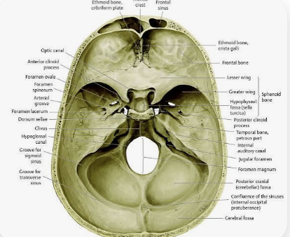
Facial
Entire face.

Frontal
Frontal bone of the skull and the Frontal lobe of the brain

Orbital
Eye/orbit region.
Nasal
Nose region.
Buccal
Cheek. 😊
Oral
Mouth.
Mental
Chin. hmm
Cervical
Neck. *instead of down there in here

Thoracic
Chest. *thor
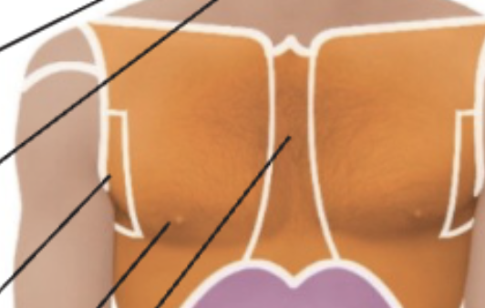
Sternal
Center of front chest

Pectoral
Chest/breastbone-side region.

Axillary
Armpit. *axe
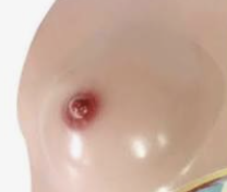
Mammary
Breast. *mama
Abdominal
Abdomen.

Pelvic
Pelvis.

Inguinal
Groin.

Pubic
Genital/pelvic region.

Deltoid
Shoulder muscle
Brachial
Upper arm.

Antecubital
Front of elbow
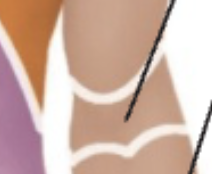
Olecranal/olecranon
Back of elbow; olecranon is the bony point.
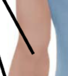
Antebrachial
Forearm.

Carpal
Wrist.

Manus/manual
Hand.
Palmar
Palm.
Digital
Fingers/toes.
Coxal
Hip.

Femoral
Thigh.
Patellar
Front of knee.
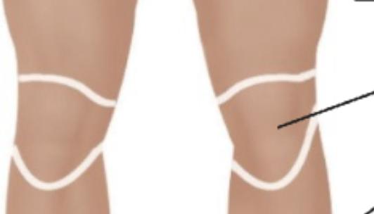
Popliteal
Back of knee.
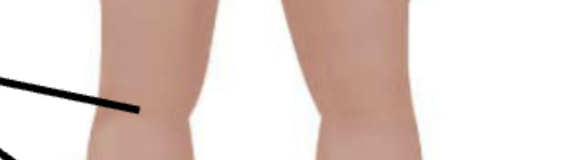
Crural
Front of lower leg.
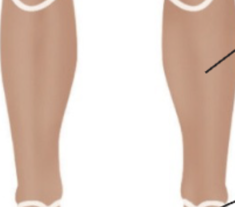
Sural
Calf/back of lower leg.

Pedal/pes
Whole Foot.
Tarsal
Ankle.
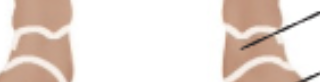
Calcaneal
Heel.

Dorsum?
Back/top surface of hand or foot.
Otic
Ear.
Occipital
Back/base of skull.
Scapular
Shoulder-blade region.

Vertebral
Spinal-column region.

Lumbar
Lower back.

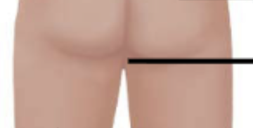
Gluteal
Buttocks.
Perineal/perineum
Area between external genitalia and anus.
Superior/Inferior
Ubove/Below
Cephalad(Cranial)/caud
Cephalad (cranial) = toward the head/
caudal = toward the tail or lower part of the body
Anterior/Posterior
Anterior=the front of the body
Posterior =the back of the body
Dorsal/ventral
Dorsal=the back or posterior portion of the body ventral= the front or anterior
Superficial/deep
Superficial= closer to the surface of the body
deep=farther away from the surface toward the inside
Proximal/distal
Proximal=Closer to the trunk of the body or the point of attachment of a limb.
Distal=Farther away from the trunk of the body or the point of attachment of a limb
Medial/lateral
Medial: Toward or closer to the midline of the body.
Lateral: Away from the midline or toward the side of the body.
intermediate
lies between a medial structure (closer to the body's midline) and a lateral structure (farther from the midline)
Supine=lying flat on your back facing up
Prone=lying flat on your stomach facing down *prone to lying down
Sagittal (Median)
Vertical plane dividing right and left portions. Side
(Midsagittal perf in the middle)
(Parasagittal not perf)
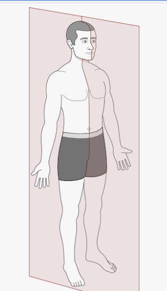
Frontal (Coronal) Plane
Divides the body into front and back half.
Transverse
Horizontal plane dividing superior and inferior portions.
Dorsal Cavity
Posterior body cavity containing cranial and vertebral cavities.

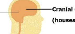
Cranial Cavity
Cavity inside the skull containing the brain.
IN DORSAL.
Vertebral/Spinal Cavity
Cavity within the vertebral column containing the spinal cord.
IN DORSAL.

Ventral Cavity
Anterior Body cavity containing thoracic and abdominopelvic cavities.
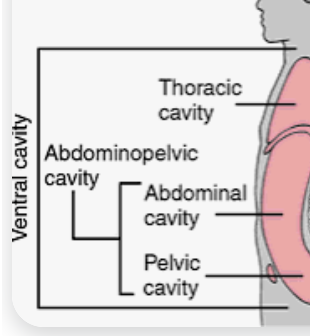
Thoracic Cavity
Chest cavity containing the lungs and heart.
IN VENTRAL
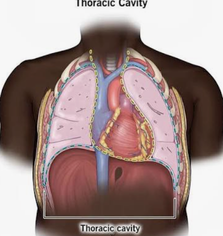
Pleural Cavity
Fluid filled space surrounding each lung.
Mediastinum
Central compartment of the thoracic region between the 2 lungs.
Pericardial Cavity
Fluid filled space surrounding the heart.
Abdominopelvic Cavity
Containing abdominal and pelvic cavities.
Abdominal Cavity
Upper abdominopelvic cavity containing the digestive organs, kidneys, and ureters
Pelvic Cavity
Lower abdominopelvic cavity Contains the urinary bladder, internal reproductive organs, and the rectum.
1 Right upper quadrant
Upper-right abdominal quadrant (patients perspective)
2 Left upper quadrant
Upper-left abdominal
3 Right lower quadrant
Lower-right abdominal quadrant
4 Left lower quadrant
Lower-left abdominal quadrant
Right hypochondriac
Upper-right abdominopelvic region. (patient perspective)

Epigastric
Upper-middle abdominopelvic region.

Left hypochondriac
Upper-left abdominopelvic region.

Right lumbar
Middle-right abdominopelvic region.

Umbilical
Middle region around the navel.

Left lumbar
Middle-left abdominopelvic region.
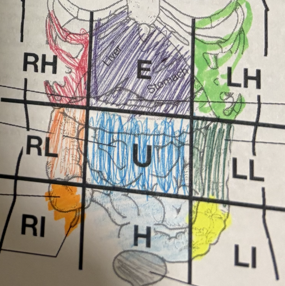
Right iliac (inguinal)
Lower-right abdominopelvic region.

Hypogastric (pubic)
Lower-middle abdominopelvic region.

Left iliac (inguinal)
Lower-left abdominopelvic region.
Total magnification calculation
Eyepiece magnification × objective magnification
If eyepiece is 10x and you are using a 4x objective, the total magnification is 40x.
If eyepiece is 10x and you are using a 10x objective, the total magnification is 100x.
If eyepiece is 10x and you are using a 40x objective, the total magnification is 400x.
Resolution
Ability of a microscope to distinguish two close objects as separate.
Parfocal
Object in center stays focused when changing objective lenses.
Diameter of field
Width of the circular area visible through the microscope. measure how wide the cell is
Depth of field
Thickness of the specimen that remains in focus. how deep you can see into the cell before it goes blurry
Scanning power lens
Lowest-power objective used to locate the specimen.
Low power lens
the objective lens on a microscope that provides low magnification
High power lens
microscope lens that magnifies an object 40 times
Oil immersion lens
high-power objective used with immersion oil to improve resolution provides a 100x magnification
Diameter of Field Calculation
Use D1 × M1 = D2 × M2 when comparing field diameter and magnification. See the professor's drawing/model/slide for this structure.
Cell
Basic structural and functional unit of life. Use a labeled cell diagram/microscope image and locate the structure.
Nucleus
Membrane-bound organelle containing most cellular DNA. Use a labeled cell diagram/microscope image and locate the structure.
Nuclear pore
Opening that regulates movement into/out of the nucleus. See the professor's drawing/model/slide for this structure.
Nucleoli
Nuclear regions where ribosomal subunits are assembled. See the professor's drawing/model/slide for this structure.
Chromatin
DNA plus proteins in its less-condensed form. Use a labeled cell diagram/microscope image and locate the structure.
Chromosome
Highly condensed DNA-protein structure visible during cell division. Use a labeled cell diagram/microscope image and locate the structure.
Centrosome
Microtubule-organizing center that helps form the mitotic spindle. Use a labeled cell diagram/microscope image and locate the structure.
Ribosome
Site of protein synthesis. Use the required bone model/specimen and identify the landmark by its shape and position.
Endoplasmic reticulum (smooth and rough)
Membrane network; rough ER is associated with protein synthesis, while smooth ER makes lipids/steroids, helps detoxify, and stores calcium. Use a labeled cell diagram/microscope image and locate the structure.