PSYC 110: Visual & Hearing System; Motor Control
1/67
There's no tags or description
Looks like no tags are added yet.
Name | Mastery | Learn | Test | Matching | Spaced |
|---|
No study sessions yet.
68 Terms
Sensation
Process of detecting sensory stimuli through sensory organs.
Perception
Conscious experience and interpretation of sensory stimuli.
Transduction
Process of converting sensory stimuli into coded neural signals.
Specialized for all our senses and is the basis for all of the sensory experiences.
Light Wavelength
The distance between 2 peaks of light waves, decoded by our visual system as color/shades of gray.
Light Amplitude
The height of the waves, decoded as brightness.
Tall amplitude = Brighter.
Short Amplitude = Dimmer.
Pupil
The dark opening that allows light into our eyes.
Iris
The colored region of our eyes; muscles that allow the eye to contract.
Cornea
The glassy surface that protects the eye.
Optic Nerves
Axons that exit the eye and connect it to the brain.
Retina
A layer in the back of the eye that is part of the brain.
Specialized to detect differences in light intensities.
Image processing starts here: left-right reversed visual field.
3 Layers of Retina Cells
Photoreceptors, bipolar cells, retinal ganglion cells.
Photoreceptors
The “darkness detectors” responsible for visual sensory transduction.
Contains rods & cones.
Rods
Highly sensitive to light: Responsible for vision in dim light and unresponsive in bright light.
Achromatic; low acuity.
Only exists outside the fovea.
Cones
Low sensitivity to light: Inoperative in dim light.
Concentrated in the fovea.
Sensitive to color (RGB wavelengths); high acuity.
Bipolar Cells
Interneurons that transfer visual information from the photoreceptors to retinal ganglion cells (RGCs).
Fovea
Small depression in the retina where visual acuity is the highest.
Center of the field of vision is focused here.
Retinal ganglion cells (RGCs)
Cells that fire action potentials through the optic nerve to the brain.
Phototransduction
The process in which light is converted into a change in membrane potential in photoreceptors by photopigments.
Phototransduction: “Dark current”
Detecting darkness: Photoreceptors depolarize (NA+ channels open) and release glutamate.
Detecting light: Photoreceptors hyperpolarize (K+ channels open) and stop glutamate release.
Young-Helmholtz Trichromatic Theory
Theory in which 3 retinal cones absorb light at a specific wavelength (red, blue, green) that combine to form a myriad of colors when stimulated.
Applies to photoreceptors in the retina.
Cones located in the fovea: Color vision is strongest in the middle of the visual field.
Rods located in the periphery: We only see light/dark on the periphery of the visual field.
Opponent-Process Theory
3 sets of opponent retinal processes enable color vision:
Red/Green; Black/White; Yellow/Blue
Some cells are excited by one of the opponent colors and inhibited by the other
Different RGCs (Red/Green; Black/White; Yellow/Blue) are excited/inhibited with one color, and signal another color
On-Center Ganglion Cells (O-P Color Theory)
Responds to a bright center and dark surroundings.
Off-Center Ganglion Cells (O-P Color Theory)
Responds to a dark center or bright surroundings.
Rhodopsin
Photo pigment that absorbs the electromagnetic radiation of light.
This causes a change in the retinal protein (Vitamin A) and
activates the opsin → opsin changes color from purple to yellow.
Rebound Effect (Afterimage)
The adaptation (overcompensation) of firing previously inhibited RGCs.
Color Blindness
A genetic variation that causes the presence of 2 cones, rather than 3.
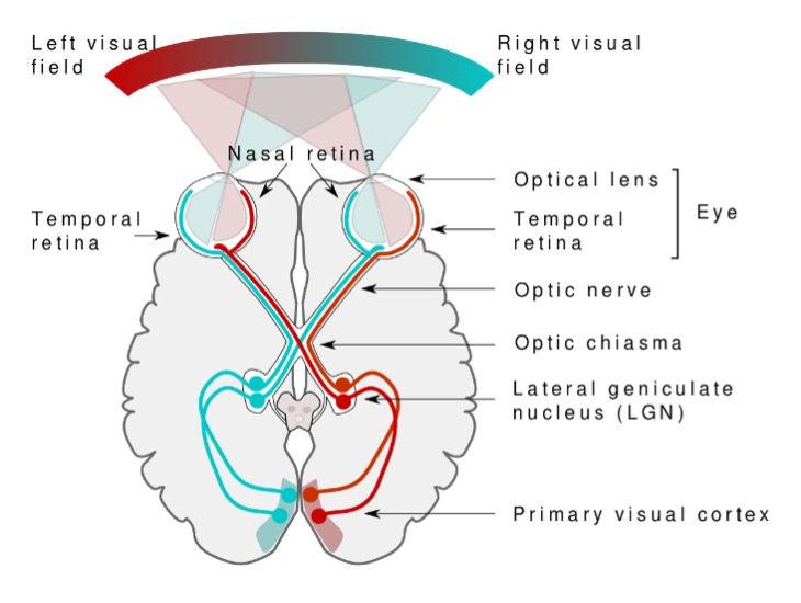
Retinal Ganglion Cells Visual Pathway
Cells from the outer halves of the retina project ipsilateral (to the same side of the brain).
Cells from the inner halves of the retina project contralateral (to the opposite side of the brain).
Lateral Geniculate Nucleus
6 layers of neurons in the thalamus that are responsible for processing different aspects of the visual image.
Lateral Geniculate Nucleus Layers 1-2
Layers of neurons that respond to movement and are achromatic.
Lateral Geniculate Nucleus Layers 3-6
Layers of neurons that are color sensitive and high acuity.
Visual Pathway
Left visual field reaches the right portion of the retina, and vice versa. The lens inverts the image.
Optic nerve axons from the nasal half of the retina cross at the optic chiasm.
Separates the visual information by visual field:
Left visual field information from both eyes is now in the right hemisphere
Most optic tract axons terminate in the lateral geniculate nucleus,
Some terminate in the superior colliculus,
LGN neurons project via optic radiation to the primary visual cortex (V1).
Cortical Modules
Hypothetical constructs with a physiological basis; each module analyzes a different aspect of a visual image.
~300,000 neurons in each module; neurons in a single module all respond to visual input from the same particular location in the visual field.
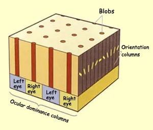
Blobs
The cortical module that analyses the color of an object.
Orientation Columns
The cortical module that analyses the shape/form of an object.
All neurons in an orientation column respond to a line of the same orientation.
Neurons in adjacent orientation columns respond to lines with a 10% difference.
Ocular Dominance Columns
The cortical module that differentiates left eye-right eye.
David Hubel & Torsten Wiesel (1959)
Recorded electrical activity from individual neurons in the brains of cats.
Specific patterns stimulated activity in specific neurons in the visual cortex.
Systematically created a map of the visual cortex.
Received the Nobel Prize in 1981.
Extrastriate Cortex (V2)
Where visual information pathways diverge into a “what” and “where/how” streams.
V2’s “WHAT” Pathway
Dorsal stream: Object recognition, identification, and processing.
Color, texture, pictorial detail, shape, size
Occipital lobe ⇒ Temporal lobe ⇒ Frontal lobe
V2’s “WHERE/HOW” Pathway
Ventral stream: Location in space and how one might interact with a visual stimulus
Location, movement, spatial transformations, relationships
Occipital lobe ⇒ Parietal lobe ⇒ Frontal lobe
Inferior Temporal Cortex
Part of the ventral stream that is involved in recognizing visual patterns and identifying objects.
Successive region following V1 that analyzes more complex characteristics
Cues
Unintentional auditory sounds.
Signals
Intentional auditory sounds.
Speech
Human acoustic communication.
Echolocation
Using sound waves that reflect from solid objects & liquids to locate objects and sound sources.
The delay between the original signal and the reflection helps determine the distance & type of object.
Hertz (Hz)
Cycles of vibrations per second. Humans can hear ~20 to 20,000 Hz.
Infrasound
Under 20 Hz (Elephants can hear/generate infrasonic frequencies).
Ultrasound
Higher frequency sounds (Cats & dogs can hear them).
Frequency (Sound Wavelength)
The distance between 2 peaks of sound waves, decoded by our auditory system as pitch.
Higher frequency = Higher-pitched
Lower frequency = Lower-pitched
Sound Amplitude
The height of the waves, decoded as volume.
Tall amplitude = Louder.
Short amplitude = Quieter.
Outer Ear
Where sound is funneled into the ear; composed of the auricles (pinna) and ear canal.
Auricles (Pinna)
Outer shell of the ear that funnels acoustic waves into the ear canal.
Ear Canal
Amplifies the sound and transmits vibrations to the tympanic membrane (ear drum).
Middle Ear
Moves sound waves in the air into fluid (inner ear); composed of the tympanic membrane (ear drum) and ossicles.
Ossicles
3 of the smallest bones in our body—Malleus (Hammer), Incus (Anvil), Stapes (Stirrup).
Move when subjected to auditory inputs.
Inner Ear
The ear region where signal transduction occurs; consists of the cochlea and auditory nerve.
Waves enter the cochlea through the oval window.
Cochlea
A fluid-filled, spiral shaped structure divided into 3 fluid-filled chambers: Scala Vestibuli, Scala Media, and Scala Tympani.
Contains the Organ of Corti.
Organ of Corti
Part of the cochlea that is made of 3 layers: Basilar membrane, hair cells (cilia), and tectorial membrane.
Basilar Membrane
A long, membrane piece that is thick/narrow at the base (resonates high frequencies) and thin/wide at the apex (resonates low frequencies).
Sound wave vibrations cause it to flex and activate specific hair cells (cilia).
Oval Window
The “window” that sound waves travel through, in between the middle ear and the cochlea.
Place Coding Theory
Location along the cochlear spiral corresponds to the sound frequency.
Tonotopical map of frequency.
Hair Cells (Cilia)
Cells that are attached to certain areas of the basilar membrane.
Tip link: The tip of the hair cell
Signal Transduction
The process of turning sound waves into neural signals.
When the basilar membrane vibrates, mechanical energy moves the cilia.
Movement influences the amount of potassium (K+) and calcium (CA2+) that enter the ion channels.
Influx of ions opens calcium channels on the body of the cilia.
Releases neurotransmitters to auditory nerves.
Auditory Nerve Fibers - Cochlear Nerve
Part of the vestibulocochlear nerve, which is a cranial nerve in the brainstem.
Encodes sound information and transmits it from the cochlea (inner ear) to the cochlear nucleus (brainstem).
Auditory Pathway
The cochlea sends a signal to the auditory nerve.
Information enters the pons first.
Crossing-over event occurs.
Then, sent up to the medial geniculate nucleus (thalamus).
Finally, to the auditory cortex (A1).
Auditory Nerve
A nerve that connects the inner ear to the brain and consists of the cochlear nerve and vestibular nerve.
Auditory Pathway Crossing-Over Event
An event that occurs at the pons, where some left auditory input is sent to the right, and vice versa.
Primary Auditory Cortex (A1)
Located on the superior
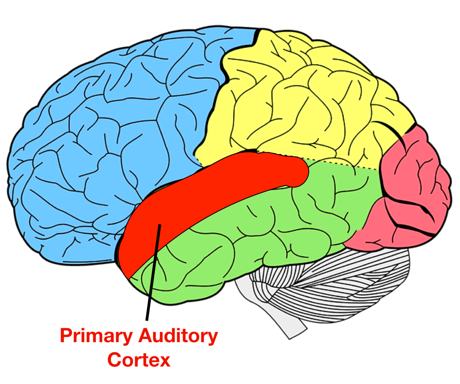
left on slide 31 of hearing