Section 3.1 DNA Damage and Repair
 Call Kai
 Learn
Learn Practice Test
 Spaced Repetition
 Match
Match Flashcards
 Knowt Play
1/16

There's no tags or description
Looks like no tags are added yet.
Name | Mastery | Learn | Test | Matching | Spaced | Call with Kai | Chat |
|---|
No analytics yet
Send a link to your students to track their progress
17 Terms
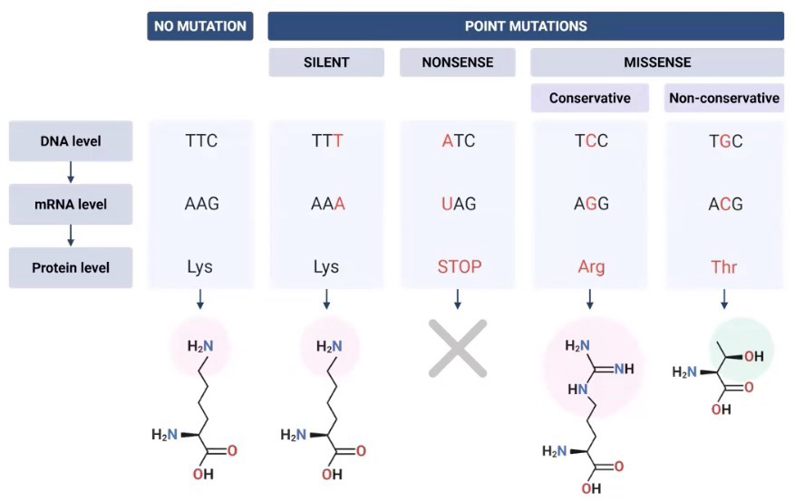
Mutation; Silent, nonsense, and missense
An accidental, permanent change in a DNA sequence; Rarely advantageous
Silent: Mutation only has an effect on the DNA level (due to wobble effect), not on protein
Nonsense: Mutation causes a premature stop in protein production
Missense mutation:
Conservative: Change in AA likely has little effect on protein because it has similar properties to original unmutated AA
Non-conservative: Change in AA likely has large effect on protein because it does not have similar properties to original unmuted AA
Insertion vs Deletion mutations
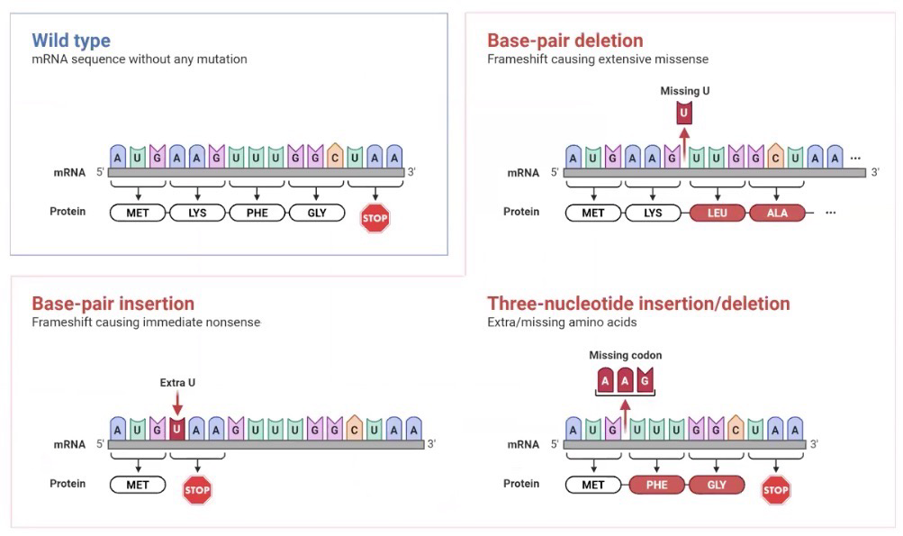
Can cause frameshift mutation: Insertion/deletion that is not a multiple of 3 bases; Often leads to an early stop codon; mutations that affect RNA splicing often generate frameshift
In-frame insertion/deletion: Addition/removal of multiple of three; typically doesn’t negatively affect protein since it’s just one AA
Not always the case though; Ex, In-frame deletion of UUC (phe) codon in CFTR is most common cystic fibrosis mutation
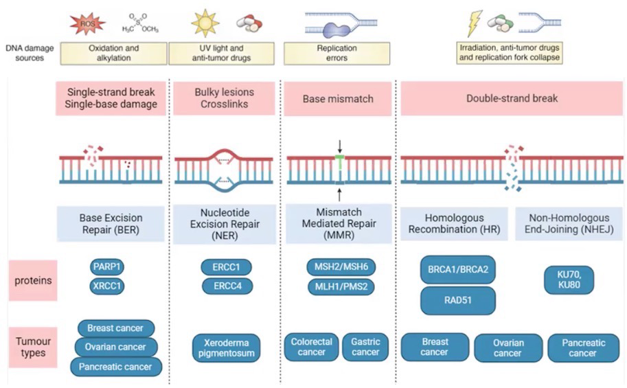
DNA damage (WILL BE ON EXAM/Specifically the types of damages)
NOT the same as mutations; DNA damage can be fixed, not mutations
Abnormal chemical structure of DNA; Can be spontaneous, Often caused by environmental factors; Can result in mutations
Very common; our cells are exposed to 70k DNA damage events per day; less than 1 in 1k become mutations; Single-strand breaks and depurination is the most common type
Cancer is the result of an accumulation of mutations

Ames Test
Mutagens are agents which promote changes in DNA sequences; They are often also classified as carcinogens (Cancer causing agents)
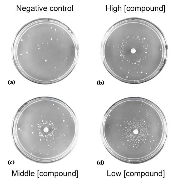
Ames test determines if a compound is a mutagen; Uses Salmonella typhimurium with an inactive enzyme of the H biosynthetic pathway; Paper disks soaked in certain concentrations of the mutagen is placed in the middle of the plate; In H-absent growth media, cell that grow without the AA had a spontaneous mutation; Level of growth indicates strength of mutagen.
Interpreting the results: This test shows positive mutagen; Carcinogen testing requires live animals
Negative control still has growth; this is considered the base-level spontaneous background mutation rate
Low concentration showed large ring of growth because it was an optimal rate of DNA damage which allowed the cell to recover but not too many as to kill the cells
Middle - High concentration showed a ring of inhibition because DNA damage rate was toxic for the cell
Recall: DNA damage can naturally occur spontaneously in the cell, which is one of the reasons why we have a negative control plate
DNA Base Damage: Deamination
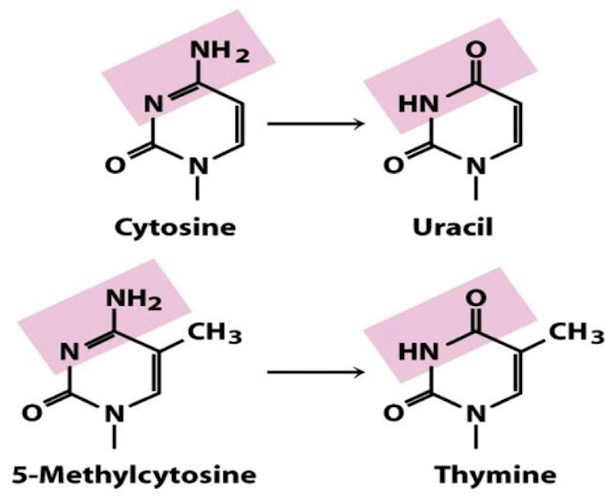
Deamination of C → U and 5-meC → T; Most common, spontaneous
Common chemicals like Sodium Nitrate/ite
Contained in preservatives, tobacco, cosmetic products, gastric juices, even veggies like spinach; Our repair systems are so good it doesn’t affect us
Recall: 5-meC can be found on CpGs (2.2), considered a mutation hotspot
DNA Base Damage: Reactive Oxygen Species (ROS)
Reactive Oxygen Species (ROS) generated as a result of cellular respiration
Most common ROS are hydroxide free radicals (·OH), which inserts into either G or T via oxidation reaction, which can result in DNA strand breaks
Damaged bases can cause Pols to add wrong bases

DNA Base Damage: Depurination
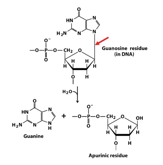
Depurination: Hydrolysis of the glycosidic bond linking a purine base to the sugar-phosphate backbone
Yields an abasic site (Site without base); Also called AP sites (apurinic/apyrimidinic site)
Much more common than depyrimidination
How DNA Damage Turns to Mutation (CpG example)
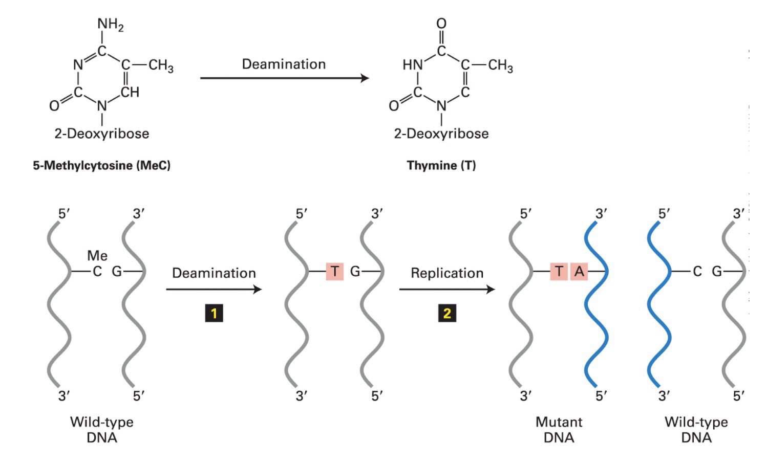
Ex. CpGs are considered mutation hotspots
Deamination DNA damage occurs → Repair system does not fix the damage → Replication occurs, one of the two replicants will contain the mutation
DNA Damage: Bulky Lesions & Crosslinking
Distorts DNA structures
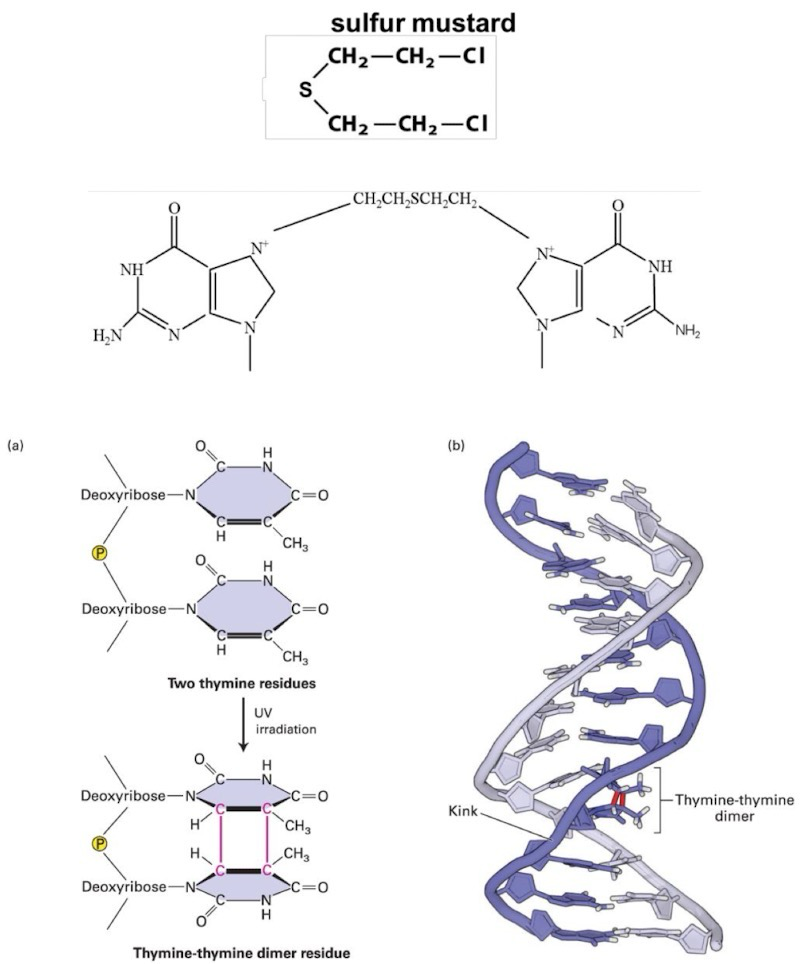
Alkylating agents covalently modify bases in DNA; Ex, sulfur mustard (mustard gas); Alkylation distorts DNA double helix
Covalent linking means the DNA sequence is stuck and can’t be separated for replication/transcription
Spontaneous alkylation by S-adenosylmethionine (SAM) of G → 7-methylguanine (Unstable and can cause depurination)
Thymine dimers: Most common DNA damage caused by ultraviolet radiation; Results in formation of cyclobutane ring between two adjacent pyrimidine rings
Common for pairs of thymines forming a thymine-dimer; Cyclobutane ring kinks the axis of the DNA helix; Confuses Pol, will either pause or add a random base
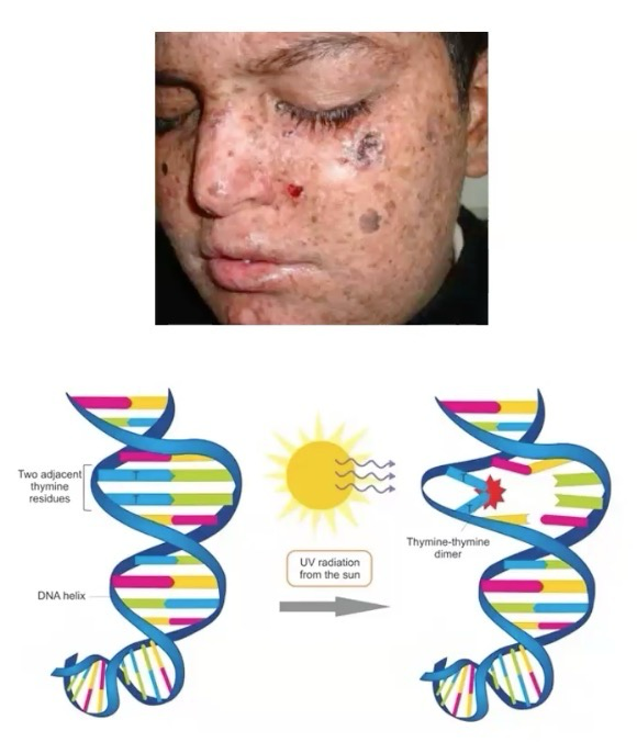
Xeroderma Pigmentosum
Caused by mutation in XPA gene encoding protein for nucleotide excision repair (NER) which removes bulky DNA lesions like thymine dimers (UV rad).
Causes accumulation of structural mutations in DNA helix. Greatly increases risk of skin cancer.
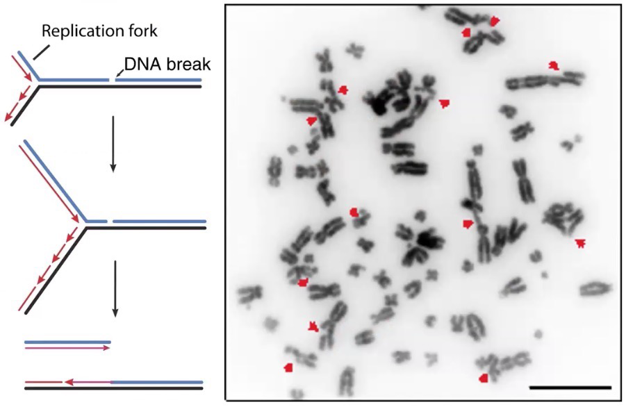
DNA Damage: DNA Strand Break
Ionizing radiation: High-energy radiation that can release electrons from atoms generating ions which can break covalent bonds (Cosmic rays, X-rays, and radioactive materials)
Single-strand break (nick)
Double-strand break (May be staggered breaks)
Also causes damage to bases at break sites, meaning DNA ligase cannot process the breaks back together (All damaged bases must be removed before breaks can be reattached
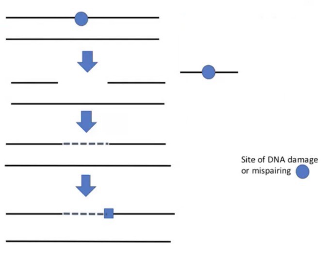
General Pathway of DNA Repair
Most DNA repair mechanisms can be broken down into four distinct phases:
Recognition of the lesion
Excision of the lesion
Resynthesis of the DNA
Ligation of loose ends
Note: We’re taught the prokaryotic version (Aka prokaryotic proteins), but the Eukaryotic version follows the same general pathway (Just more complex when looked at in detail)
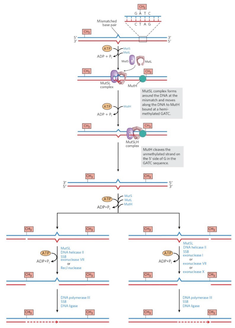
Mismatch Repair: Recognition and Removal of Mismatch
Recognition:
This pathway follows DNA pol during replication to double-check its work
Mismatch Repair (MMR) corrects mismatched base pairs mainly from errors in replication; Genetic defects in eukaryotic MMR are associated with Hereditary Non-Polyposis Colorectal Cancer
Nonfunctioning/mutated NMR pathways are ALWAYS associated with a diseased state
MutL-MutS complex recognizes the mismatch
MutH identifies parental GAmeTC strand and newly synthesized strand; MutH endonuclease nicks the unmethylated strand
Removal:
Nick can be 5’ or 3’ of the mismatch
Exonuclease digests from nick through mismatch
DNA Pol III fills the gap
DNA ligase seals the nick
Base Excision Repair: Removal and Resynthesis
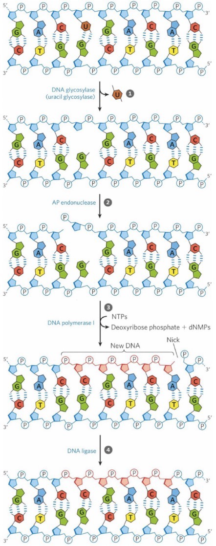
Base excision repair (BER): Repairs mismatches due to damaged bases, depurination, and single-strand breaks in DNA
DNA glycosylase cleaves glycosidic bond making an abasic site (AP site); Or the site is already abasic due to depurination; Eukaryotes have many DNA glycosylases, including uracil-, 8-oxoguanine, and thymine-DNA glycosylases
AP endonuclease initiates repair of abasic sites by making a single strand break in DNA at the abasic site
DNA Pol I has both the 5’ → 3’ exonuclease and the DNA synthesis activities; DNA Pol I is a high-fidelity polymerase; In Eukaryotes DNA Pol β serves this purpose
DNA ligase seals the nick
Nucleotide Excision Repair
Primarily used to fix damage that distorts DNA structure; especially when involving more than one nitrogenous base
Proteins are named after the disease study (UV radiation and XP)
Nucleotide Excision Repair (NER) repairs lesions that distort the DNA double helix, such as thymine dimers or alkylation
Excinuclease: Hydrolyzes two phosphodiester bonds, one on either side of the distortion
Prokaryotes: UvrABC excinuclease
Eukaryotes: XP exonuclease is a large complex that contains XP-A through XP=G subunits plus TFIIH helicase
XP: Xeroderma pigmentosum
Helicase excises the damaged DNA leaving a gap
Prokaryotes: UvrD helicase
Eukaryotes: TFIIH (NER and Transcription is interconnected; Increases accuracy of transcription)
Cockayne syndrome (One of the proteins involved in interconnection is nonfunctional; associated with higher risk of developing mutations due to less surveillance of genome)
DNA Pol I (E. coli) or DNA polymerase ε (humans) fills the gap
DNA ligase seals the nick

Non-Homologous End Joining in Eukaryotes
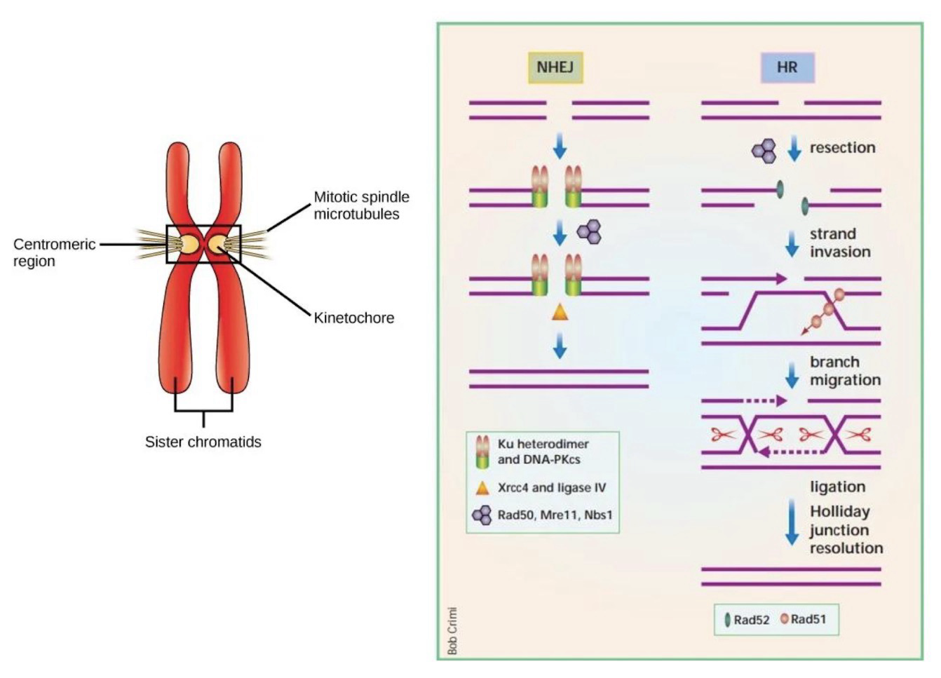
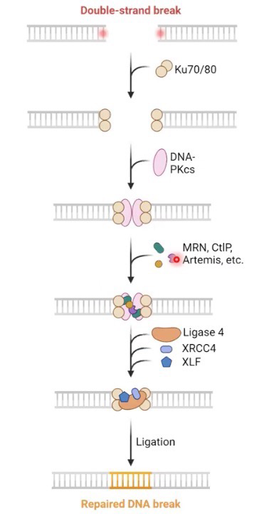
A process used to repair double-strand breaks
Non-Homologous End Joining (NHEJ) is the predominant mechanism in G0 and G1 (1.2, S28) for the repair of double-strand breaks in mammalian cells; Error prone repair
Ku70/80 complex binds loose ends of DNA
PKcs is a kinase that initiates bridging of the broken ends
Other factors bind to and process the loose ends to produce an area of “micro-homology” (sticky ends): Exonuclease will remove bases (including damaged ones); DNA pol µ and λ add bases (Error prone terminal transferases); Artemis endonuclease removes any overhanging flaps
DNA ligase seals the nicks
Since only 1.5% of our genome encode for protein, the cell is willing to risk the error. If unlucky, NHEJ ends up destroying the protein or affecting transcription
Homologous Recombination in Eukaryotes
Homologous Recombination Repair (HRR) can also repair a double-strand break in DNA after DNA has been replicated
HRR only can be used during S and G2 phases because the Template for this reaction is a sister chromatid; BRCA1/2 involved in strand invasion; Mutation in BRCA1/2 is highly correlated to cancer development (Especially Breast and Uterine cancer)
Strand invasion: Detecting, grabbing and bringing broken chromosome to sister chromatid for repair
No genetic information is lost as a result of HRR, so this is an error-free repair mechanism