PSY396 Lecture 9: Brain imaging
 Call Kai
 Learn
Learn Practice Test
 Spaced Repetition
 Match
Match Flashcards
 Knowt Play
1/25

There's no tags or description
Looks like no tags are added yet.
Name | Mastery | Learn | Test | Matching | Spaced | Call with Kai | Chat |
|---|
No analytics yet
Send a link to your students to track their progress
26 Terms
Methods in cognitive neuroscience - Electrophysiological methods
• EEG
• MEG
➢Measure changes in electric/magnetic fields = Direct measurement of neural activity
Methods in cognitive neuroscience - Haemodynamic methods
• PET
• (f)MRI
• (f)TCD (Transcranial Doppler)
• NIRS (Near-infrared spectroscopy)
➢ Measure changes in blood flow/oxygen to meet metabolic needs = Indirect measurement of neural activity
Magnetic Resonance Imaging (MRI)
Consists of
• a very strong magnet that builds up a stable and intense magnetic field
• Three weak(er) gradient magnets that build variable magnetic fields
• Radio frequency coils that transmit radio frequency waves into the body

Types of imaging - Structural imaging
static maps of the physical structure of brain
e.g., computerised tomography (CT) and magnetic resonance imaging (MRI)
Types of imaging - Functional imaging
dynamic maps of the moment-to-moment activity of the brain when engaged in cognitive tasks
e.g., functional MRI, c.f., resting state fMRI for functional connectivity
Advantage of MRI over CT
• Does not use ionizing radiation
• Better spatial resolution (e.g. folds of individual gyri)
• Better discrimination between white matter and gray matter
• Adapted for use in fMRI
Relaxation times and weights - T1-weighted imaging
• fat (white matter) gives a strong signal (bright); cerebrospinal fluid (CSF) gives a weak signal (dark)
• Good for demonstrating anatomy

Relaxation times and weights - T2-weighted imaging
• fat (white matter) gives a weak signal (dark); cerebrospinal fluid (CSF) gives a strong signal (bright)
• Good for demonstrating pathology: - Lesions and white matter alterations are often associated with an increased water content

Basic of MRI Physics
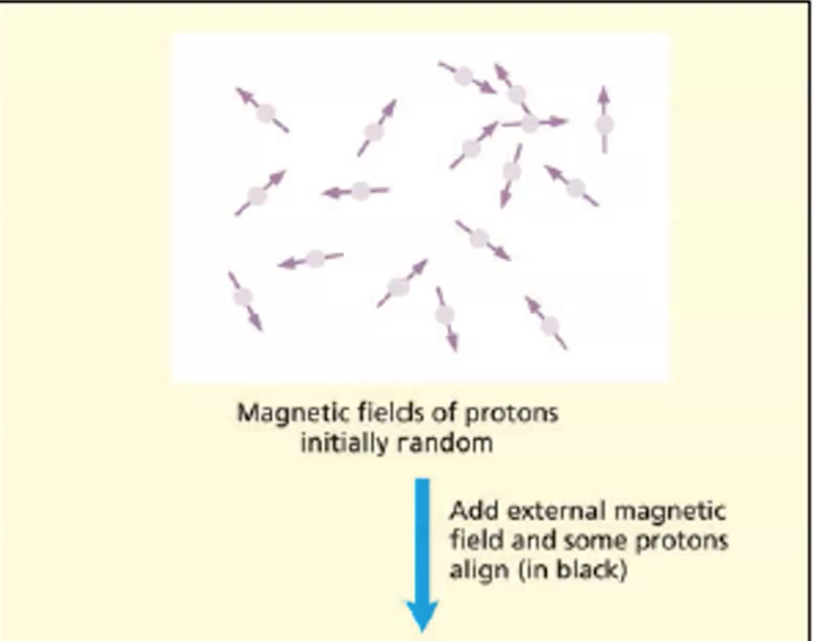
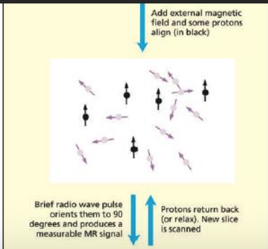
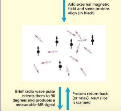
• Protons: subatomic particle, classified as nucleons, which is contained in the atoms in the water in the brain
• Water consists of two atoms of hydrogen

Functional Imaging
• Neural activity consumes oxygen as well as generating electrical signals
• In order to compensate for increased oxygen consumption, more blood is pumped into the active region
• PET measures the blood flow in a region, whereas fMRI measures the blood oxygenation
• The time taken for this response is slow (several seconds) and so functional imaging has a poor temporal resolution, but a good spatial resolution
• This is the complementary profile to ERPs
fMRI in a nutshell
• Brain activity requires oxygen
• Increased brain activity results in increased blood flow to the areas that are active
• More oxygenated blood → less magnetic disturbances
• Protons spins longer period of time
• Signal MRI can detect stay longer
• can detect changes in blood oxygenation levels that correlate with neural activity with high spatial resolution - Temporal resolution is very poor though.
• The measurement of blood flow, blood volume and oxygen use is called the blood-oxygen-leveldependent (BOLD) signal.
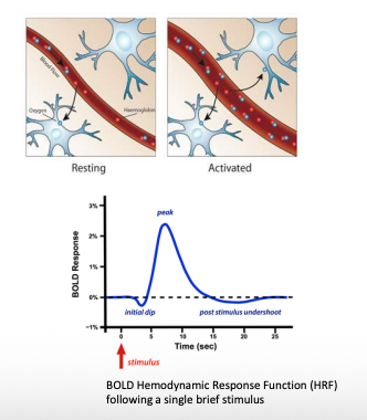
fMRI - Hemodynamic response function (change in BOLD signal over time)

fMRI - When several stimuli are presented close in time the different HRFs summate

How fMRI works
Although the physics of MRI is well understood (leading to at least two Nobel Prizes), there is an explanatory gap between neural activity and what we see in fMRI images
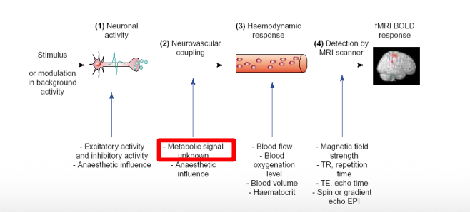
The neurovascular unit (NVU)
How it works is still a mystery

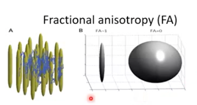
Diffusion weighted imaging and tractography
Diffusion Tensor Imaging (DTI)
Constrained spherical deconvolution (CSD)
Measures white matter organization
based on limited diffusion of water molecules in axons

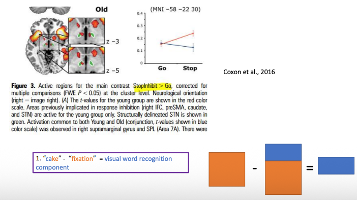
Study - Functional Brain Activation Associated with Inhibitory Control Deficits in Older Adults
1. Only in young, supramarginal gyrus, anterior insula, rIFC, and preSMA showed increased BOLD response successful Stop compared to Go
2. Greater WM structural integrity is associated with greater activation in preSMA
How to run psychological experiments using fMRI
• Head cage (with mirror): stable participant’s head
• Monitor display: stimulus presentation
• Headphones: stimulus, communications with experimenters
• Response pad
• Panic button
Peterson et al. (1988) : cognitive subtraction

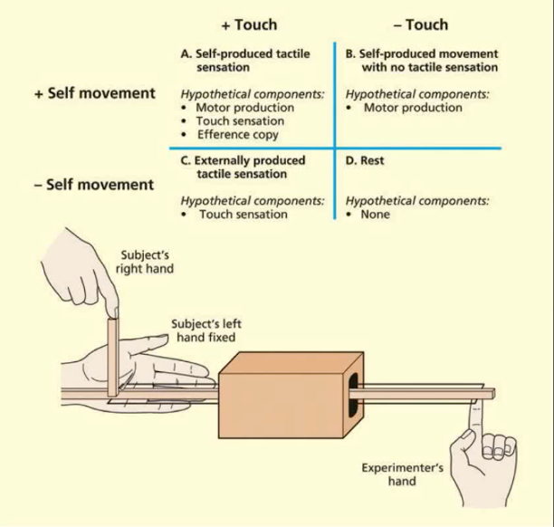
factorial design
• One can look for regions of activation that are shared across several different subtractions rather than relying on a single subtraction.
• A baseline task (or tasks) is still required
A-B = touch sensation (efference copy)
C-D= touch sensation
→ Isolate the regions involved in tactile sensation
A-C = motor production
B-D = motor production
→ Isolate the regions involved in motor production
(A-B)- (C-D) = touch sensation (efference copy)- touch sensation = efference copy
→ Isolate the regions involved in tactile sensation induced by self movement
parametric design
regional physiology will vary monotonically and systematically with the amount of cognitive or sensorimotor processing

Block design
more power, detect small effects because the signal from the hemodynamic response function of each impulse are assumed to sum linearly, the BOLD signal evoked by the block of impulses will be much larger than the signal evoked by a single brief stimulus presentation

Event-related:
enable a much wider range of experimental designs and are more closely related to the typical design structure of most cognitive psychology experiments

Things to consider - BLOCK vs EVENT design
• Predictability
Block design: go go go go stop stop stop stop stop
Even-related design: go go stop go go go stop go go
• Time on task (you want your subject to do what you want them to do)
“3s picture viewing”
fMRI Caution
Bennett, Baird, Miller, and Wolford (2010) reported differences in BOLD signals between socially inclusive images and socially exclusive images in the brain and spinal cord of a dead Atlantic Salmon
Limitations and cautions for MRI studies
• Causation cannot be established
• The exact neurophysiological cannot be revealed, i.e., inhibition or excitation
• Difficult to present stimuli to participant (no metal)
• Virtually all results are for supine participants
• Claustrophobic experience for some participants
• Some side effects, such as vertigo, “pressure around the head”, nausea, metallic taste, light flashes, twitching, and headache
• Some risks for:
Hearing
Health (no pacemakers, artificial joints, cochlear implants—any metal in the body can cause burns)
Life: At least one death from MRI (from an oxygen tank becoming a projectile near the magnet [Chen, 2001, July 31] and at least one non-fatal accident [Colletti, 2004])