Synaptic transmission
1/37
There's no tags or description
Looks like no tags are added yet.
Name | Mastery | Learn | Test | Matching | Spaced |
|---|
No study sessions yet.
38 Terms
What is a synapse?
A synapse is a junction where a neuron communicates with another cell—either another neuron or an effector like a muscle or gland.
Synapse (CNS vs PNS)
CNS: The target cell is typically another neuron.
PNS: The target cell can be a neuron, muscle, or gland.
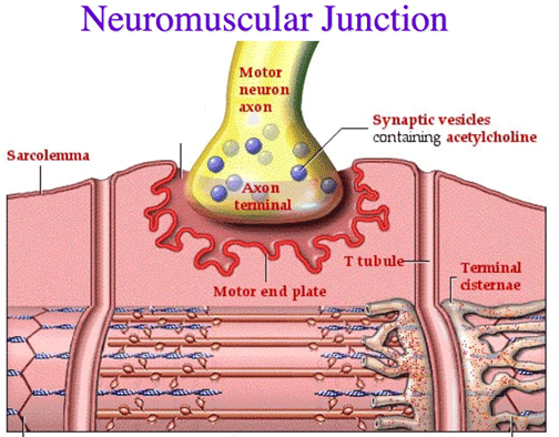
Neuromuscular junction

A specific type of synapse where a motor neuron communicates with a muscle fiber to initiate contraction.
How do most synapses transmit signals?
Most use chemical transmission, where neurotransmitters are released by the presynaptic neuron and affect the membrane potential of the postsynaptic cell.
Neurotransmitter
Chemical messenger released at a synapse that can excite or inhibit the postsynaptic cell depending on the type of receptor it binds to.
What is an electrical synapse and where is it found?
An electrical synapse directly passes current via gap junctions. It is found in:
Some brain neurons
Cardiac muscle
Smooth muscle
Glial cells
Gap junctions
Specialized protein channels made of connexins that allow ions to pass directly between adjacent cells, enabling fast electrical communication.
Gap junctions can be opened or closed by phosphorylation (adding a phosphate group) or dephosphorylation (removing a phosphate group) of connexin proteins.
What are the types of synapses based on signal transmission?
Chemical synapses: Use neurotransmitters to send signals.
Electrical synapses: Use direct electrical currents via gap junctions.
Chemical Synapse
A type of synapse where neurotransmitters are released from the presynaptic neuron’s terminal boutons to influence the postsynaptic cell.
Electrical Synapse
A type of synapse where ions flow directly between adjacent cells through gap junctions, allowing rapid signal transmission.
What molecules help hold presynaptic and postsynaptic cells together at chemical synapses?
Cell adhesion molecules (CAMs).
What are the three types of neuron-to-neuron synapses?
Axodendritic: Axon to dendrite
Axosomatic: Axon to cell body
Axoaxonic: Axon to another axon
Is signal transmission at neuron-neuron synapses usually bidirectional?
No, it is almost always one-way: from the presynaptic neuron's axon to the postsynaptic neuron.
Where are neurotransmitters stored before release?
In small, membrane-bound vesicles inside the presynaptic boutons (axon terminals).
What triggers neurotransmitter release into the synaptic cleft?
The vesicle fuses with the presynaptic membrane, releasing neurotransmitters via exocytosis.
Why can't neurotransmitters cross the membrane on their own?
Because they are hydrophilic and cannot pass through the lipid bilayer without vesicle fusion and exocytosis.
What is a miniature postsynaptic potential?
The small postsynaptic response caused by the release of a single vesicle of neurotransmitter.
How is synaptic strength related to firing rate?
More frequent action potentials cause more vesicles to release neurotransmitters, producing a stronger effect on the postsynaptic neuron.
What happens to vesicle membranes after neurotransmitter release?
They are recycled back into the presynaptic neuron for reuse.
What triggers neurotransmitter release at the axon terminal?
The arrival of an action potential at the axon terminal triggers the opening of voltage-gated calcium channels, allowing Ca²⁺ to enter and initiate neurotransmitter release.
Docked vesicles
Synaptic vesicles that are already positioned at the presynaptic membrane, ready for rapid fusion when an action potential arrives.
Why is neurotransmitter release so fast (under 100 μs)?
Because many synaptic vesicles are already docked near voltage-gated Ca²⁺ channels, allowing immediate fusion and release once calcium enters the terminal.
What role does calcium play in neurotransmitter release?
Ca²⁺ acts as a direct signal that triggers vesicle fusion with the axon terminal membrane, leading to neurotransmitter exocytosis.
Calmodulin
A calcium-binding protein that, when activated by Ca²⁺, stimulates protein kinases that phosphorylate other proteins involved in vesicle release.
What happens after Ca²⁺ binds to calmodulin?
The Ca²⁺-calmodulin complex activates protein kinases, which phosphorylate regulatory proteins, triggering a cascade that supports vesicle exocytosis.
Synaptotagmin-1
A calcium sensor protein on synaptic vesicles that, upon binding Ca²⁺, promotes vesicle fusion by interacting with the SNARE complex.
What are the roles of the C2A and C2B domains in synaptotagmin-1?
These domains bind calcium and insert into the presynaptic membrane, pulling the SNARE complex to drive vesicle fusion and neurotransmitter release.
What type of ion channels are found on the postsynaptic membrane and how are they activated?
hey are chemically regulated ion channels (neurotransmitter receptors) that open when a neurotransmitter binds to them, not in response to voltage changes
Chemically regulated ion channel
A type of ion channel on the postsynaptic membrane that opens in response to neurotransmitter binding, unlike voltage-gated channels.
What is an excitatory postsynaptic potential (EPSP)?
A depolarization caused by positive ions (like Na⁺) entering the cell, making it more likely to reach the threshold for an action potential.
What is an inhibitory postsynaptic potential (IPSP)?
A hyperpolarization caused by negative ions (like Cl⁻) entering or positive ions (like K⁺) leaving the cell, making it less likely to fire an action potential.
Where are neutotransmitter receptors found?
on the dendrites and cell body of a neuron, which allow these parts of the neuron to respond to neurotransmitters released from potentially thousands of different synapses onto the cell’s surface.

Decremental spread
The gradual weakening of a graded potential (like an EPSP) as it spreads from the dendrites/cell body toward the axon hillock.
What happens when neurotransmitters bind to receptors and cause an EPSP?
A depolarization occurs, which spreads toward the axon hillock with decreasing strength (decremental spread).
What must happen for an action potential to be generated at the axon hillock?
The EPSP must be strong enough to reach the threshold and open voltage-gated channels at the axon hillock.
Axon hillock
The part of the neuron where graded potentials are summed, and if threshold is reached, action potentials are initiated.
What kind of channels are found at the axon hillock, and what opens them?
Voltage-gated channels that open when the membrane reaches threshold depolarization.
4 major classes of neurotransmitters