Learn: ANSC 222 Exam 1
 Call Kai
 Learn
Learn Practice Test
 Spaced Repetition
 Match
Match Flashcards
 Knowt Play
1/439

There's no tags or description
Looks like no tags are added yet.
Name | Mastery | Learn | Test | Matching | Spaced | Call with Kai | Chat |
|---|
No analytics yet
Send a link to your students to track their progress
440 Terms
Atomical Position
Feet/hooves on the ground, looking forward
Frontal/Dorsal
divides into dorsal and ventral halves
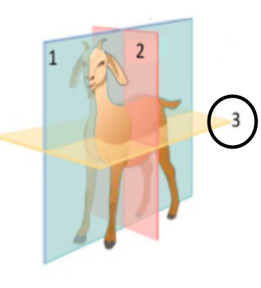
Medial/Sagittal
divides into left and right halves
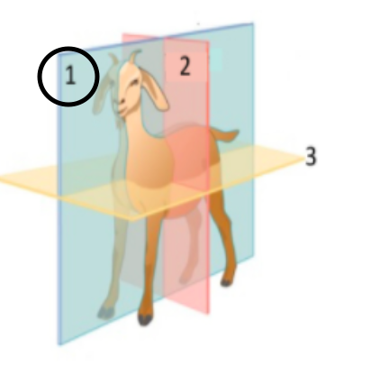
Transverse
divides into cranial and caudal halves

Proximal
closer to the point of attachment
Distal
further from the point of attachment
Cranial/Caudal
directional terms for a point above the knee
Dorsal/Palmer
directional terms for a point below the knee on the front foot
Dorsal/Planter
directional terms for a point below the knee on the back foot
Rostral
toward the nose
Body Cavities
Thoracic, Abdominal, Pelvic
Thoracic
heart and lungs
Abdominal
digestive system
Pelvic
exception and reproductive system
Cells and Collagen Fibers
Bone is composed of:
Calcium and Phosphate
Bones store:
Levers for locomotion
Bones are:
Tubercular (Spongey, Cancellous) and Cortical (Compact)
Bone structures
Tubercular Bone
High volume, Low weight
Trabecular-cavity walls
Cortical Bones
Low volume, High weight
Blood vessels - run in the Haversian canal
Osteoblasts, Osteocytes, Osteoclasts
Types of Bone Cells
Osteoblasts
Bone forming
Osteocytes
Form osteoblasts "trapped" in the matrix
Osteoclasts
Resorb Bone and Recycles Calcium for use Elsewhere
Intramembranous Formation
Bone condensation, skull and facial bones, thickens long bones
Endochonral Ossification
Cartilage model, primary ossification center, lengthens long bones
Growth Plate Fuses
When do animals stop growing
Bone Remodeling Function
Blood mineral homeostasis, repairs cracks, adapts to stress
Bone Remodeling
1. Osteoclasts demineralize and digest bone
2. Osteoblasts recruited
- lay down osteoid
- osteoid in mineralized into bone
3. Osteocytes within new bone
- connected by cell extensions
Bone Repair
1. Blood clots, damaged bone removed by macrophages
2. Proliferation of connective tissue surrounding fracture
3. Bone callus formation
- Both endochondral and intramembraneous bone formation unite broken ends
4. Remodeling produces normal tissue
- gradual return to activity
Skeletal, Cardiac, Smooth
Types of Muscles
Skeletal Muscles
Voluntary, striated, long fibers
Multinucleated - fusion of myoblasts
Exercise induces hypertrophy
Cardiac Muscles
Involuntary, striated
Mono-nucleated
Exercise induces hypertrophy
Intercalated Disks
Does not normally divide
Smooth Muscles
Involuntary, not striated
Mon-nucleated
Strengthen through hyperplasia
Hyperplasia
rapid cell division
Hypertrophy
Cell volume increases
Neuromuscular Junction
What is the circled structure
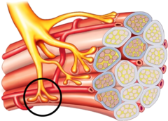
Smooth Muscle
Wall of organs, blood vessels, eyes, glands, skin
Many functions including food moving through tract, and regulation of blood vessel diameter
Skeletal Muscle
Bone
Body Movement
Cardiac Muscle
Heart
Pumps blood
Simple Squamous
What is A

Simple Cuboidal
What is B

Simple Columnar
What is C

Pseudo-stratified Ciliated Columnar with Goblet Cells
What is D

Transitional
What is E
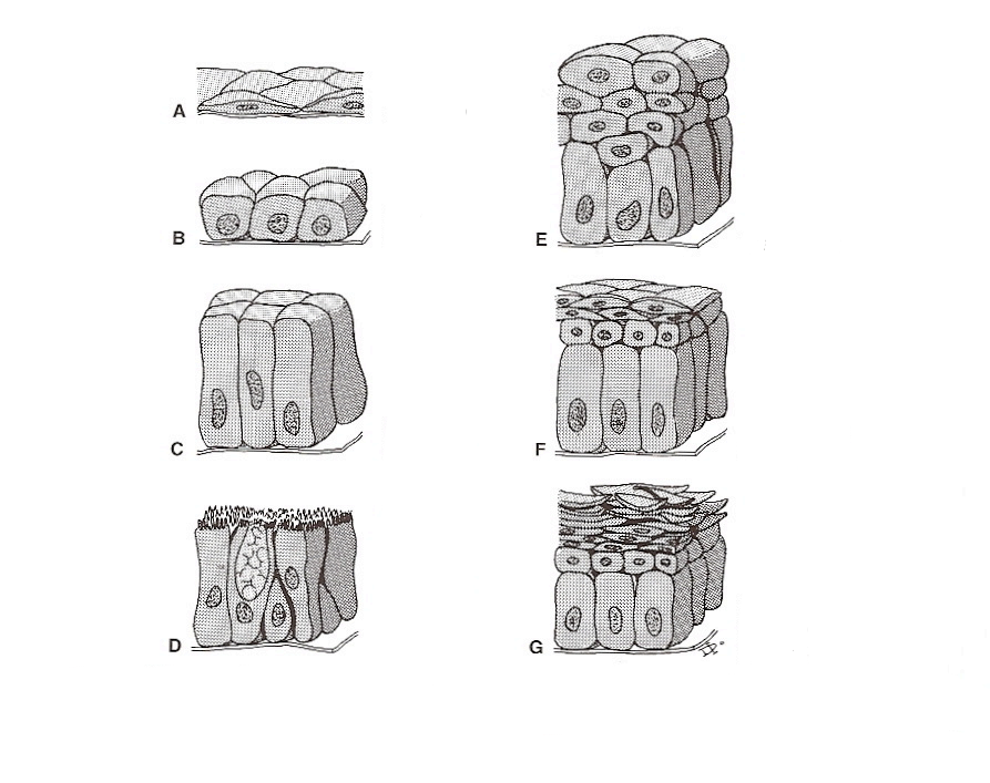
Stratified Squamous Nonkeratinizing
What is F
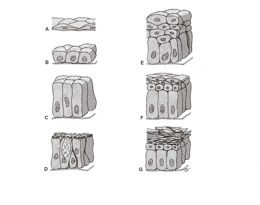
Stratified Squamous Keratinizing
What is G

Diaphysis
What is the red line pointing to

Proximal Epiphysis
What is the blue line pointing to

Distal Epiphysis
What is the green line pointing to

Spongy Bone
What is the blue line pointing to

Trabeculae
What is the green line pointing to
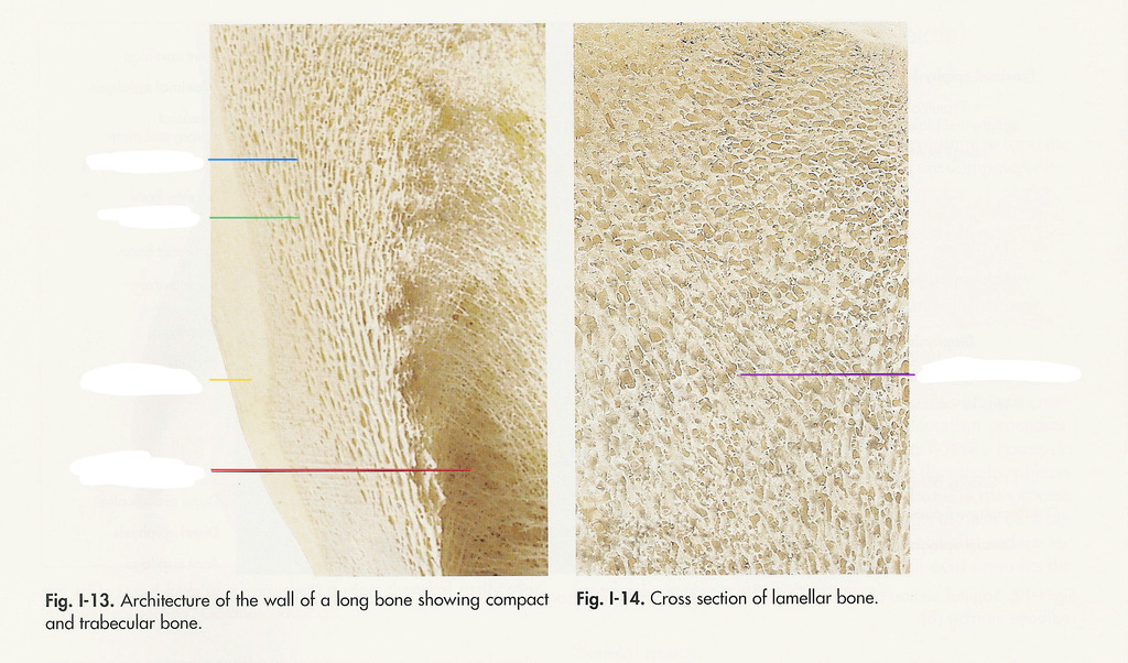
Compact Bone
What is the yellow line pointing to
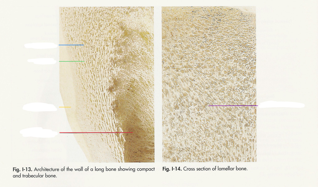
Medullary Cavity
What is the red line pointing to

Tubules and Lamellae
What is the purple line pointing to

Bone Forming the Proximal Part of the Joint
What is the black line pointing to

Synovial Villi
What is the blue line pointing to
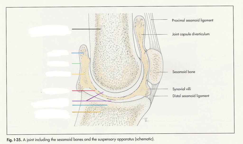
Joint Capsule
What is the green line pointing to

Synovial Fluid
What is the yellow line point to

Articular Process
What is the red line pointing to
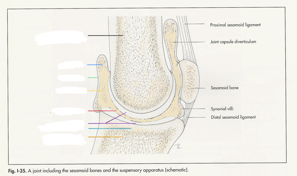
Joint Cartilage
What is the purple line pointing to
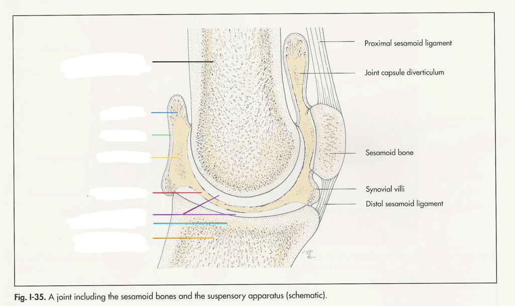
Subchondral Bone Plate
What is the light blue line pointing to

Bone Forming the Distal Part of the Joint
What is the orange line pointing to

Appendicular Skeleton
What is the blue part

Axial Skeleton
What is the white part

8
How many cranial bone are in humans
14
How many facial bones are in humans
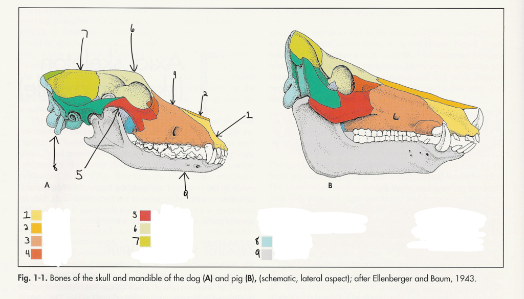
Incisive
1

Nasal
2

Maxilla
3

Lacrimal
4

Zygomatic
5
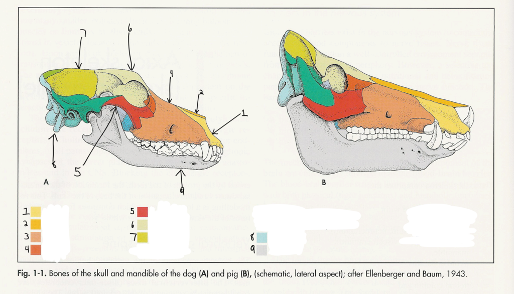
Frontal
6

Parietal
7

Occipital
8
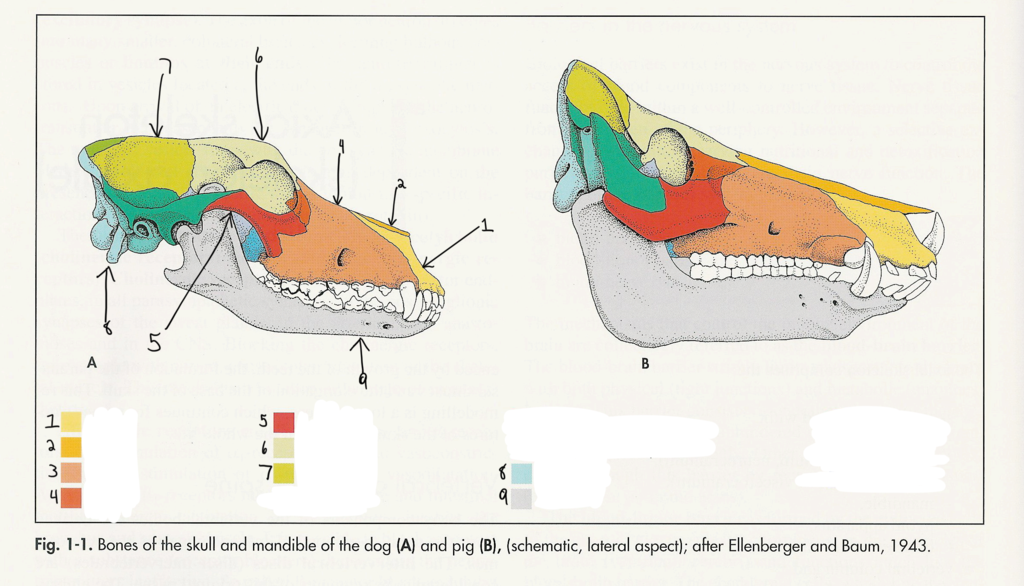
Mandible
9
Cranial Cavity
A

Frontal Sinus
B

Endoturbinate I
C

Maxillotrubinate
D

Maxilla
E
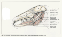
Incisive
F
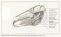
Mandible
G

Rostral
What is the blue line pointing to

Body
What is the red line pointing to

Articular Process
What is the blue line pointing to

Ramus
What is the green line pointing to

Frontal
What is outlined in blue

Orbit
What is outlined in green

Nasal
What is outlined in light green

Incisive
What is outlined in yellow

Maxilla
What is outlined in orange
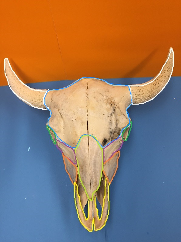
Lacrimal
What is outlined in purple

I-Band, H-Band, Z-Line, A-Band
Types of Bands
Know what is happening (Lecture 3)

Relaxed muscle

Contracting muscle

Fully contracted muscle

Z-Disk
What is the black areas

A-Band
What is the blue area
