HUMB1004 Week 5: Cervical spine and sacrum
 Call Kai
 Learn
Learn Practice Test
 Spaced Repetition
 Match
Match Flashcards
 Knowt Play
1/254

There's no tags or description
Looks like no tags are added yet.
Name | Mastery | Learn | Test | Matching | Spaced | Call with Kai | Chat |
|---|
No analytics yet
Send a link to your students to track their progress
255 Terms
Relative to the average vertebra, is the cervical vertebral body smaller or larger?
Small
What feature do cervical vertebra have that allow for muscle attachment in addition to transverse processes?
Uncinate processes
Where are the uncinate processes located?
Superolaterally
True or false: cervical spinous processes are bifid (forked)
True
What feature is unique to cervical vertebra transverse processes?
They have a transverse foramen
What is the purpose of the transverse foramen of cervical vertebra?
Allows passage of vertebral artery, vertebral veins, and some sympathetic nerves
What feature is found on the lateral end of the transverse processes of cervical vertebra?
Tubercles (anterior and posterior)
What is the articular pillar?
Fused superior and inferior articular processes of cervical vertebra only
Describe the vertebral foramen of a cervical vertebra
Large and triangular
How are the facets of cervical vertebra oriented?
At 45 degrees to the transverse plane
True or false: the facets of cervical vertebra lie much closer to the transverse plane than any other vertebra
True
True or false: intervertebral joints in cervical region are true synovial
True
What are uncovertebral joints?
Joints between the uncinate processes and vertebral bodies of the cervical vertebrae
What structure do the intervertebral foramina passage?
Spinal nerves
In cervical vertebrae, what structures form the intervertebral foramen?
The uncovertebral joint and the articular pillar
Where does the vertebral artery V1 arise?
From the subclavian artery
Why is V1 named pre-foraminal?
Because it arises at the lateral aspect of cervical vertebra then usually enters the transverse foramen at C6. Prior to this, does not course through vertebral foramen
How often do V1/pre-foraminal arteries to enter the spinal column at C6?
70-80% of the time
True or false: V1 most commonly enters the vertebral column at C7
False, 70-80% enter at C6
Where is V2 located?
When V1 enters transverse foramen up to C2, through the transverse foramen
What is the other name for V2?
Foraminal segment of vertebral artery
Where does V3 begin?
At C2
Describe the course of V3 until it becomes V4
Torturous (lots of bends and twists) until the transverse foramen of C1, then course posteriorly to form a groove at C1, then pierces a membrane and dura
What does V3 become once it pierces the dura?
V4
Another name for V4
Intra-dural segment
What is the vertebral artery termed superior to the intra-dural segment?
The Basilar artery
Why are vertebral and basilar arteries important?
They supply the brain stem and spinal cords (commonly imaged)
True or false: Basilar arteries supply the brain
False, internal carotid arteries supply brain, basilar and vertebral arteries supply the brain stem and spinal cords
What is the relation between the common carotid artery and C6?
The CCA lies just anterior to the anterior tubercle of C6
What is the anterior tubercle of C6 also called?
The carotid tubercle
What is the importance of the carotid tubercle?
In the case of a haemorrhage, the CCA can be compressed against the carotid tubercle to stem the flow of blood
What are the arrows pointing to and what might have caused it?
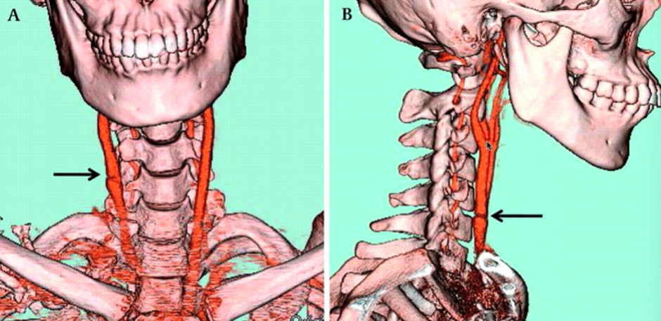
Carotid artery tear. Blunt trauma i.e. CCA pushed against the tubercle which has caused a tear
CCA and IJV travel in their own connective tissue sheath, called the _______
Carotid sheath
Other name for C1
Atlas
Why is C1 atypical?
It has no vertebral body, instead it has an anterior arch with an anterior tubercle for muscle attachment, and a posterior arch (modified laminae) with a posterior tubercle
Instead of transverse processes, what does C1 have?
Lateral masses
Where are the superior and inferior articulating processes located on C1?
On the superior and inferior surfaces of the lateral masses
How to distinguish inferior and superior surfaces of C1
Superior aspect has elongated anterior to posterior processes as it must articulate with occipital condyles, inferior only must articulate with axis, hence has a rounded projection. Additionally, there is a groove for V3 section of artery on the anterior surface of the posterior arch superiorly
Where is the Atlanto-occipital joint located?
Between superior articulating processes of Atlas and occipital condyles
What movements are available at the Atlanto-occipital joint?
Flexion and extension. No rotation or lateral flexion are available
What features of C2/axis make it atypical?
It has a dens/odontoid process, and a similar body to C1
What special articular joint does C1/C2 have? (between the dens and anterior arch of C1)
The median atlantoaxial joint
What is the name of the joints found between articulating facets of C1/2?
Latero-atlantoaxial joints
What movement are the median and lateral atlantoaxial joints responsible for? (in combination)
50% of total craniocervical rotation
True or false: the craniocervical rotation between atlas and axis are reliant on the close articulation of the dens of axis and the anterior arch of the atlas
True
What occurs when there is a loss of articulation at the median atlantoaxial joint?
Loss of stability at joint
What are the 3 ways to lose stability at the medial atlantoaxial joint?
Fracture of the odontoid process, Os odontoideum, and Os terminale
True or false: the odontoideum ossifies as a part the axis
False, it starts at its own centre of ossification and later fuses with the VB of axis
What is Os Odontoideum and what are its implications?
The failure of the odontoid process to fuse with axis, causes instability
What is the fovea Dentis?
The notch in the atlas for the dens to reside in
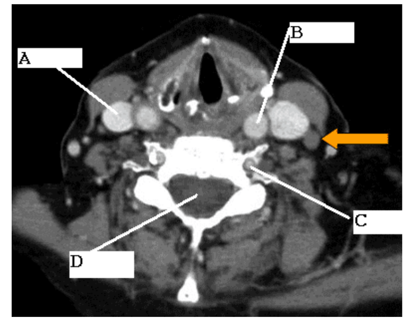
A
Internal jugular vein
What is B?
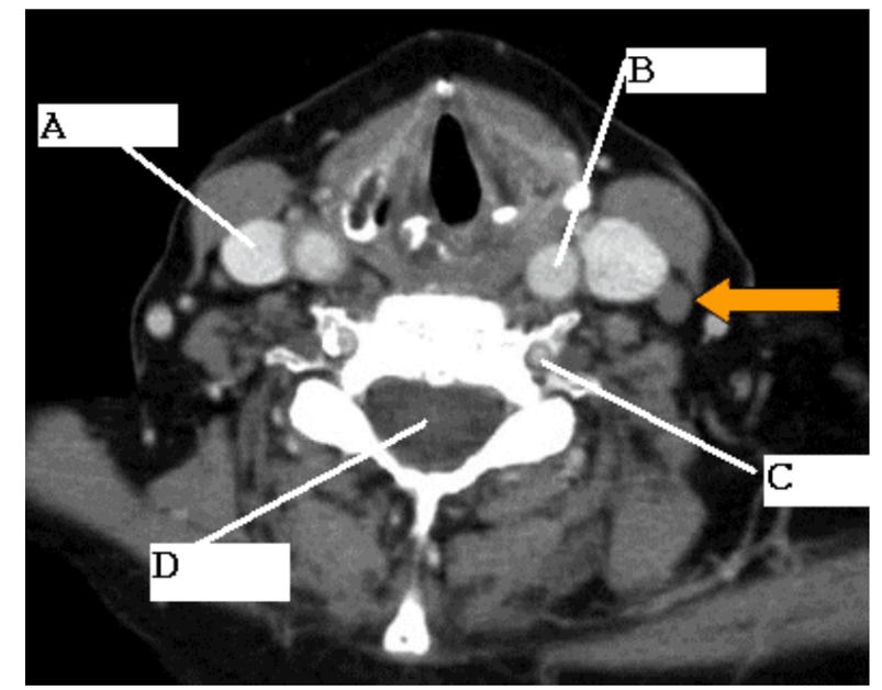
Common carotid artery

What is C?
Transverse foramen
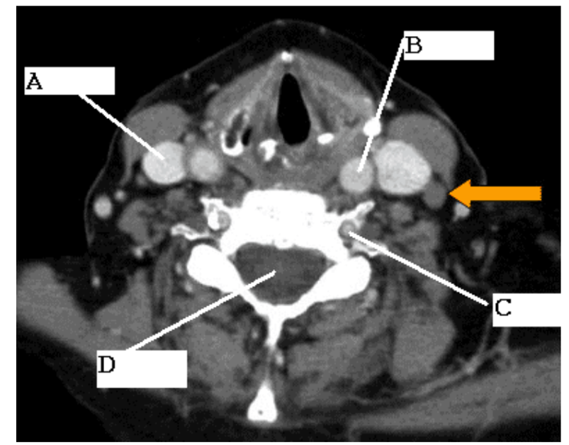
What is D?
Spinal cord in vertebral foramen
What is the carotid sheath?
The connective tissue in which the CCA and IJV travel in
What is shown here?

Os odontoideum
What is shown here
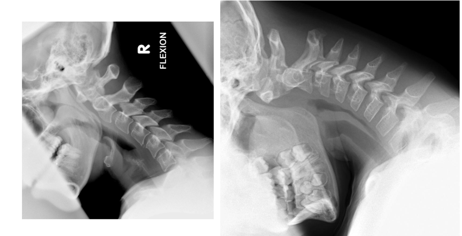
Os odontoideum
Is Os terminale a cause for concern?
No, just notable. Still stable
A typical cervical vertebra consists of body, foramen, spinous process, transverse process (describe)
Small VB, triangular vertebral foramen, bifid spinous process and modified transverse foraminae
The transverse foramen of cervical vertebrae contain:
Vertebral arteries
Bifid spinous processes are found on vertebrae _____
C2-6
C1 does not have a vertebral body. Instead, it has…
Anterior tubercle of anterior arch
C2 is not a typical vertebra as it:
Has an odontoid process on the anterior superior surface

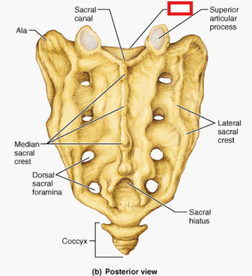
Superior articulating process
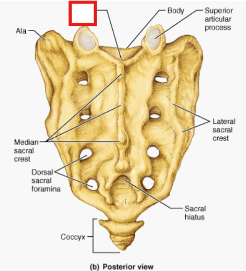
Sacral body
Sacral canal

Ala

Median sacral crest
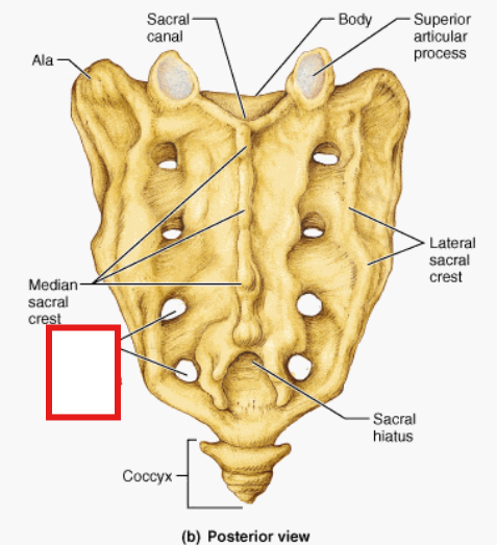
Dorsal sacral foramina
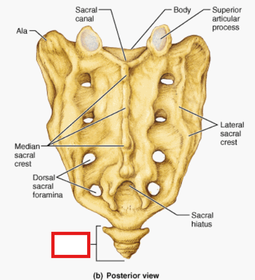
Coccyx (2-4 bones fused)

Sacral hiatus
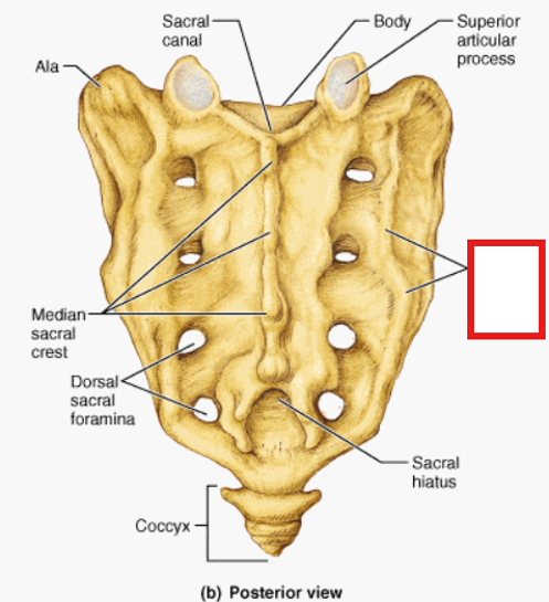
Lateral sacral crest
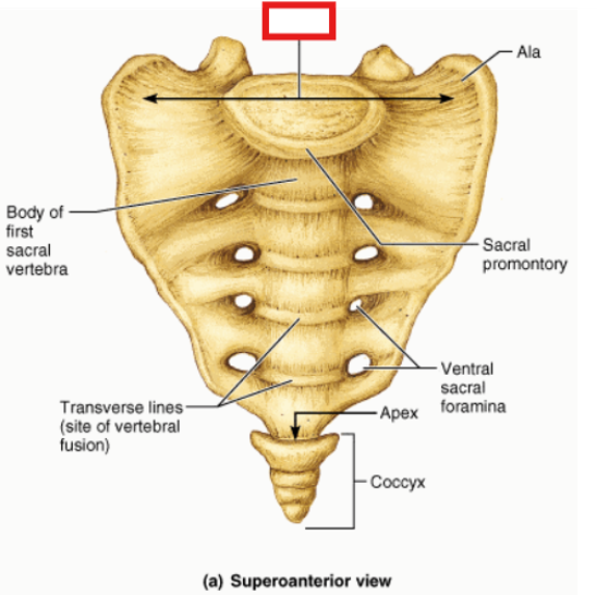
Base
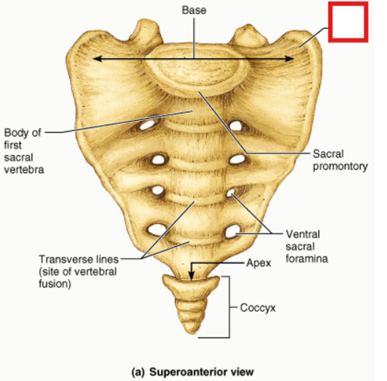
Ala
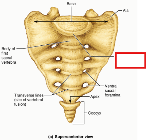
Sacral promontory
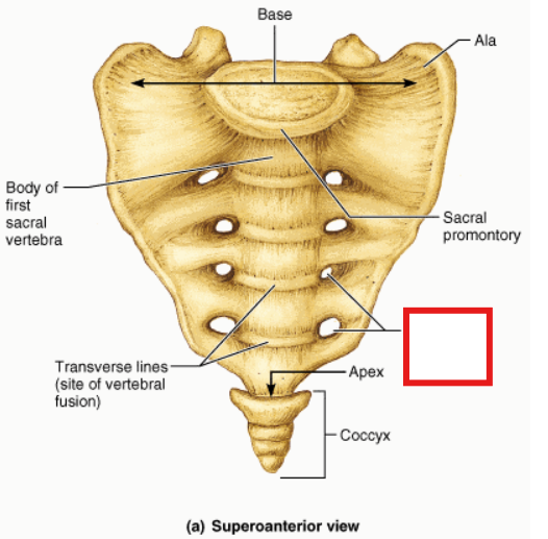
Ventral sacral foramina

Apex and coccyx
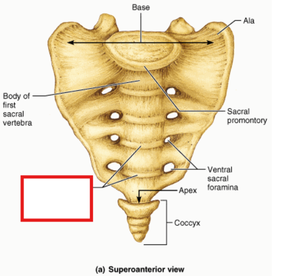
Transverse lines (site of vertebral fusion)
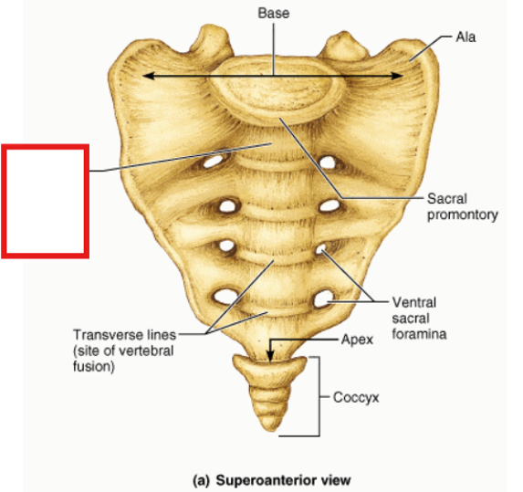
Body of first sacral body
Which direction is the base of the sacrum tilted towards?
Anteriorly
Which direction is the apex of the sacrum tilted towards?
Posteriorly
What joint is formed at the apex of the sacrum and the coccyx?
The sacrococcygeal joint

Sacroiliac joint
True or false: the sacroiliac joint has a great degree of movement
False, it is moveable but to a very limited degree
What part of the sacroiliac joint is synovial?
The first 1/3rd
What part of the sacroiliac joint is fibrous (syndesmoses)
The last 2/3rds
What type of cartilage is found on the sacral part of the sacroiliac joint?
Hyaline
What type of cartilage is found on the iliac part of the sacroiliac joint?
Fibrocartilage

Orange:
Sacrum/sacral ala
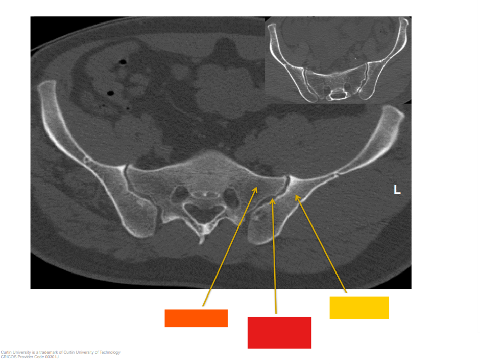
Red:
Sacroiliac joint
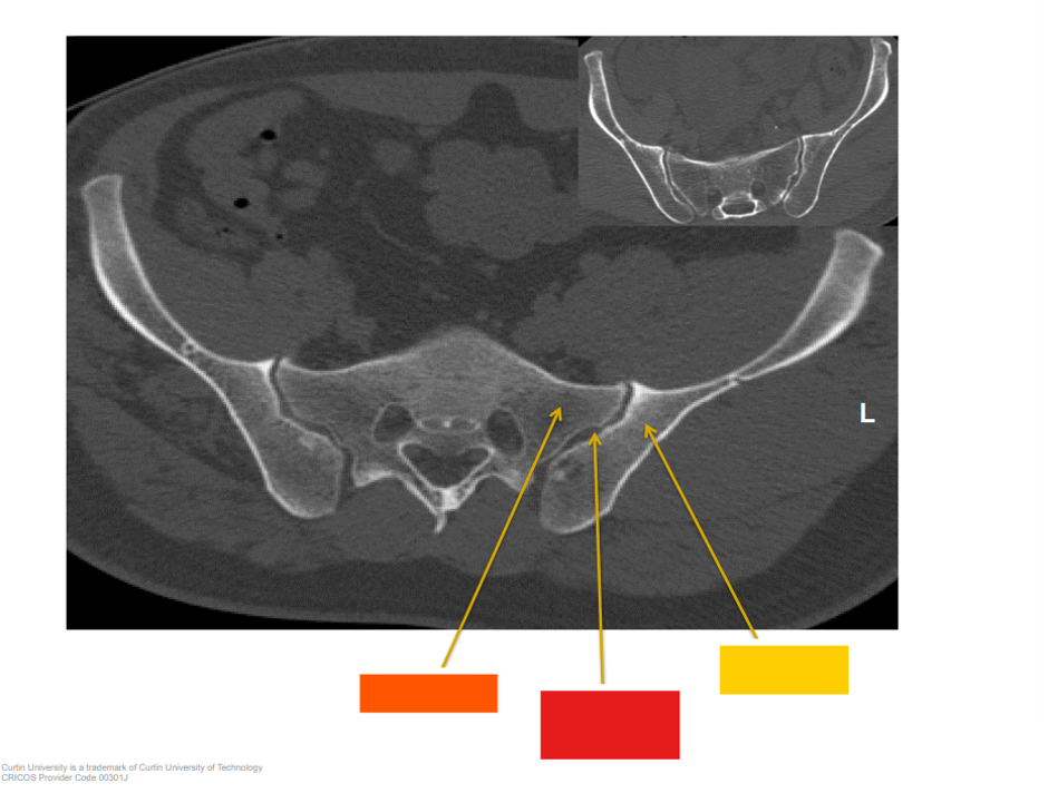
Yellow:
Ilium
True or false: All sacroiliac joints are the same shape
False, some are deeper, some are shallower
Describe the dorsal surface of the sacrum
Roughened
Describe the ventral surface of the sacrum
Smoothened
Articular processes of sacrum are articulated in what plane?
Coronal
What is the sacral hiatus?
Lack of 5th tubercle
What is the importance of the sacral hiatus?
Can be used to introduce epidural anesthesia
Where does the dura end? Why is this important?
At S2, hence anaesthesia can be introduced as high as S3 and the dura will not be perforated
How are the sacral horns and coccygeal cornu related?
The coccygeal cornu opposes the sacral horns
What is the type of joint at the sacrococcygeal joint?
Symphysis (fibrocartilaginous)