Parasitology and Physiology
 Call Kai
 Learn
Learn Practice Test
 Spaced Repetition
 Match
Match Flashcards
 Knowt Play
1/269

There's no tags or description
Looks like no tags are added yet.
Name | Mastery | Learn | Test | Matching | Spaced | Call with Kai | Chat |
|---|
No analytics yet
Send a link to your students to track their progress
270 Terms
Define the terms:
Endotherm
Ectotherm
Homeotherm
Poikilotherm
Endotherm: an animal that generates their own body heat through internal metabolic processes
Ectotherm: an animal that relies on external environmental sources to heat their body
Homeotherm: an animal that maintains a stable, constant internal body temperature within a narrow range, regardless of the surrounding environment
Poikilotherm: an animal whose internal body temperature changes and varies depending on the surrounding environment
Define the terms:
Thermoneutral zone
Lower and upper critical temperatures
Zone of thermal comfort
Thermal setpoint
Thermoneutral zone: the range of ambient environmental temperatures where an organism maintains its core body temperature using basic dry heat loss mechanisms without changing metabolic heat production
Lower and upper critical temperatures: minimum and maximum temperatures of the thermoneutral zone
Zone of thermal comfort: the range of temperatures resulting in psychological thermal satisfaction
Thermal setpoint: the target core body temperature value
Describe the process of conduction in heat exchange
Heat transfers through direct physical contact between particles
Fast-vibrating particles collide with neighbouring particles, passing kinetic energy along
Describe the process of convection in heat exchange
Heat transfers through the movement of fluids, which includes both liquids and gases
Warm fluid expands and rises, while cooler fluid sinks to replace it- creating a circulating current
Describe the process of radiation in heat exchange
Heat transfers via electromagnetic waves through space, without needing any medium or contact
Objects emit infrared waves that travel outward until they strike another object and warm it
Describe the process of evaporation in heat exchange
Heat transfers away from a surface as a liquid changes state into a gas
High-energy liquid molecules escape into the air as vapour, leaving cooler molecules behind
Describe the Q10 effect
Metabolic rate increases for every 10℃- this is the Q10 effect
A Q10 value of 2 means that metabolic rate doubles for every 10℃
A Q10 value of 3 means that metabolic rate triples for every 10℃
Give the equation linking M, CD, C, R and E- including what is represented by these letters
M: metabolic rate
CD: conduction
C: convection
R: radiation
E: evaporation
Hs = Hm - (∓Hcd ∓Hc ∓Hr - He)
Describe the action of the thermoregulatory centre, including its location
The thermoregulatory centre is located in the hypothalamus, specifically within the preoptic area
The centre uses a negative feedback mechanism to constantly balance heat gain and heat loss
Temperature receptors monitor the temperature of blood flowing through the brain
Receptors in the skin detect external changes and send nerve impulses to the hypothalamus
If the core temperature strays from the normal set point, the centre sends electrical impulses to effectors (muscles, sweat glands, blood vessels) to correct it
List the physiological mechanisms used by endotherms against low temperatures
Vasoconstriction
Shivering: involuntary rapid muscle contraction
Non-shivering thermogenesis: brown adipose tissue breaks down fats to release heat
Piloerection: erector muscles pull hair/feathers up to trap an insulating layer of warm air
Hormonal regulation: thyroxine increases the basal metabolic rate to produce more internal heat
Decreased sweating: prevents evaporative cooling
List the behavioural mechanisms used by endotherms against low temperatures
Huddling: reduces exposed surface area and shares body heat between animals
Increased activity: moving around generates friction and muscular heat
Torpor/hibernation: metabolic rate/body temperature is lowered to conserve energy
Seasonal changes in fat/fur: allows for better insulation
List the physiological mechanisms used by endotherms against high temperatures
Vasodilation
Sweating: absorbs body heat through evaporation
Panting: increases water evaporation from the respiratory surfaces of the mouth/lungs
Gular fluttering: rapidly vibrating membranes of the throat and floor of the mouth to increase evaporative cooling
Thermal windows: highly vascularised appendages are designed to radiate excess heat
List the behavioural mechanisms used by endotherms against high temperatures
Habitat selection: seeking shade, burrowing underground or entering water to avoid direct solar radiation
Stretching out: increases surface area exposed to moving air
Shifting to nocturnal or crepuscular activity to avoid the hottest parts of the day
Licking the fur/skin: utilises the cooling effect of moisture evaporation
Urohidrosis: excreting urine onto the legs so the evaporating liquid cools blood vessels in the limbs
Give the lower critical temperature range of:
Naked human
Newborn lamb (wet)
Newborn lamb (dry)
Newborn piglet
100kg pig
Arctic fox
Dairy cow producing 30L milk/day
Naked human- 28 to 29
Newborn lamb (wet)- 38 to 40
Newborn lamb (dry)- 25 to 30
Newborn piglet- 34 to 35
100kg pig- 0 to 5
Arctic fox- -40 to -70
Dairy cow producing 30L milk/day- -15 to -30
What happens during the last few weeks of pregnancy in ewes to help the lamb survive for the first few hours?
In the final weeks of pregnancy, brown fat is laid around the heart and kidneys to act as an energy source
Describe how you can recognise the different severities of hypothermia in lambs
Normal temperature: 39-40℃
Mild-moderate hypothermia: 37-39℃
Hunched posture, lowered head
Hollow flanks
Lethargy
Excessive bleating
Severe hypothermia: <37℃
Inability to hold the head up
Lying flat out and unresponsive
How could you manage hypothermia in lambs under five hours old?
Towel dry
Place in a warming box or under a heat lamp until temperature rises above 37℃
Give 50ml/kg warmed colostrum via stomach tube
Continual gradual warming to 39℃ before returning the lamb to the ewe
How could you manage hypothermia in lambs over five hours old?
Administer an intraperitoneal injection of warm 20% glucose solution (10ml/kg) into the abdomen, before attempting to warm or feed
Towel drug
Place in a warming box or under a heat lamp until temperature rises above 37℃
Give 50ml/kg warmed colostrum via stomach tube
Continual gradual warming to 39℃ before returning the lamb to the ewe
In which clinical scenarios (aside from hyperthermia) might you lower the body temperature of an animal?
Cardiac arrest: neuroprotection
Status epilepticus: neuroprotection
Acute spinal cord injury: minimises swelling
Cardiac and vascular surgeries: restricts blood flow to specific areas
Topical anaesthesia: numbs peripheral nerve endings
When might Bair Huggers be used?
FAM (forced air warming) systems such as Bair Huggers are often used to treat perioperative hypothermia, affecting up to 80% of anaesthetised cats and dogs
Define the terms:
Tidal volume
Inspiratory reserve volume
Expiratory reserve volume
Tidal volume (Vₜ): the amount of air inhaled or exhaled during one normal, quiet respiratory cycle
Inspiratory reserve volume (IRV): the extra volume of air that can be maximally inhaled after a normal tidal inhalation
Expiratory reserve volume (ERV): the extra volume of air that can be maximally exhaled after a normal tidal inhalation
Define the terms:
Residual volume
Inspiratory capacity
Viral capacity
Residual volume (RV): the volume of air remaining in the lungs after maximal forced expiration, preventing lung collapse
Inspiratory capacity (IC): the maximum amount of air that can be inspired following a normal tidal expiration
Vital capacity (VC): the maximum volume of air that can be exhaled after a maximal inhalation
Define the terms:
Functional residual capacity
Total lung capacity
Respiratory rate
Functional residual capacity (FRC): the amount of air remaining in the lungs at the end of a normal tidal expiration
Total lung capacity (TLC): the absolute maximum volume of air the lungs can hold after a maximal inspiratory effort
Respiratory rate (f): the number of breaths taken per minute
Define the terms:
Minute ventilation
Dead space
Alveolar exchange
Minute ventilation (Vₑ): the total volume of air inspired or expired per minute
Dead space (Vd): the volume of the respiratory tract that does not participate in gas exchange
Alveolar ventilation (Vₐ): the volume of air per minute that reaches the alveoli and takes place in gas exchange
Give the equation for inspiratory capacity
Inspiratory capacity (IC) = Tidal volume (Vₜ) + Inspiratory reserve volume (IRV)
Give the equation for vital capacity
Vital capacity (VC) = Tidal volume (Vₜ) + Inspiratory reserve volume (IRV) + Expiratory reserve volume (ERV)
Give the equation for functional residual capacity
Functional residual capacity (FRC) = Expiratory reserve volume (ERV) + Residual volume (RV)
Give the equation for total lung capacity
Total lung capacity (TLC) = Vital capacity (VC) + Residual volume (RV)
Give the equation for minute ventilation
Minute ventilation (Vₑ) = Tidal volume (Vₜ) x Respiratory rate (f)
Give the equation for alveolar ventilation
Alveolar ventilation (Vₐ) = (Tidal volume (Vₜ) - Dead space (Vd)) x Respiratory rate (f)
Draw a diagram to show:
Inspiratory reserve volume
Tidal volume
Expiratory reserve volume
Residual volume
Inspiratory capacity
Functional residual capacity
Vital capacity
Residual volume
Total lung capacity

When does a ventilation/perfusion mismatch occur?
A V/Q mismatch occurs when the normal ratio of air entering the alveoli (V) to the blood flow in the capillaries (Q) is disrupted
What is a high V/Q mismatch? What is it caused by?
Ventilation exceeds perfusion, meaning there is airflow to an area of the lung, but blood flow through the capillaries is reduced/blocked
Causes: pulmonary embolism, pulmonary hypertension, decreased cardiac output
What is a low V/Q mismatch? What is it caused by?
Perfusion exceeds ventilation, meaning blood flows through the capillaries but the matching alveoli receive little to no air
Causes: chronic obstructive pulmonary disease, pneumonia, atelectasis, acute respiratory distress syndrome, pulmonary fibrosis
List the two endoparasite groups of veterinary importance
Nemathelminths (threadworms)
Nematoda (roundworms)
Platyhelminths (flatworms)
Cestoda (tapeworms)
Trematoda (flukes)
Describe 3 examples of canine respiratory parasites
Nematodes (lungworms): Angiostrongylus vasorum, Crenosoma vulpis, Eucoleus aerophilus
Infects the airways and lung tissue
Transmitted by ingesting infected slugs/snails
Lung flukes: Paragonimus sp.
Forms cysts inside the lung tissue
Transmitted by ingesting raw/undercooked intermediate hosts (crayfish, crabs, rodents)
Nasal mites: Pneumonyssoides caninum
Live inside the nasal passages and sinuses
Transmitted by nose-to-nose contact with another infected animal
Describe 3 examples of canine cardiac parasites
Nematodes (lungworms): Angiostrongylus vasorum
Also known as French Heartworm
Adults live in the heart and pulmonary arteries
Transmitted by ingesting infected slugs/snails
Heartworm: Dirofilaria immitis
Live in the pulmonary arteries and the right side of the heart
Transmitted by mosquito bites
Parasitic kinetoplastid: Trypanosoma cruzi
Attacks the heart muscle
A protozoan parasite carried by ‘kissing bugs’
Define the terms:
Parasitism
Mutualism
Commensalism
Parasitism: only one organism benefits from the interaction, at a detriment to the other
Mutualism: organisms of different species both benefit from the interaction
Commensalism: only one organism benefits from the interaction, without causing any harm to the other
Define the terms:
Facultative parasite
Obligate parasite
Endoparasite
Ectoparasite
Facultative: may survive in the absence of the host
Obligate: at some or all stages of the life cycle cannot survive in the absence of the host
Endoparasite: lives internally inside the host
Ectoparasite: lives in/on the outer body surfaces of the host
Define the terms:
Definitive host
Intermediate host
Permissive host
Non-permissive host
Reservoir host
Paratenic host
Vector (include both types)
Definitive: the host in which parasite sexual reproduction occurs
Intermediate: the host in which parasite asexual reproduction occurs
Permissive: not usually used by the parasite, but still favours life cycle completion
Non-permissive: the host in which the parasite cannot complete the life cycle
Reservoir: temporary host used in the absence of the natural host
Paratenic: will host the parasite without any further development
Vectors:
Mechanical: a vector which mechanically spreads the parasite, and is not utilised in the life cycle
Biological: a vector needed by the parasite to complete part of the life cycle
How can parasites be transmitted?
Food
Water
Soil
Direct contact
Airborne
Vector-borne
Mother-to-fetus
What is the difference between a direct and indirect parasite life cycle?
Direct life cycle: parasitic stages develop in/on one host
Indirect life cycle: intermediate host(s) is needed for the development of some stages of the parasite
What kind of information should you know about the life cycle of a parasite?
What is the type of life cycle?
Who serves as a definitive host?
Who serves as an intermediate host (if any)?
What is the infective stage?
What is the route of infection?
Why is it useful to study the parasite life cycle?
Determine an appropriate therapeutic window
Manage disease properly
Protect other animals from cross-infection
Protect the public from zoonotic infections
How can you control the spread of parasites?
Bovine lungworm is the only vaccine-preventable parasitic disease
Hygiene
Nutrition
Disruption of transmission
Describe the 3 different ways of detecting parasites
Microscopic
Coproscopic faecal samples using Baermann’s larval migration assay
Cytological profile of bronchoalveolar lavage fluid
Serological
Antigen-based assays
Antibody-based assays
Molecular
Conventional PCR: inhibitors in faeces can limit sensitivity
Real-time PCR: for detection of infection in definitive and intermediate hosts, and can detect DNA circulating in canine blood
How can you differentiate between nematode sp.?
Size differences
Species specificity
Morphological differences
Describe the life cycle of dipylidium caninum
Infected adult flea is ingested
Adult worms develop in the small intestine
Posterior segments of the adult worms are passed in the faeces
Segments and egg packets are found in faeces and on fur
Eggs are ingested by larvae flea
Larvae flea develops into an adult flea, and contains an immature tapeworm
Describe the life cycle of fasciola hepatica
Metacercaeriae cysts in grass are ingested
Fluke develops into an adult within the host
Eggs are passed in faeces
Miracidium larvae form
Intermediate snail host
Cercaeriae develops into Metacercaeriae cysts in grass
Describe the life cycle of d.viviparus
Host ingests L3 whilst grazing on contaminated pasture
L3 larvae leave their protective sheath, and pierce the wall of the host’s intestine
Larvae move to the local lymph nodes and molt into L4
They travel through the lymphatic system and blood vessels to the heart and lungs
Once in the alveoli/bronchioles, they break through into the airways and molt into L5
L5 move into the trachea and bronchi, where they grow into full adults
Females lay eggs containing L1, which are passed through the digestive tract and into the environment via faeces
Once outside, L1 molt twice in warm, damp weather to become infective L3 larvae in 5-7 days
Describe the life cycle of angiostrongylus vasorum
A dog/fox becomes infected by ingesting an infected slug or snail, containing L3
The L3 larvae burrow through the intestinal wall and travel to nearby lymph nodes, where they molt into L4 and L5
The L5 travel to the pulmonary arteries and the right side of the heart, where they grow into adult worms and lay eggs
The eggs move to the lung capillaries, where they hatch into L1
L1 break through the alveolar walls, move into the digestive tract and exit the body through faeces
Once in the environment, L1 is either eaten by a snail/slug, or will actively penetrate the foot
Inside, they grow and molt twice- until they reach the infective L3 stage
Describe the life cycle of aelurostrongylus abstrusus
Cat becomes infected by eating an infected paratenic host
The L3 larvae burrow through the intestinal wall and travel to nearby lymph nodes
They travel via the lymphatic/circulatory system to the lungs, where they develop into adults
Adult worms live in the terminal respiratory bronchioles and alveolar ducts
Female worms lay eggs in the lung parenchyma, which hatch into L1
L1 break through the alveolar walls, move into the digestive tract and exit the body through faeces
Once in the environment, L1 is either eaten by a snail/slug, or will actively penetrate the foot
Inside, they grow and molt twice- until they reach the infective L3 stage
Describe the life cycle of ascaris suum
Pigs ingest eggs containing L3 from contaminated soil, food or water
The larvae hatch inside the small intestine, and penetrate the intestinal wall
Larvae travel to the liver, where they burrow through tissue leaving fibrous scars (milk spots)
The blood carries larvae to the heart and lung capillaries, where they break through the alveolar walls, and move into the digestive tract
Back in the small intestine, they undergo molts to become L4 and L5
By 6 weeks, they reach adulthood and live in the lumen of the small intestine
Females produce eggs, which are passed via faeces into the environment
Fertilized eggs develop into L3 in the soil within 3-4 weeks under warm, moist conditions
Describe the life cycle of toxocara Canis
A puppy is infected by ingesting eggs from contaminated soil/objects
Eggs hatch in the small intestine, and larvae pierce the gut wall
Larvae travel via the blood stream to the heart and lungs
The blood carries larvae to the heart and lung capillaries, where they break through the alveolar walls, and move into the digestive tract
Back in the small intestine, they undergo molts to become L4 and L5
By 6 weeks, they reach adulthood and live in the lumen of the small intestine
Females produce eggs, which are passed via faeces into the environment
Environmental development takes 2-6 weeks, where the eggs embryonate and become infectious
Describe the different types of arthropod
Pentastoma: tongue worms
Insecta: diptera (flies), phthiraptera (lice), siphonaptera (fleas)
Lice can either be biting (mallophaga) or sucking (anoplura)
Arachnida: astigmata (mites), prostigmata (mites), mesostigmata (mites), metastigmata (ticks)
Ticks can either be hard (ixodidae) or soft (argasidae)
How do diptera affect animals?
Lucilia sericata- green bottle/blow fly
Attracted by dirty wool due to scouring or foot rot wounds
Fly strike
Eggs are deposited on skin
Larvae lacerate skin and burrow
This is a distressing condition, and may be fatal due to secondary infection/septicaemia
Describe the pulmonary parasites that commonly affect ruminants and equids
Dictyocaulus sp.
D.viviparus (cattle, deer and camelids)
D.filaria (sheep and goats)
D.arnfledi (donkeys and horses)
Adult lungworms live in the trachea and bronchi, causing parasitic bronchitis (‘husk’)
Deposition of eggs or newly hatched larvae may lead to parasitic pneumonia
The male has a copulatory bursa
How can you detect d.viviparus?
Baermann apparatus is used to detect L1 larvae in faeces
ELISA testing
Postmortem of lungs
Histopathology
Describe the differences between mild, moderate and severe infection of d.viviparus
Clinical Sign | Mild Infection | Moderate Infection | Severe Infection |
Coughing | Intermittent, more pronounced on exercise | Frequent even at rest | Deep, harsh cough |
Respiratory rate | Often normal (30 bpm) | Tachpnoea (40-60 bpm) | Severe tachypnea (>80 bpm) |
Lung auscultation | Often normal, some occasional squeaks | Pronounced squeaks and crackles | Pronounced squeaks and crackles |
Other signs | None | Usually none | Dyspnoea, salivation, fever |
Why has cattle lungworm increased?
Increased reliance on anthelmintics instead of vaccination comprises the build-up of immunological responses
Survival of L3 on pasture may have improved due to climatic change
Describe the clinical signs of d.arnfieldi
Persistent cough
Not responding to certain drugs
Lung sounds on auscultation
How does d.arnfieldi differ between donkeys and horses?
Donkeys
Infection has a pre-patent period of 13 weeks
Adult parasites are found in small bronchi
Patent infection with eggs in faeces
Horses
Horses are most likely infected when kept on pasture with donkeys
Not patent, very few (if any) eggs in faeces
How can you diagnose d.arnfieldi?
A bronchoalveolar lavage or a tracheal wash may reveal worms, and larger numbers of eosinophils
Which pulmonary parasites commonly affect dogs?
Angiostrongylus vasorum
Oslerus osleri
Eucoleus aerophilus
Filaroides sp.
Crenosoma vulpis
Which pulmonary parasites affect cats?
Aelurostrongylus abstrusus
Which pulmonary parasites affect sheep and goats?
Muellerius capillaris
Protostrongylus rufescens
Which pulmonary parasites affect swine?
Metastrongylus apri
What is the type of life cycle of angiostrongylus vasorum?
Life cycle is indirect
Dog is the definitive host
Slug/snail is the intermediate host
What do the antigens of angiostrongylus vasorum cause?
Type III hypersensitivity (immune complex deposition)
Complement activation
Immune infiltrate in lungs and other tissues
What does L1 of angiostrongylus vasorum cause?
Pulmonary inflammation/granuloma
Pulmonary arteriolar vasoconstriction
How can you detect angiostrongylus vasorum?
Radiography (interstitial pulmonary patterning, right ventricular/atrial enlargement
Detection of L1 in faeces
Detection of L1 in BAL fluid
How can you treat angiostrongylus vasorum?
Fenbendazole
Moxidectin
Milbemycin
What is the type of life cycle of aelurostrongylus abstrusus?
Life cycle is indirect
Cat is the definitive host
Slug/snail/rodent/bird is the intermediate host
How can you detect aelurostrongylus abstrusus?
Radiography (bronchiointerstitial patterning)
Detection of L1 in faeces
Detection of L1 in BAL fluid
How can you treat aelurostrongylus abstrusus?
Fenbendazole
Ivermectin
Levamisole
What are nontrue lungworms?
Nontrue lungworms only have a lung phase during their migration
The residing tissue for adults is not the lungs (commonly the intestine)
How does toxocara Canis affect all types of dog?
In older/adult dogs, larvae bypass the lungs and undergo somatic migration- spreading through the blood stream to go dormant in skeletal muscles and organs
In pregnant females, the dormant larvae ‘wake up’ due to hormonal shifts in the final trimester
Puppies can be infected through the mother’s milk
What are the different types of larva migrans?
Visceral larva migrans: migration through organs
Ocular larva migrans: migration through the eye
Cutaneous larva migrans: migration through the skin
Describe how the different species of dilofilaria affect different animals
Dogs/cats (cardiac/pulmonary): D.immitis
Dogs/cats (ocular): D.repens
Raccoon (subcutaneous): D.tenius
Bear (subcutaneous): D.ursi
Porcupine (subcutaneous): D.subdermata
Describe the factors affecting d.immitis distribution
Maturation of microfilariae to infective L3 requires 2 weeks at 27℃, and the temperature should not be below 14℃
Mosquito breeding on water (all juvenile stages are aquatic)
Mosquito species which support the microfilariae
The presence of susceptible hosts
Describe the life cycle of d.immitis
A mosquito carrying L3 bites a dog and deposits the larvae onto the skin, where they actively enter through the wound into the body
Within 3 days, L3 molt into L4 in the skin and connective/muscle tissues
Over the next 2 months, L4 larvae migrate to the pulmonary arteries and the right side of the heart, where they undergo their final molt
Over several months in the pulmonary vessels, they grow significantly and release microfilariae into the bloodstream
A mosquito bites the infected dog, and ingests the circulating microfilariae
Inside the mosquito’s Malpighian tubes, the microfilariae molt twice, progressing to L3
The L3 larvae migrate to the mouthparts (proboscis), ready to infect a new dog
Describe how cardiopulmonary disease can progress following d.immitis
Narrowing of pulmonary artery
Disruption of intima leads to platelet influx and production of platelet-derived growth factor (PDGF), which increases proliferation of medial smooth muscle cells and fibroblasts
Exercise intolerance
Hydrothorax
Hydroperitoneum
Chronic cough
Dyspnoea
Haemoptysis
Eosinophilia in lung
Oedema in lung parenchyma
Describe the roles of different immune cells in parasitism
Eosinophils: important in parasitic and allergic diseases (migrate from the blood into the tissue)
Eosinophilia occurs in blood and tracheal washes during cardiorespiratory parasitism
Basophils: degranulate at the surface of blood-borne parasites (microfilariae)
Mast cells: the tissue version of basophils- activate and produce amines, such as histamine
Histamine: an important vasodilator, which increases the number of immune cells in tissues
Neutrophils: D.immitis carries Wolbachia, which plays a role in neutrophil-induced inflammation seen in heartworm infection
Describe the roles of:
Large granules
Small granules
Cytokines
In parasitism
Large specific granules
Major basic protein
Eosinophil cationic protein
Eosinophil derived neurotoxin
Eosinophil peroxidase
Small granules
Aryl sulfate
Acid phosphatase
Gelatinase
Cytokines
IL3, IL5 (eosinophil growth factors)
IL1, IL6, IL8, TNF-Alpha
Describe caval syndrome
Large worm burden in the right atrium and posterior vena cava
Increased venous pressure damages liver parenchyma
Increased cholesterol content of erythrocyte membranes, leading to fragility and haemolysis
Severe anaemia may be seen in blood as erythrocytes are replaced
Erythrocyte haemolysis leads to haemoglobinaemia, bilirubinaemia, haemoglobinuria and jaundice
Describe how you can diagnose heart worm
Complete blood count (CBC)
Some other diseases will give similar results
Knotts test/Millipore filter (acidic phosphatase staining or methylene blue) to enrich the microfilariae
Microfilariae may not be seen if few are present
Occult infections are not detected
Thoracic radiographs
Some other diseases will give similar results
Electrocardiogram/echocardiogram (double line= heartworm)
Difficult to interpret
Arteriogram
Indirect fluorescent antibody
ELISA
Only adult female uterine antigens are detected
False negatives occur if immature females are present, there is a male-only infection, or if there is an issue with the kit
Tracheal wash
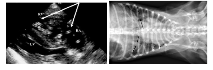
What is shown?
Heartworm
How would you treat:
Adult heartworm
Microfilariae
Adult worms
Melarsomine dihydrochloride (ImmiticideⓇ)
Microfilariae
Ivermectin (HeartguardⓇ)
Milbemycin (InterceptorⓇ)
Both can be used as preventative for prophylaxis
Describe the use of adulticides in heart worm infection
Dead worms may induce significant inflammatory immune reactions
This can lead to pulmonary thromboembolism (clot in the pulmonary artery, blocking blood flow)
Disseminated intravascular coagulation (DIC)- microcoagulation, high platelet counts, endothelial damage
With increased activity, blood flow to the blocked vessels will increase, and cause capillary rupture- further increasing pulmonary vascular resistance and right-sided heart failure
Draw a table to show the differences between feline and canine heart worm
Feature | Feline Heartworm | Canine Heartworm |
Susceptibility | 60% | 80-100% |
Worm lifespan | 2-3 years | 5-7 years |
Number of worms | 1-6 | Can be >30 |
Ectopic infection | Common | Uncommon |
Microfilaraemia | 1 month (<20%) | Persistent (80-90%) |
Organ pathology | Lung | Heart and lung |
What are the complications in diagnosing feline d.immitis?
Usually amicrofilaraemia, therefore blood smears may give false negatives
Diagnosis may be improved by analysing blood samples acquired in the evening, when parasites are more active (nocturnal periodicity)
A clinically infected cat may give very low worm burdens- therefore ELISA analysis to detect the antigen may give false negatives
Single-sex infections occur more in cats: no microfilariae may be produced
Describe how ELISA can be used to diagnose feline heart worm
In cats, very few worms can cause significant pathology
ELISA which detects the antigen (used in dogs) may not be sensitive enough
The ELISA developed for use in cats instead measured antibody to parasite (rather than parasite antigen)
Antigen is attached to well
Test sample is added
Labelled antibody which detects feline IgG is added
Substrate is added
Colour develops, result is read on a plate reader
Describe the limitations of using ELISA to diagnose feline heart worm
Positive antibody results may not tell you the current status, since the antibody can be present in cats who have previously be infected
Although antibody titre can be raised in ongoing infection, it is difficult to correlate antibody titre with worm burden or current/previous infection
It is a less appropriate screening test, and should interpreted in relation to clinical signs and other diagnostic findings
What is the main function of the cardiovascular system?
The function of the cardiovascular system is to deliver oxygenated blood to the tissues, and remove carbon dioxide
What is reflected by oxygenation of arterial blood in the tissue?
Oxygenation of arterial blood in the tissue reflects blood pressure, heart function, breathing and lung function
How can you measure oxygen delivery?
To measure tissue oxygen delivery, we can measure: tissue perfusion and blood-oxygen content
What is tissue perfusion driven by?
Tissue perfusion is driven by mean arterial blood pressure (MAP)
How can MAP be measured?
Invasive measurement
Non-invasive measurement
Feeling the pulse (very difficult)
Invasive measurement
Gives real-time pressure
Gives more information from a trace
More accurate
Insert a cannula into a peripheral artery
Connect cannula to transducer
Reported as systolic/diastolic (mean)
Non-invasive measurement
Sphygmomanometry
Oscillometric
Doppler
Describe what CVP (central venous pressure) measures, and give the normal values
CVP measures the pressure of blood returning to the right side of the heart
This reflects the volume of blood returning
Normal values
Cats: 0-5cm water
Dogs: 0-10cm water
To convert from mmHg to cm water, multiply by 1.36
How can you measure cardiac output? What is cardiac output?
Rarely used
Can use Fick method, dye, thermodilution, bioimpedance, pulse contour analysis, lithium
Cardiac output is the product of stroke volume (SV) and heart rate (HR)