A & P 314 Lab Exam 1 Quiz 3
1/22
There's no tags or description
Looks like no tags are added yet.
Name | Mastery | Learn | Test | Matching | Spaced |
|---|
No study sessions yet.
23 Terms
Blood pressure
force of blood against the walls of the arteries as it circulates through the body, in millimeters of mercury (mmHg)
systolic pressure
pressure in the arteries when the left ventricle of the heart contracts and pushes blood into the aorta and the systemic circulatory system, higher number
Diastolic pressure
pressure in the arteries when the left ventricle is at rest, lowest number
Hypotension
blood pressure below 90 mmHg over 60 mmHg, symptoms include blurry vision, confusion, dizziness, and weakness
hypertension
blood pressure above 130 mmHg over 80 mmHg, contributes to burst blood vessels, blood clotting, stroke, or heart attack
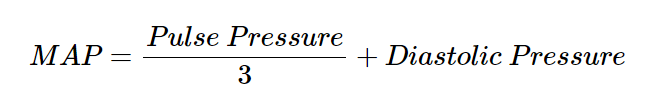
Pulse pressure
difference between the systolic and diastolic pressures
mean arterial pressure
Sphygmomanometer
medical device used to measure blood pressure. It consists of an inflatable cuff that is wrapped around the upper arm and connected to a gauge that displays the pressure.
Korotkoff sounds
series of sounds heard through a stethoscope when measuring blood pressure with a sphygmomanometer. These sounds are produced by the turbulent flow of blood through the arteries as the pressure in the cuff is gradually released
cardiac conduction system
responsible for generating and coordinating the action potentials that control the heartbeat
sinoatrial node
small cluster of specialized cardiomyocytes, in the superior margin of the right atrium, natural pacemaker of the heart, generates electrical impulses that initiate each heartbeat
atrioventricular node
cluster of specialized cardiomyocytes located in the inferior margin of the right atrium, near the opening of the coronary sinus. Serves as a gatekeeper and delays the action potential from the SA node, allowing time for the atria to contract and fill the ventricles with blood before the ventricles contract
bundle of His
bundle of specialized cardiomyocytes that carries the action potential inferiorly through the interventricular septum to the apex of the heart and splits into the Purkinje fibers
Purkinje fibers
specialized muscle fibers that branch out from the bundle of His and spread throughout the ventricles, deliver the action potential to the muscle cells of the ventricles, causing simultaneous ventricle contraction
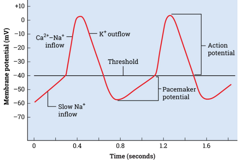
Pacemaker Potential
slow influx of Na+ ions resulting in a gradual depolarization
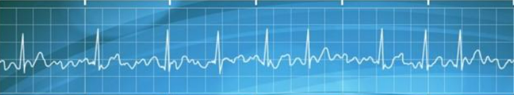
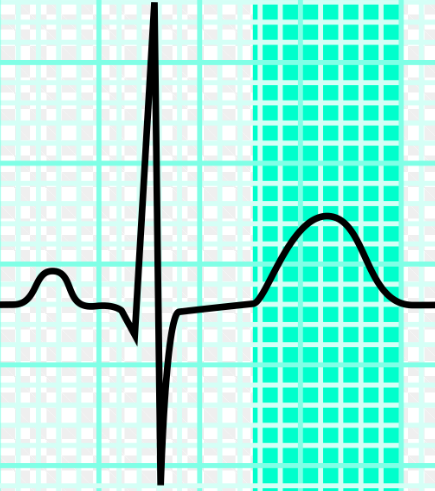
electrocardiogram
composite image of the change in cardiac voltage over time
P wave
first small peak, atrial depolarization
QRS complex
sharp downward, upward, downward spike, ventricular depolarization
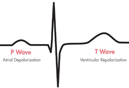
T wave
final small peak, ventricular repolarization, highlighted region
Tachycardia
a heart rate above 100, often caused by physical exertion, sympathetic stimulation, and fever
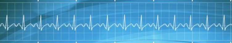
regular sinus rhythm
most common ECG tracing in healthy adults, BMP from 60 to 100 BPM. The QRS complex will be narrow, and the P wave and T wave will be upright
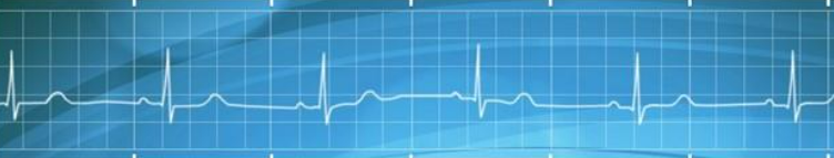
Bradycardia
heart rate of <60 BPM. In healthy adults, it typically is not a concern until the heart rate drops below 50 BPM
Atrial fibrillation
an irregular, chaotic rhythm with no discernable P waves, but does have a discernable QRS complex, from the atria firing out of sync quivers and jiggles instead of pumping blood increasing risk of blood clots