L1, L2, L7, L14, L15 - Introduction to Metabolism, Oxidation States, Vitamins, Inheritance Patters, Population Genetics
 Call Kai
 Learn
Learn Practice Test
 Spaced Repetition
 Match
Match Flashcards
 Knowt Play
1/134

There's no tags or description
Looks like no tags are added yet.
Name | Mastery | Learn | Test | Matching | Spaced | Call with Kai | Chat |
|---|
No analytics yet
Send a link to your students to track their progress
135 Terms
Metabolism
the process of ENERGY generation and use.
1. Includes all aspects of biological processes
2. Such as: nutrition, digestion, absorption, elimination, respiration, circulation, and temperature regulation
3. Also defined as the organized release or capture of small amounts of energy in processes whose overall change in energy is large.
The combustion of glucose to 6 CO2 + 6 H2O
1. In a bomb calorimeter, would yield ~ 2,870 kJ of energy as heat.
2. In an aerobic cell, would yield approximately 30 – 38 ATP with some loss of energy as heat (which contributes to the maintenance of body temperature).
Catabolism
oxidative, exergonic, breakdown
Catabolism is a __________ that goes from ____________ to ___________. Generates 3 things:_________, _________, ________ .
destructive process; energy yielding nutrients; energy poor end products; energy (i.e. ATP), waste products (CO2, H2O, NH4+) and/or building blocks for anabolic reactions, reducing equivalents (i.e. NADH & FADH2) for more ATP synthesis
Anabolism
reductive, endergonic, synthesis
Anabolism is a _________ that goes from ____________ to ____________ using the _________ and possible ____________ generated by ___________. _________is the form of _______ power needed for anabolic pathways.
constructive process; precursor molecules; cell macromolecules; energy; intermediates/end products; catabolic processes; NADPH; reducing
Complicating factors: Everyone consumes ______________; and body has varying demand for _________ along with _________ with _________!
varying types and amounts of food DAILY; energy DAILY; different tissues; different needs
Catabolism — Stage 1
The various kinds of proteins, polysaccharides, and fats are broken down into their building blocks.
Catabolism — Stage 2
The various building blocks are degraded into a common product, the acetyl unit of acetyl CoA; producing some ATP and reducing power (NADH, FADH2), as well.
Catabolism — Stage 3
Catabolism converges via the TCA cycle (which also produces more NADH & FADH2) to 3 principal end products that may be excreted: carbon dioxide, water, and ammonia. ATP is generated by the electron transport chain, which uses the reducing power (NADH, FADH2) as the electron donors and oxygen as the terminal electron acceptor.
**NADPH —> does NOT feed into ETC

Central to metabolic control is…
…blood glucose level maintenance.
Maintenance of blood glucose levels, which serves as a ready energy supply for ATP production, is important. The brain prefers glucose for energy production, so establishing a constant blood supply is critical.
** glucose —> water soluble food source, only fuel for RBCs!
The ______ is the primary organ of metabolism and plays an essential role in _____________. It acts as a major producer and storage compartment for _____________ (in the form of ______ and _______)
Liver; maintaining blood glucose levels; ready cellular energy sources; glycogen and triacylglycerols
_______ regulate responses to metabolic needs. ______ and _______ stimulate glycogen_________. ________ stimulates glycogen _________and ________. Insulin increases ______________ and inhibits ____________.
Hormones; Glucagon and epinephrine; breakdown; Insulin; synthesis and other synthetic pathways; cellular glucose uptake; GNG and glycogen breakdown
What are biomolecules?
polymers made of basic building blocks
input of energy needed to build polymers (activated precursor)
(table) Four Classes Biomolecules (functioning biomolecule, activated precursor, and basic building block)
Carbohydrates and Lipids —> no template required
Nucleic Acids (and therefore proteins) —> template IS required
dNTPs —> used to make DNA
NTPs —> used for energy (i.e. ATP, GTP)

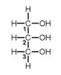
glycerol
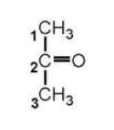
acetone
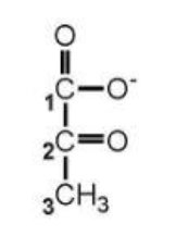
pyruvate
D-glucose (linear)

alpha-D-glucose
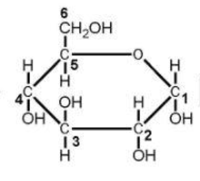
beta-D-glucose
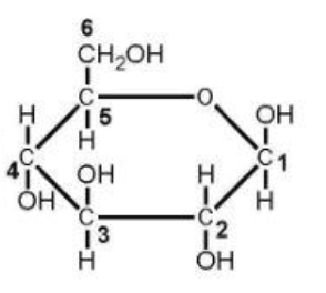
D-fructose (linear)

alpha-D-fructose

beta-D-fructose

Major Dietary Carbohydrates
starch
cellulose
lactose
sucrose
Starch
(common plant storage form)—exists in 2 forms:
a. amylose (~30%): linear glucose polymer with α(1→4) linkages;
b. amylopectin (~70%): branched glucose polymer with linear α(1→4) linkages and branching α(1→6) linkages
Cellulose
(structural building block of plant cells walls)
glucose polymer with β(1→4) linkages
cellulose is not digested and thus constitutes roughage because humans do not have enzymes that can cleave β(1→4) glycosidic linkages (except for lactose)
Lactose
(galactose-β-1,4-glucose): milk sugar
Sucrose
(glucose-α-1,2-fructose): table sugar
Digestion: Mouth
salivary α-amylase: specific cleavage of α-glycosidic bonds
Result: digest dietary carbohydrates to dextrins and disaccharides
dextrins: mixture of shorter branched and unbranched polymers of glucose linked by α(1→4) or α(1→6) glycosidic bonds
Digestion: Stomach
No carbohydrate digestion—HCl stops amylase
Digestion: Intestine
a. Pancreatic α-amylase
b. Result—continued digestion of dextrins to produce more disaccharides
i. isomaltose: disaccharide of glucose with α(1→6) linkage
ii. maltose: disaccharide of glucose with α(1→4) linkages
Digestion: Mucosal Cells
a. disaccharide hydrolysis
b. disaccharidases (at luminal surface of enterocytes):
isomaltase (cleaves isomaltose)
maltase (cleaves maltose)
lactase (cleaves lactose)
sucrase (cleaves sucrose)
v. trehalase (cleaves trehalose)
**trehalose: a disaccharide of glucose linked by an α(1→1) bond; found in mushrooms and other fungi
c. Result: yields monosaccharides, which can now be absorbed by the enterocytes—then released into portal circulation to liver.
i. glucose
ii. fructose
iii. galactose
Digestion Diagram
alpha-Amalyase —> secreted by saliva, digests starch
pancreatic alpha-amalyase —> continues to digest disaccharides
mucosal cell membrane-bound enzymes break down disaccharides into monosaccharides (glucose, fructose, galactose)
What about cellulose? —> unable to digest, cholesterol sticks to it and it is excreted as-is

Na+ dependent absorption
1. glucose
2. galactose
Na+ independent absorption
fructose: driven by [fructose] gradient
Figure: Absorption of dietary monosaccharides by an enterocyte
(a.k.a. intestinal mucosal cell)
Figure Notes: Glucose (glc) and galactose (gal) are at lower concentrations in the lumen and are moving “against” a concentration gradient into the enterocyte through the SGLT (Na+-dependent glucose transporter), which has a higher concentration of glucose and galactose building up within the cell. Movement of a glucose or galactose molecule through the SGLT is called secondary active transport as it requires co-transport of a Na+ ion (described above).
The Na+ and K+ gradients are maintained using a Na+/K+ ATPase, which requires ATP and is called primary active transport. The Na+ gradient is maintained with high [Na+] outside cell and low [Na+] inside cell. The K+ gradient is maintained with high [K+] inside the cell and low [K+] outside the cell.
Once the glucose and galactose build-up inside the enterocyte, they can flow down their concentration gradients from the cell into the portal blood.
Fructose (frc) can only move with its concentration gradient, so must build up to a high concentration in the lumen before it can flow by passive diffusion into the enterocyte.
Once it builds up in the enterocyte, it flows from the cell into the portal blood.
![<p><strong>Figure Notes:</strong> Glucose (glc) and galactose (gal) are at lower concentrations in the lumen and are moving “against” a concentration gradient into the enterocyte through the SGLT (Na<span style="font-family: Arial; line-height: normal; font-size: 8px;">+</span>-dependent glucose transporter), which has a higher concentration of glucose and galactose building up within the cell. Movement of a glucose or galactose molecule through the SGLT is called secondary active transport as it requires co-transport of a Na<span style="font-family: Arial; line-height: normal; font-size: 8px;">+</span> ion (described above).</p><p>The Na<span style="font-family: Arial; line-height: normal; font-size: 8px;">+</span> and K<span style="font-family: Arial; line-height: normal; font-size: 8px;">+</span> gradients are maintained using a Na<span style="font-family: Arial; line-height: normal; font-size: 8px;">+</span>/K<span style="font-family: Arial; line-height: normal; font-size: 8px;">+</span> ATPase, which requires ATP and is called primary active transport. The Na<span style="font-family: Arial; line-height: normal; font-size: 8px;">+</span> gradient is maintained with high [Na<span style="font-family: Arial; line-height: normal; font-size: 8px;">+</span>] outside cell and low [Na<span style="font-family: Arial; line-height: normal; font-size: 8px;">+</span>] inside cell. The K<span style="font-family: Arial; line-height: normal; font-size: 8px;">+</span> gradient is maintained with high [K<span style="font-family: Arial; line-height: normal; font-size: 8px;">+</span>] inside the cell and low [K<span style="font-family: Arial; line-height: normal; font-size: 8px;">+</span>] outside the cell.</p><p>Once the glucose and galactose build-up inside the enterocyte, they can flow down their concentration gradients from the cell into the portal blood.</p><p>Fructose (frc) can only move with its concentration gradient, so must build up to a high concentration in the lumen before it can flow by passive diffusion into the enterocyte.</p><p>Once it builds up in the enterocyte, it flows from the cell into the portal blood.</p>](https://assets.knowt.com/user-attachments/65a137df-25b8-4ea4-a0ef-888ec27bb0f0.png)
The scientific numbering system
uses 1, 2, 3, 4, …
The carbon with the highest oxidation state is designated “Carbon #1”
For molecules containing a carboxylic acid group, like the fatty acid drawn above, this is the carbon with the highest oxidation state, thus is carbon #1. The remaining carbons are numbered sequentially until the end carbon (the methyl carbon in this case).
Greek letters
Typically used for biological molecules having a carboxylic acid group.
The alpha (α) carbon is always the carbon NEXT TO the carboxylic acid carbon
Then designate the subsequent carbons as beta (β), gamma (γ), and so forth.
Note: for fatty acids, regardless of fatty acid chain length, the last carbon (the methyl carbon) is always designated the omega (ω) carbon.
Nomenclature hints
“ic acid”: indicates the molecule has the protonated form of an acid group(s) [i.e. COOH]
“ate”: indicates the molecule has the deprotonated form of an acid group(s) [i.e. COO-]
“yl” ending: indicates a functional group attached to another functional group. For example when a fatty acid is attached to another molecule, it is now designated in the name of the molecule as “fatty acyl” or more commonly “acyl”. So a “triacylglycerol” is a molecule with three fatty acids attached to glycerol.
Oxidation (a.k.a. dehydrogenation)
loss of electrons; or typically for organic molecules—the loss of hydrogens
usually in biological reactions it is a loss of 2 electrons via 2 hydrogens
Reduction (a.k.a. hydrogenation)
gain of electrons; or typically in organic molecules—the gain of hydrogens
usually in biological reactions it is a gain of 2 electrons via 2 hydrogens
Note: Oxidation and reduction reactions are ALWAYS coupled in organic chemistry—those electrons/hydrogens have to go “somewhere” (i.e., living organisms are not made of copper wires).
Hydration
gain of a water molecule
Dehydration
loss of a water molecule
Hydration/dehydration reactions involve the addition or loss of ____________, thus are NOT ____________.
entire water molecule to a compound; oxidation- reduction reactions
Protonate
gain of a proton (H+)
Deprotonate
loss of a proton (H+)
Protonation/deprotonation are _____________, not ___________.
acid- base reactions; oxidation-reduction reactions

Hydrogen terminology

Electron transfer of hydrogens, and coenzymes involved in oxidation-reduction reactions.
NADP+/NADPH is used primarily for synthetic reactions
NADH and FADH2 are important reducing agents in the body
Primary, secondary and tertiary alcohols

Relationship between α-keto acids and α-amino acids
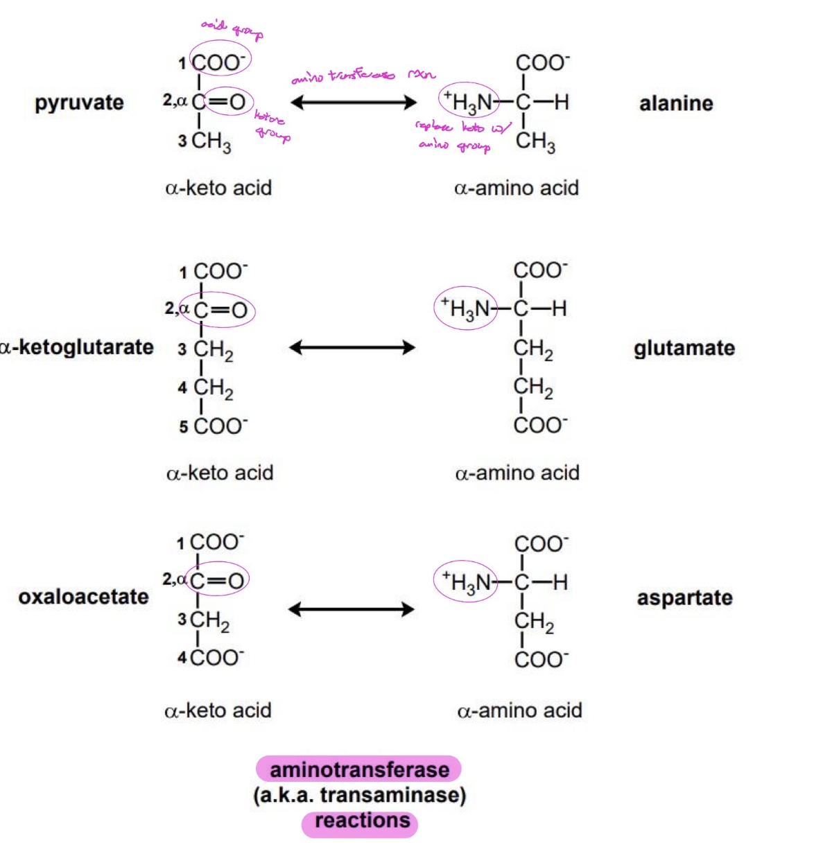
Nomenclature of oxidation states and suffixes

Oxidation States Flow Chart

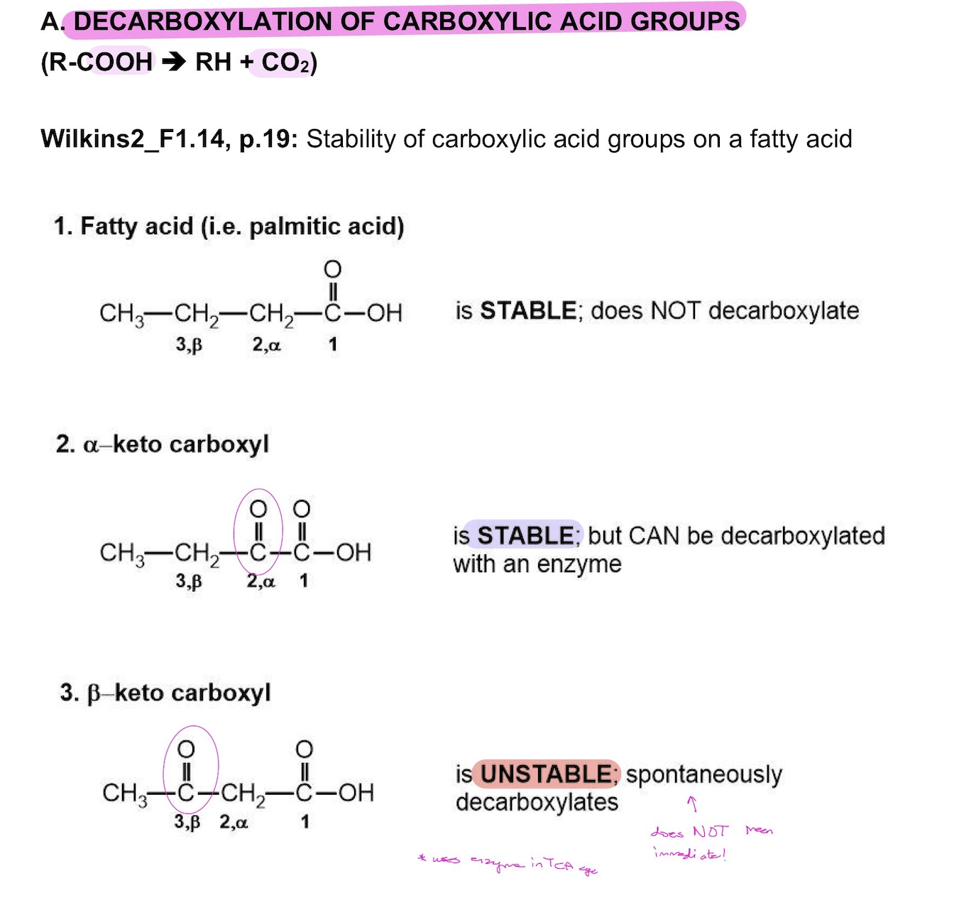
DECARBOXYLATION OF CARBOXYLIC ACID GROUPS
(R-COOH ➔ RH + CO2)
KEY STRATEGIES FOR MAKING ENERGY (ATP)
GOAL: Make ATP, NADH, and FADH2
1. Oxidize by removing hydrogens (dehydrogenation)
2. Oxidize:
Alkanes—use FAD because removing hydrogens from 2 carbons on the molecule being oxidized.
Alcohols—use NAD+ because removing hydrogen from 1 carbon
Aldehydes—use NAD+ because removing hydrogen from 1 carbon
**Also note: the oxidation of aldehydes generally yields a high energy bond “S—P” which can be used for substrate level phosphorylation to yield ATP (or an energy equivalent).
FADH2 and NADH go to the electron transport chain—more ATP ultimately made.
3. Use H2O to add oxygens [note oxygen in CO2 from fuel degradation comes from H2O, not molecular oxygen (O2)].
4. Arrange to produce α- or β-keto acids, so can decarboxylate and get rid of fully oxidized carbons.
Vitamins Definition
Vitamins are compounds that are required for normal growth and are obtained from our diet. They are NOT synthesized, for all practical purposes, by the human body.
Nutritional Considerations
1. Fat-soluble vitamins are not easily removed - can overdose.
2. Water-soluble vitamins are easily cleared. Thus, resupply and deficiencies can become a big issue.
3. Because vitamins play an essential role in many biological reactions, deficiencies lead to a broad range of problems.
This is particularly true in rapid growing and/or metabolizing tissues such as skin, mucosa, red blood cells, and neural tissue.
Developing embryos are also very susceptible to vitamin deficiencies.
Recommended dietary allowance (RDA) [a.k.a. RDI or DRI]
1. RDA:
Defined as the amount of a nutrient that is needed to meet the requirements of nearly all (97-98%) of the healthy population.
RDAs are specific for age and gender - including separate values for pregnant/lactating women.
2. Developed by the Food and Nutrition Board of the National Academy of Sciences/National Research Council.
They are continually updated as new research findings surface.
3. Also called:
Reference Daily Intake (RDI) - This or RDA is what will be currently found on nutrition labels (though this may change!)
Dietary Reference Intake (DRI). These values are the most current and may eventually replace the RDI nomenclature in general.
4. In a “western diet”, deficiencies are rare.
There are populations, however, who are susceptible to vitamin deficiencies (infants, those with infections, alcoholics, etc.). —> damage to gastrointestinal tract can affect vitamin reabsorption in these cases
It is important as clinicians that you be aware of these groups!
Lipid soluble vitamins
Are hydrophobic and are not readily excreted
Are stored in tissues, so one can overdose taking too much of these vitamins
Have complex functions, not all of which are known
The four lipid soluble vitamins are:
Retinol (vitamin A)
functions in the visual cascade (part of rhodopsin);
deficiencies in adults lead to night blindness
Structure (as example)
b. Cholecalciferol (vitamin D)
functions in bone metabolism;
deficiencies in children lead to rickets
c. α-Tocopherol (vitamin E)
functions as an antioxidant;
deficiencies lead to hyporeflexia and ataxia
d. Phylloquinone (vitamin K)
functions in blood clotting cascade;
deficiencies lead to hypothrombinemia and hemorrhagic disease

Water soluble vitamins
The “active” coenzyme (a.k.a. cofactor) forms of the water soluble vitamins are derivatives of the vitamin forms obtained in the diet, except for vitamin C. —> we don’t get the active form in our diet, the vitamins activate in the body
These coenzymes play key roles in metabolism as the carriers of carbon, hydrogen, and nitrogen in many enzymatic reactions.
There are nine water soluble enzymes: 8 “B” vitamins and Vitamin C, which are covered in more detail below.
Note: the DRI values shown below are for reference only, do NOT memorize these values as you will not be tested on the DRI values.
Riboflavin (vit. B2) [DRI – 1.3 mg]
1. Coenzyme derivatives: Flavin Mononucleotide (FMN) and Flavin Adenine Dinucleotide (FAD)
2. Carrier of: two hydrogen atoms (NOT protons)
3. Structures of flavin mononucleotide (FMN, oxidized form; FMNH2, reduced form) and flavin adenine dinucleotide (FAD, oxidized form). [FADH2, the reduced form is not shown.]—See figures below
A nucleotide consists of a nitrogenous base, a 5-carbon sugar (for FMN the sugar is in a linear configuration), and one or more phosphate groups.
Note that the same two nitrogen atoms in the flavin ring are the points where hydrogen atoms are attached in oxidation/reduction (a.k.a. redox) reactions for both FMN and FAD.
4. Enzymes requiring FMN or FAD
a. Dehydrogenase enzymes catalyzing reactions oxidizing an alkane to an alkene always reduce FAD to FADH2, such as succinate dehydrogenase (TCA cycle) and acyl CoA dehydrogenase (fatty acid oxidation). Note that FADH2 is a substrate of the ETC.
b. Cytochrome P450s (CYPs)
a family of heme-containing monooxygenase enzymes (P450 refers to the absorbance at 450 nm of the protein)
important in detoxification of foreign compounds (xenobiotics) such as in acetaminophen overdose.
CYPs use FAD and FMN in redox reactions.
5. Deficiency:
Not associated with any major human disease.
Symptoms of deficiency show up as scaly dermatitis, glossitis of the tongue (tongue appearing smooth and purplish), and cheilosis (breaking and chapping of skin around the mouth).
![<p>1. <em>Coenzyme derivatives</em>: Flavin Mononucleotide (FMN) and Flavin Adenine Dinucleotide (FAD)</p><p class="p1">2. <em>Carrier of</em>: two hydrogen atoms (NOT protons)</p><p class="p1">3. Structures of flavin mononucleotide (FMN, oxidized form; FMNH2, reduced form) and flavin adenine dinucleotide (FAD, oxidized form). [FADH2, the reduced form is not shown.]—<strong><em>See figures below</em></strong></p><ul><li><p class="p1">A nucleotide consists of a nitrogenous base, a 5-carbon sugar (for FMN the sugar is in a linear configuration), and one or more phosphate groups.</p></li><li><p class="p1">Note that the <strong>same</strong> two nitrogen atoms in the flavin ring are the points where hydrogen atoms are attached in oxidation/reduction (a.k.a. redox) reactions for both FMN and FAD.</p></li></ul><p class="p1">4. Enzymes requiring FMN or FAD</p><p class="p1">a. Dehydrogenase enzymes catalyzing reactions oxidizing an alkane to an alkene always reduce FAD to FADH2, such as <em>succinate dehydrogenase </em>(TCA cycle) and <em>acyl CoA dehydrogenase</em> (fatty acid oxidation). Note that FADH2 is a substrate of the ETC.</p><p class="p1">b. Cytochrome P450s (CYPs)</p><ul><li><p class="p1">a family of heme-containing monooxygenase enzymes (P450 refers to the absorbance at 450 nm of the protein)</p></li><li><p class="p1">important in detoxification of foreign compounds (xenobiotics) such as in acetaminophen overdose.</p></li><li><p class="p1">CYPs use FAD and FMN in redox reactions.</p></li></ul><p class="p1">5. Deficiency:</p><ul><li><p class="p1">Not associated with any major human disease.</p></li><li><p class="p1">Symptoms of deficiency show up as scaly dermatitis, glossitis of the tongue (tongue appearing smooth and purplish), and cheilosis (breaking and chapping of skin around the mouth).</p></li></ul><p></p>](https://assets.knowt.com/user-attachments/5879e8e5-e437-4f8d-9bf4-c166edafd053.png)
Niacin (vit. B3) (a.k.a. nicotinic acid) [DRI – 16 mg]
1. Coenzyme derivative: Nicotinamide Adenine Dinucleotide (NAD+)
2. Carrier of: Hydride ion (H-, a hydrogen atom with 2 electrons)
3. Structure of nicotinamide adenine dinucleotide (NAD+)—See figure below
The para position of the nicotin-amide ring is the site of hydride attachment in redox reactions. NAD+ (oxidized form); NADH (reduced form)
Can be phosphorylated forming NADP+ (oxidized form); NADPH (reduced form)
4. NAD+/NADH and NADP+/NADPH are NOT metabolically interchangeable
NAD+/NADH, ~1000/1, favoring metabolite oxidation to make ATP [e.g. key enzyme in glycolysis: glyceraldehyde 3-phosphate dehydrogenase (GAPDH)]; NADH is a substrate of the ETC. —> more NAD+ in the body so typically an oxidizing agent (i.e. it wants to be reduced
NADP+/NADPH, 1/10 to1/100, favoring metabolite reduction, thus NADPH is the form of reducing power used in reductive biosynthesis (i.e., reductase enzymes used for fatty acid synthesis) —> more NADPH in body so typically a reducing agent
5. Deficiency: Pellagra (from Italian meaning “sour skin”)
Characterized by 4 Ds (Dermatitis, Diarrhea, Dementia, and Death).
b. Deficiency found:
In populations whose staple diet is unprocessed corn (the nicotinate in corn needs to be released by alkali extraction before it can be absorbed through the GI tract).
Chronic alcoholics also show deficiency due to absorption problems.
6. Niacin synthesis:
Niacin can be synthesized from the amino acid tryptophan.
However, in all practicality, most people require a dietary source.
![<p>1. <em>Coenzyme derivative</em>: Nicotinamide Adenine Dinucleotide (NAD+)</p><p class="p1">2. <em>Carrier of</em>: Hydride ion (H-, a hydrogen atom with 2 electrons)</p><p class="p1"><em>3. </em>Structure of nicotinamide adenine dinucleotide (NAD+)—<strong><em>See figure below</em></strong></p><ul><li><p class="p1">The <em>para</em> position of the nicotin-amide ring is the site of hydride attachment in redox reactions. NAD+ (oxidized form); NADH (reduced form)</p></li><li><p class="p1">Can be phosphorylated forming NADP+ (oxidized form); NADPH (reduced form)</p></li></ul><p>4. NAD+/NADH and NADP+/NADPH are <strong>NOT</strong> metabolically interchangeable</p><ul><li><p class="p1">NAD+/NADH, ~1000/1, favoring metabolite <em>oxidation</em> to make ATP [e.g. key enzyme in glycolysis: glyceraldehyde 3-phosphate <em>dehydrogenase </em>(GAPDH)]; NADH is a substrate of the ETC. —> more NAD+ in the body so typically an oxidizing agent (i.e. it wants to be reduced </p></li><li><p class="p1">NADP+/NADPH, 1/10 to1/100, favoring metabolite reduction, thus NADPH is the form of reducing power used in reductive biosynthesis (i.e., <em>reductase </em>enzymes used for fatty acid synthesis) —> more NADPH in body so typically a reducing agent </p></li></ul><p class="p1">5. Deficiency: Pellagra (from Italian meaning “sour skin”)</p><ul><li><p class="p1">Characterized by 4 Ds (Dermatitis, Diarrhea, Dementia, and Death).</p></li></ul><p class="p1">b. Deficiency found:</p><ul><li><p class="p1">In populations whose staple diet is unprocessed corn (the nicotinate in corn needs to be released by alkali extraction before it can be absorbed through the GI tract).</p></li><li><p class="p1">Chronic alcoholics also show deficiency due to absorption problems.</p></li></ul><p class="p1">6. Niacin synthesis:</p><ul><li><p class="p1">Niacin <em>can</em> be synthesized from the amino acid tryptophan.</p></li><li><p class="p1">However, in all practicality, most people require a dietary source.</p></li></ul><p></p>](https://assets.knowt.com/user-attachments/e65d5cf1-715d-4943-b078-18769056df81.png)
Niacin: A clinical case illustrating an example of new challenges to physicians as a result of behavioral changes in society
A previously healthy 14-year-old boy presented to the Emergency Department with nausea, vomiting, upper abdominal pain, palpitations, and dizziness for about 6 hours. There was no history of chest pain, shortness of breath, fever, cough, nasal congestion, diarrhea, or headache. Vital signs: temperature, 95.2ºF; blood pressure 111/48 mm Hg; pulse rate, 105 beats/min; and respiratory rate 18 breaths/min. Other laboratory data indicated hepatotoxicity, metabolic acidosis, and hypoglycemia evolving into hyperglycemia.
Upon questioning, he admitted to smoking marijuana for the preceding few weeks. He was due to meet with his parole officer on the day he became sick. To beat his urine drug test, he had ingested eleven 500-mg tablets of timed- release niacin, totaling 5.5 g during 36 hours before the onset of symptoms. He had toxicity from use of niacin to beat urine drug screening!
Ascorbate (vit. C) [DRI – 90 mg]
1. Coenzyme derivative: NONE (form ingested is the active form)
2. Carrier of: two hydrogen atoms (NOT protons)
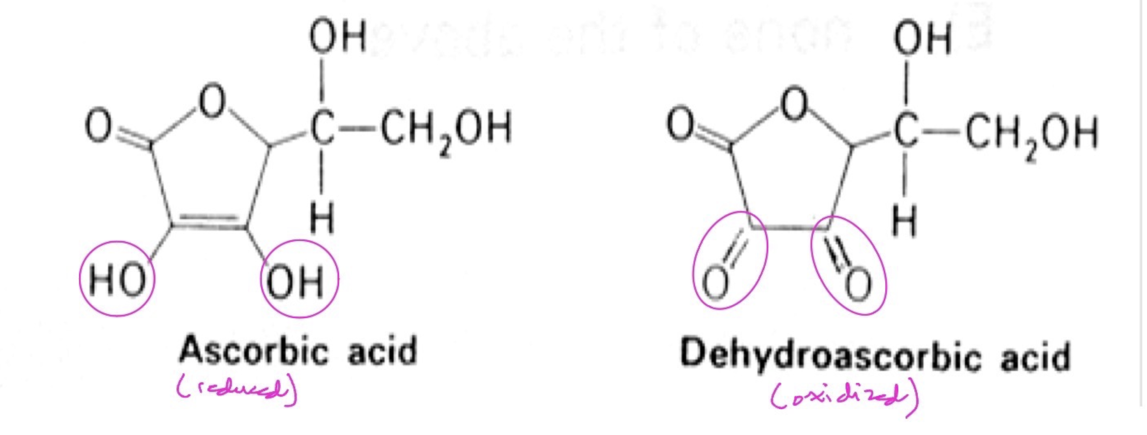
3. Structures of ascorbic acid (reduced form) and dehydroascorbic acid (oxidized form)—See figure below
4. Function and enzymes requiring vitamin C: acts as an antioxidant (reducing agent); important in hydroxylation reactions, such as the action of prolyl and lysyl hydroxylases on collagen.
5. Deficiency: Scurvy
Vitamin C is essential for the formation of functional collagen (which contains hydroxylated amino acids)
Symptoms (like easy bleeding and bruising, oral gum regression (see figure below), and joint pain) are directly related to the weakening of collagen-containing tissues such as blood vessels, connective tissue, and bone.
6. Fun fact: Most mammals can synthesize ascorbate from glucose. However, humans, other primates, guinea pigs, and bats lack the terminal enzymes. We still have the gene, but it is mutated so that no protein is produced (happened about 1 million years ago)!
Thiamine (vit. B1) [DRI – 1.2 mg]
1. Coenzyme derivative: Thiamine pyrophosphate (TPP)
2. Carrier of: Carbon chains (active site is the transfer point)
3. Structure of thiamine pyrophosphate—See figure below
4. Enzymes requiring TPP
pyruvate dehydrogenase complex: pyruvate to acetyl CoA for entry into the TCA cycle
α-ketoglutarate dehydrogenase complex: TCA cycle enzyme
transketolase: pentose phosphate pathway enzyme
5. Deficiency: Wernicke-Korsakoff syndrome; a.k.a. Beri Beri (“I cannot, I cannot”)
a. Clinical manifestations
Wernicke encephalopathy: mental status changes (apathy, mild confusion, to complete coma); ocular abnormalities such as nystagmus (involuntary eye movement); gait ataxia (unsteady, uncoordinated walk like a "drunken sailor")
Korsakoff syndrome: persistent state of mental dysfunction; memory impairment associated with confabulation (giving blatantly false information but with no real intent to deceive)
b. Biochemistry and pathology of disease
thiamine deficiency affects cerebral energy use, initiating tissue injury in brain regions with high metabolic requirements;
lactic acidosis due to accumulation of pyruvate and lactate
c. Presentation by malnourished individuals, or undernourished, such as alcoholics, and post-surgical patients, particularly those with gastric bypass. [Another new challenge to physicians resulting from "advances in medicine" in the form of bariatric surgery.]
![<p>1. <em>Coenzyme derivative</em>: Thiamine pyrophosphate (TPP)</p><p class="p1">2. <em>Carrier of</em>: Carbon chains (active site is the transfer point)</p><p class="p1">3. Structure of thiamine pyrophosphate—<strong><em>See figure below</em></strong></p><p class="p1">4. Enzymes requiring TPP</p><ul><li><p class="p1">pyruvate dehydrogenase complex: pyruvate to acetyl CoA for entry into the TCA cycle</p></li><li><p class="p1">α-ketoglutarate dehydrogenase complex: TCA cycle enzyme</p></li><li><p class="p1">transketolase: pentose phosphate pathway enzyme</p></li></ul><p class="p1">5. Deficiency: Wernicke-Korsakoff syndrome; a.k.a. Beri Beri (“I cannot, I cannot”)</p><p class="p1">a. Clinical manifestations</p><ul><li><p class="p1">Wernicke encephalopathy: mental status changes (apathy, mild confusion, to complete coma); ocular abnormalities such as nystagmus (involuntary eye movement); gait ataxia (unsteady, uncoordinated walk like a "drunken sailor")</p></li><li><p class="p1">Korsakoff syndrome: persistent state of mental dysfunction; memory impairment associated with confabulation (giving blatantly false information but with no real intent to deceive)</p></li></ul><p class="p1">b. Biochemistry and pathology of disease</p><ul><li><p class="p1">thiamine deficiency affects cerebral energy use, initiating tissue injury in brain regions with high metabolic requirements;</p></li><li><p class="p1">lactic acidosis due to accumulation of pyruvate and lactate</p></li></ul><p class="p1">c. Presentation by malnourished individuals, or undernourished, such as alcoholics, and post-surgical patients, particularly those with gastric bypass. [Another new challenge to physicians resulting from "advances in medicine" in the form of bariatric surgery.]</p>](https://assets.knowt.com/user-attachments/40218205-c271-4d0e-b254-238ec89d2b64.png)
Pantothenic acid (vit. B5) [DRI – 5 mg]
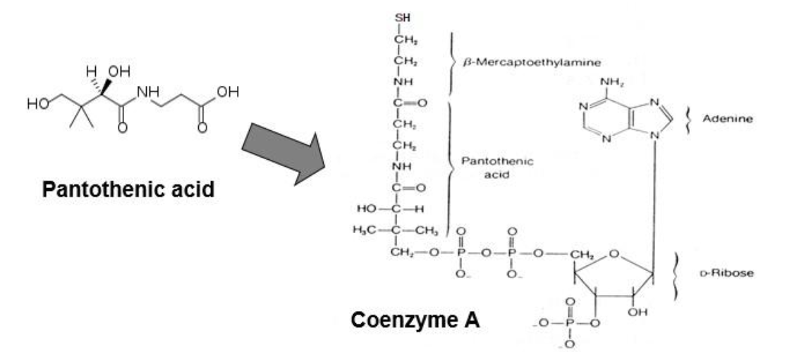
1. Coenzyme derivative: Coenzyme A (CoA), and is part of the acyl carrier protein (ACP) of the fatty acid synthase complex
2. Carrier of: carbon chains (acyl groups)
3. Structure of pantothenic acid and coenzyme A (See figure below). Active site is a thiol group (-SH) that carries acyl compounds as activated thiol esters.
4. Examples of acyl compounds carried by coenzyme A
acetyl CoA
succinyl CoA
fatty acyl CoA
5. Deficiency: not well characterized; is very rare, and all reported are reversible with addition of pantothenate.
For our purposes, no true deficiency has been characterized and therefore, opinions regarding DRI range widely.
The word pantothenate comes from the Greek and means “from everywhere”. Pantothenic acid is found ubiquitously in natural foods.
And it’s a good thing too because CoA is required for the metabolism of all fat, protein, and carboh
Biotin (vit. B7 or vit. H) [DRI – 30 μg]
1. Coenzyme derivative: covalently attached to carboxylase enzymes
2. Carrier of: one-carbon group, specifically carboxyl (CO2) groups
3. Structure of biotin and its attachment to an enzyme (see figure below)
4. Enzymes requiring biotin:
All carboxylases
Entry into 2 key metabolic pathways involve carboxylases: pyruvate carboxylase in gluconeogenesis and acetyl CoA carboxylase in fatty acid synthesis.
5. Deficiency: Rare (the vitamin is present in a wide variety of foods and in intestinal bacteria).
Groups at an increased risk of deficiency include pregnant women and anyone who eats a lot of raw eggs! —> **Egg white protein avidin binds tightly to biotin (so much so, that it is used as a tool in the research setting) preventing its absorption. Cooking denatures avidin and releases the biotin.
Deficiency is characterized by hair loss, dermatitis, and neurological symptoms (muscle pain, hallucinations, depression).
Subclinical biotin deficiency has been demonstrated in over half of pregnant women in several studies presumably due to high fetal demand.
Genetic disease(s) in which holocarboxylase synthetase (HLCS), the enzyme that attaches biotin to lysine residue of carboxylases is deficient/defective leads to an apparent biotin deficiency, even if the vitamin is plentiful.
6. Note: Biotin is sometimes called vitamin H, from German words meaning hair and skin

Folate (vit. B9) [DRI – 400 μg; pregnant/lactating women – 600 μg]
1. Coenzyme derivative: Tetrahydrofolate (THF)
2. Carrier of: one-carbon groups; can carry one-carbon groups at different oxidation states: methyl, methylene, methenyl, and formyl
3. Structure of folic acid and one form of it as a carrier of a one-carbon group, N5, N10-methylene-tetrahydrofolate (see figure below)
4. Enzymes requiring THF:
Enzymes that catalyze one-carbon transfer reactions that are particularly important in nucleotide synthesis (purine synthesis and thymidine synthesis)
Enzymes that catalyze one-carbon transfer reactions involved in amino acid synthesis, such as methionine.
5. Deficiency: Absolutely critical to neural tube development.
Neural tube defects and spontaneous abortion (miscarriage) frequency increases with folate deficiency.

Cobalamin (vit. B12) [DRI – 2.4 μg]
1. Coenzyme derivative: Cobamide coenzymes
The only known reason that cobalt is required as a trace mineral in our diets!
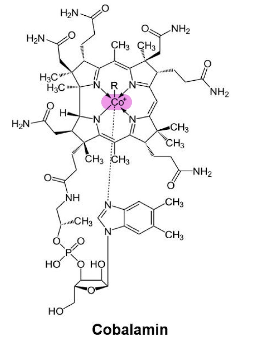
2. Carrier of: one-carbon group, specifically methyl (-CH3) groups
3. Structure of cobalamin (see figure below)
4. Two essential enzymes require cobalamin:
Homocysteine methyltransferase, which remethylates homocysteine to methionine.
Methylmalonyl CoA mutase, which is involved in catabolism of odd- numbered carbon fatty acids and some amino acids.
5. Deficiency: Pernicious anemia
Vitamin B12 is required in the process of making red blood cells.
A lack of this vitamin leads to this form of megaloblastic anemia.
Pyridoxal (vit. B6) [DRI – 1.7 mg]
1. Coenzyme derivative: Pyridoxal phosphate
2. Carrier of: Nitrogen group (amine group in transamination reactions)
3. Structures of pyridoxal phosphate (PLP and PMP) in a transamination reaction (see figure below)
4. Enzymes requiring pyridoxal phosphate:
All aminotransferases (a.k.a. transaminases), many of them
Examples: alanine aminotransferase (ALT) and aspartate aminotransferase (AST)
5. Deficiency: fairly rare except for drug induced - isoniazids (used to treat TB) induce a Vitamin B6 deficiency, though the mechanism is not clear.

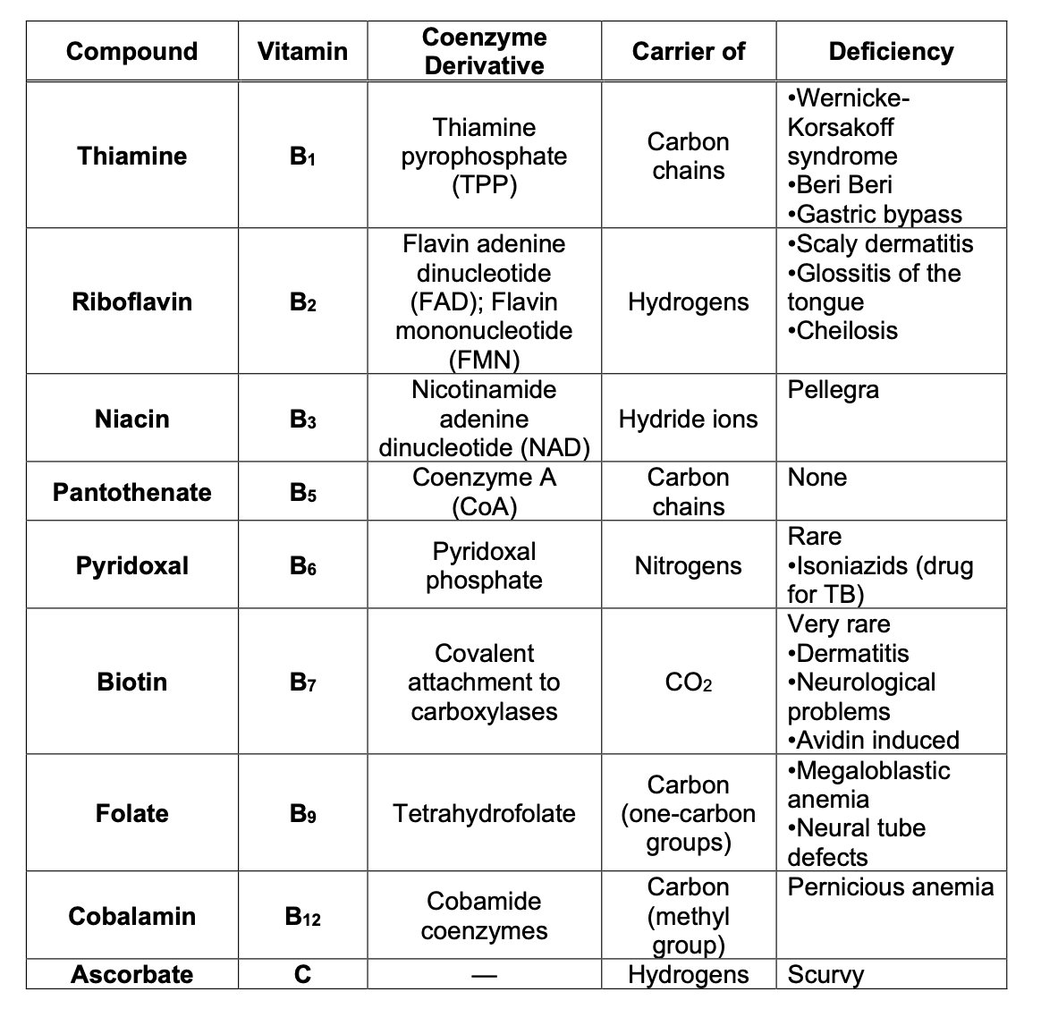
Summary of the Water Soluble Vitamins
Food Sources of the Water Soluble Vitamins

Penetrance
The probability that an individual with a specific genotype will express a phenotype to any degree.
Expressed as a percentage, i.e., the number of affected people with the genotype over the total number of people with the genotype.
**just because you have the same genotype doesn’t mean the phenotype will present
Complete penetrance
Everyone with a given genotype will express the phenotype to some degree.
All or nothing concept. Either the disease manifests itself or it does not.
**most common
Reduced penetrance
Not everyone with a particular genotype will express the phenotype.
For example, a person who is an obligate carrier (i.e., heterozygous) of an autosomal dominant disease gene may show no symptoms.
Condition may appear to ‘skip generations’. —> they have the genotype but don’t express the phenotype
A condition with 90% penetrance: means that only 90% of individuals who inherit the mutation will actually express phenotype.
Examples:
a. Split hand deformity: ~70% penetrant.
b. Hereditary cancer syndromes have reduced/variable penetrance.
Example: Retinoblastoma
i. Malignant tumor of the retina
ii. Presents in early childhood
iii. 60% sporadic: unifocal, avg. age of dx- 24 months
iv. Hereditary retinoblastoma: ~90% penetrant. (See figures next page)
Not everyone with the genotype will develop the condition.
40% hereditary: may be bilateral or multifocal, avg. age of dx-15 months
If family history present, may be diagnosed in first month of life.
The effect of reduced penetrance on recurrence risk
a. Dominant conditions: unaffected person can have an affected child
b. Risk of inheriting the mutation does not change, but the risk of developing the disease does.
c. Unknown if person is at 50% risk or no risk to have an affected child.
So, what is the chance that a child of an individual with hereditary retinoblastoma will also develop retinoblastoma?
a. ½ = chance offspring will inherit the mutant allele
b. 9/10 = chance the offspring will express the condition if inherits the mutant allele (because hereditary retinoblastoma is 90% penetrant).
c. Thus, ½ X 9/10 = 9/20 or ~45% chance
Variable expressivity
1. Definition: Manifestation of a disease is variable amongst individuals that have the same genotype.
a. Differences in:
what features are present.
how severely an affected an individual is (can range from mild, possibly even unnoticed, to severe).
b. Difficult to predict phenotype in families based on presentation in affected relatives.
c. Affected individuals that are mildly affected may not have come to clinical attention
2. The majority of genetic conditions exhibit variable expressivity!
3. Example: Tuberous Sclerosis (TS) [See example pedigree below]; Common features include:
Skin findings (facial angiofibromas, hypomelanotic macules, shagreen patch, ungual or periungual fibromas)
CNS (subependymal glial nodules, cortical tubers, seizures, cognitive impairment)
Renal and retinal harmartomas
Cardiac rhabdomyoma
![<p>1. Definition: Manifestation of a disease is variable amongst individuals that have the same genotype.</p><p>a. Differences in:</p><ul><li><p>what features are present.</p></li><li><p>how severely an affected an individual is (can range from mild, possibly even unnoticed, to severe).</p></li></ul><p>b. Difficult to predict phenotype in families based on presentation in affected relatives.</p><p>c. Affected individuals that are mildly affected may not have come to clinical attention</p><p>2. The majority of genetic conditions exhibit variable expressivity!</p><p>3. Example: <strong><em>Tuberous Sclerosis</em></strong> (TS) [<strong><em>See example pedigree below]</em></strong>; Common features include:</p><ul><li><p>Skin findings (facial angiofibromas, hypomelanotic macules, shagreen patch, ungual or periungual fibromas)</p></li><li><p>CNS (subependymal glial nodules, cortical tubers, seizures, cognitive impairment)</p></li><li><p>Renal and retinal harmartomas</p></li><li><p>Cardiac rhabdomyoma</p></li></ul><p></p>](https://assets.knowt.com/user-attachments/a869432a-8270-4edb-836e-98fe9014bf9c.png)
Allelic heterogeneity
1. Definition: Several different alleles (mutations) of one gene that all cause the same disease
The expression of the disease may vary depending on which disease allele(s) are present in an individual (variable expressivity).
Example: Cystic fibrosis
a. Over 1000 known mutations at the CFTR gene at locus 7q31 with varying severity.
b. 70% of mutations in the Caucasian population are: ∆F508 —> deletion of phenylalanine at 508
c. Median age of survival: 30s
Death usually due to end-stage lung disease
d. Cystic fibrosis also demonstrates:
Variable expressivity: most have respiratory and digestive, others only respiratory
Pleiotropy: effects on multiple different organ systems
Locus heterogeneity
Definition: Mutations at different loci (i.e., in different genes) causing the same disease or phenotype.
Often occurs when two different genes are responsible for creating a compound gene product.
***PDH Complex —> E1, E2, E3 subunits coded by different genes, and a mutation in any of these genes can cause the whole complex not to work; they may be different mutations but present the same clinically
Example: Hearing loss
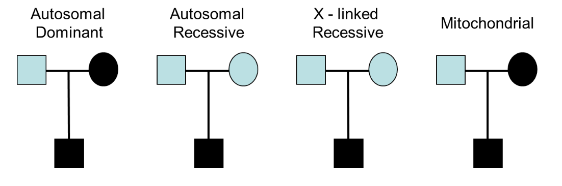
a. several genes involved in non-syndromic hearing loss
b. different inheritance patterns (see figure below)
Pleiotropy
Definition: A single gene producing diverse phenotypic effects
A pleiotropic disease gene could have multiple adverse effects in different organ systems/body areas.
So one gene mutation can cause a lot of other effects depending on severity of condition
Example: Cystic fibrosis
Definition: All heritable conditions are present at birth, but not all are expressed at birth.
a. Difficult to predict recurrence risks in children of at-risk adults, as it is unknown whether or not the parent will be affected
b. Genetic testing may be available to predict who will develop disease.
Example: Huntington disease
a. Progressive cognitive, psychiatric and motor decline
b. Symptoms generally not seen until age 30 or older

De novo mutations
1. Definition: A mutation that occurs spontaneously, i.e., is not inherited.
2. De novo mutation occurs first in proband (spontaneously).
When it occurs in the egg or sperm cell involved in conception of proband, the mutation is present in all cells of the proband.
3. Recurrence risks (See pedigree below)
a. Parents are not at increased risk in future pregnancies
b. However, proband does have increased risk of passing mutation to child.
Risk would be the same for someone who inherited the mutation
4. Frequent in some diseases
5. More commonly seen for autosomal dominant or X-linked conditions, less commonly seen in autosomal recessive conditions —> it would be really rare for a sperm and an egg cell to both spontaneously mutate at the same time
6. Examples:
a. Neurofibromatosis: 50%
b. Tuberous sclerosis: 67% (2/3 of time is due to NEW mutation)
c. Duchenne muscular dystrophy —> prevents males from having children, so it MUST arise spontaneously
Genetically lethal, new mutation rate must = 1/3 to maintain disease frequency

Mosaicism
Presence of two different cell lines in the body
Somatic mosaicism
Mutation occurs after fertilization, affecting a subpopulation of cells.
Only a percentage of cells have the mutation.
1. If the proportion of affected cells is high enough, mosaic individual will manifest disease.
2. The expression of the disease may be less severe due to presence of normal cells.
3. May also result in expressing only specific features or affecting only certain regions of the body.
4. Difficult to determine accurate risk of recurrence (because do not know if the germ cells are affected)
there is NO WAY to say whether this will be passed down or not because it is determined by whether or not the affected fetal stem cells went on to make gametes

Example: Segmental neurofibromatosis type I
May only see café-au-lait spots, neurofibromas, axillary and inguinal freckling limited to a specific area of the body, and they do not cross the midline.

What is important to keep in mind about mosaicism?
It only is in the individual! —> if the issue affects germ line cells the child will go on to inherit the ENTIRE MUTATION i.e. in very cell of their body (goes to classic inheritance patterns)
Germline mosaicism
Definition: Mosaicism present in germ cells only.
Due to a mutation that occurs after fertilization, during mitosis (not meiosis) of germ cells.
Results in a portion of the germ cells having a mutation that the parent does not express. (See graphic representation below)
1. Can lead to an unaffected parent having several affected children, since the abnormal cells are present in a proportion of the gametes.
2. With only one affected child in the family, cannot determine if this is due to a de novo mutation in child (with no increased risk for future sibs) or parental germline mosaicism (with an increased risk)
3. The majority of genetic conditions are thought to have a rate of 1% or less for germline mosaicism.
2 Examples: Duchenne muscular dystrophy & Osteogenesis imperfecta
Duchenne muscular dystrophy
• 15% risk of germline mosaicism in the mother when proband is only affected family member.
Osteogenesis imperfecta
5-6% chance of germline mosaicism
Abnormality of collagen formation, with increased risk of fractures.
Different types—spectrum of mild (few fractures) to lethal in the newborn period.
Skewed X-inactivation
1. Definition: One X chromosome appears to be “preferentially” inactivated.
Recall that all females are mosaic for their expression of their two X chromosomes, with normally about 50% of a female’s cells having the maternal X inactivated and 50% having the paternal X inactivated.
2. Note that in reality, at the cell stage when an X chromosome is inactivated, the process is still random. However, we only see “the survivors”—the females in which the majority of cells inactivated an X chromosome that allows for survival, though the female may still be severely affected.
3. Can be minimally skewed or as high at 100% (See figure below)
The higher the skewed percentage, the more likely to have an effect.

X-linked recessive (XLR)
Female carriers may express phenotype
If the majority of cells have the active abnormal X, females may express some or all features of the condition (depending on the degree of skewing).
Example: Duchenne muscular dystrophy: 1/3 of females may have muscle weakness.
X-linked dominant (XLD)
a. Females with mutations may have milder expression than males
b. Females who carry mutation may not exhibit expected phenotype.
If normal X is more often inactivated (mutation is expressed in most cells): more severe phenotype.
If mutated X is more often inactivated (normal gene is expressed in most cells): milder than typical, possibly appear unaffected.
c. Example: Rett syndrome
Females:
Normal growth/development until 6-18 months.
Then, enter a period of developmental stagnation and rapid regression: progressive microcephaly, loss of purposeful hand movements (hand wringing), progressive cognitive and developmental regression, seizures, etc.
Variation in severity occurs.
Rarely, a female with no or mild clinical expression may have child who is severely affected.
Males:
Severe neonatal encephalopathy, seizures, abnormal tone, and breathing abnormalities.
Rarely survive infancy.
Incomplete dominance
1. Definition: Expression of heterozygote (Aa) is different from and intermediate to both homozygotes (AA, aa).
2. Heterozygotes are affected, but homozygotes are more severely (often lethally) affected.
Example: Achondroplasia
a. Most common form of inherited dwarfism.
b. Short stature with disproportionately short arms and legs (rhizomelic shortening).
c. Intelligence and life span are usually normal.
d. Common for both parents to have condition (Aa x Aa)—See Punnett square below.
