MUSCULOSKELETAL SYSTEM
 Call Kai
 Learn
Learn Practice Test
 Spaced Repetition
 Match
Match Flashcards
 Knowt Play
1/105

There's no tags or description
Looks like no tags are added yet.
Name | Mastery | Learn | Test | Matching | Spaced | Call with Kai | Chat |
|---|
No analytics yet
Send a link to your students to track their progress
106 Terms
Muscle Atrophy
Reduction in muscle mass or decreased myofiber diameter, caused by physiologic processes, metabolic disorders, or denervation, key phrase is smaller muscle fibers due to decreased use, disease, or loss of nerve supply.
Physiologic Muscle Atrophy
Normal reduction in muscle size caused by disuse, cachexia, or aging, key phrase is disuse atrophy, occurs without primary muscle disease.
Endocrine Muscle Atrophy
Muscle atrophy secondary to hypothyroidism, hypercortisolism (Cushing's disease), or pituitary dysfunction or tumors in aging horses, commonly seen in dogs and older horses.
Denervation Atrophy
Progressive muscle atrophy due to loss of nerve supply from peripheral nerve injury or neurologic disease, myofibers progressively decrease in diameter, classic example is equine laryngeal hemiplegia.
Muscle Hypertrophy
Increase in myofiber size by addition of myofilaments, caused by increased workload or compensatory response, characterized by larger muscle fibers rather than increased fiber number.
Physiologic Hypertrophy
Normal increase in muscle size caused by exercise or increased functional demand, normal adaptive response.
Compensatory Hypertrophy
Enlargement of remaining functional myofibers after loss of functional fibers or disruption of normal cellular processes, remaining fibers enlarge to compensate for damaged or lost muscle fibers.
Rhabdomyolysis
Necrosis of skeletal muscle fibers caused by skeletal muscle injury, characterized by myoglobinuria and increased Creatine Phosphokinase (CK/CPK), key phrase is muscle necrosis.
Myoglobinuria
Presence of myoglobin in urine due to muscle fiber breakdown following rhabdomyolysis, hallmark finding and clinically detectable in urine.
↑ Creatine Phosphokinase (CK/CPK)
Elevated serum muscle enzyme indicating skeletal muscle damage due to leakage from injured muscle, one of the most important diagnostic criteria for rhabdomyolysis.
Ischemic Myonecrosis (Ischemic Necrosis)
Necrosis of skeletal muscle due to inadequate blood supply caused by major blood vessel occlusion, external pressure, compartment syndrome, or vasculitis or vasculopathy, downer cows develop localized pale muscle from prolonged recumbency and arterial compression, myofibers are most sensitive to anoxia followed by satellite cells then fibroblasts, severity depends on vessel size and duration of ischemia.
Nutritional Myopathy (White Muscle Disease)
Degeneration and necrosis of skeletal and cardiac muscle caused primarily by selenium deficiency with or without vitamin E deficiency, characterized by white streaks, chalky calcification, and pale muscles, affects cattle, horses, sheep, and goats, especially neonates, selenium is required for glutathione peroxidase.
Toxic Myopathy
Muscle necrosis caused by toxic plants, contaminated feed, hay, or ionophore antibiotics including monensin, lasalocid, narasin, and maduramicin, ionophores cause calcium overload leading to skeletal and cardiac muscle necrosis, clinical signs include muscle weakness, recumbency, and increased CK and AST, diagnosis requires toxin identification, horses are extremely sensitive while ruminants tolerate lower doses.
Exertional Myopathy (Exertional Rhabdomyolysis)
Exercise-induced rhabdomyolysis caused by overexertion, selenium deficiency, muscular dystrophy, electrolyte depletion, or glycogen storage disease, known as capture myopathy in wild animals, tying-up syndrome in horses, and also occurs in working dogs such as sled dogs and greyhounds.
Traumatic Myopathy
Muscle injury and necrosis caused by crush injuries, lacerations, excessive stretching, burns, injections, or fractures, heals mainly by fibrosis resulting in scar tissue and reduced muscle function, may cause muscle rupture especially in the diaphragm of dogs, cats, and horses.
Myositis Ossificans
Fibrosis with osseous metaplasia causing bone formation within muscle following chronic muscle trauma, key phrase is bone forms inside injured muscle.
Musculoaponeurotic Fibromatosis
Progressive fibrous tissue proliferation causing muscle atrophy due to chronic fibrous proliferation after injury, mainly affects horses.
Gastrocnemius Muscle Rupture (Horse)
Rupture of the gastrocnemius muscle origin caused by exercise injury or struggling to rise, species specific to horses.
Adductor Muscle Tear (Splits)
Tearing of hindlimb adductor muscles caused by slipping on smooth or slippery floors, species specific to cattle, key phrase is splits.
Myositis
Inflammation of skeletal muscle with or without fluid exudate caused by infectious agents, parasites, foreign bodies, or immune-mediated causes, inflammatory patterns include perivascular, perimysial, endomysial, and granulomatous.
Perivascular Myositis
Inflammation surrounding blood vessels caused by perivascular inflammatory infiltration, affects perivascular tissues.
Perimysial Myositis
Inflammation of the perimysium caused by inflammatory infiltration of connective tissue surrounding muscle fascicles, key phrase is between muscle fascicles.
Endomysial Myositis
Inflammation surrounding individual muscle fibers, usually macrophage-mediated, key phrase is inflammation around individual myofibers.
Granulomatous Myositis
Formation of granulomas within muscle caused by foreign bodies, parasite encystment, fungi, or bacteria.
Clostridial Myositis (Gas Gangrene, Malignant Edema, Blackleg)
Acute hemorrhagic myonecrosis with gas production caused by Clostridium chauvoei, Clostridium perfringens, Clostridium septicum, or Clostridium novyi, toxins damage myofibers and intramuscular vessels, producing hemorrhagic myonecrosis with pseudocystic gas spaces, toxemia is common and often fatal, blackleg occurs in cattle, gas gangrene or malignant edema in horses, organisms are Gram-positive bacilli.
Pyogenic Bacterial Myositis
Suppurative muscle inflammation with myofiber necrosis caused by pyogenic bacteria introduced through wounds or trauma, may produce localized abscesses or spread along fascial planes.
Pasteurella Myositis (Cats)
Cellulitis extending into adjacent muscle caused by Pasteurella multocida after cat bite wounds, species specific to cats.
Chronic Fibrosing Myositis
Chronic fibrosing nodular inflammation of the tongue muscles caused by Actinobacillus lignieresii, Actinomyces bovis, or Staphylococcus aureus, mainly affects cattle, includes wooden tongue, lumpy jaw, and botryomycosis in horses and pigs.
Viral Myositis
Inflammation and necrosis of muscle caused by viral infection, gross lesions are often absent or appear as small poorly defined foci, injury occurs through vasculitis-induced infarction such as bluetongue in sheep or direct viral myofiber necrosis.
Parasitic Myositis
Muscle inflammation caused by Neospora caninum, Hepatozoon americanum, Trypanosoma cruzi, Trichinella spiralis, or Sarcocystis spp., most infections are clinically insignificant, important pathogens include Neospora, Hepatozoon, and T. cruzi in dogs, and Trichinella in pigs.
Sarcocystosis
Intramyofiber protozoal cysts caused by Sarcocystis spp., characterized by dark purple intramyofiber cysts in cattle, also occurs in horses, sheep, and goats, usually incidental with minimal inflammation because cysts are intracellular.
Trichinosis
Encysted larvae within skeletal muscle caused by Trichinella spiralis, characterized by pale elongated gray-white cysts in the diaphragm, pigs are the common reservoir and bears are also affected, inflammation is minimal until larvae die.
Malignant Hyperthermia (Porcine Stress Syndrome, PSS)
Inherited metabolic myopathy caused by an autosomal recessive genetic mutation, most severe in homozygous pigs, triggered by stress or anesthesia, characterized by Pale Soft Exudative (PSE) pork, pale pink moist swollen lumbar epispinal muscles, predisposes to death, postmortem muscle deterioration, and poor pork quality, common in Landrace, Pietrain, Hampshire, and Yorkshire pigs.
Pale Soft Exudative (PSE) Pork
Postmortem muscle degeneration caused by Porcine Stress Syndrome, grossly appears pale, soft, wet, and edematous, major cause of poor pork quality.
Rhabdomyoma
Benign tumor of striated skeletal muscle caused by neoplastic proliferation of skeletal muscle cells, occurs in dogs, horses, and cats, commonly affects the heart and lungs.
Rhabdomyosarcoma
Malignant tumor of striated skeletal muscle arising from primitive mesenchymal cells, occurs in dogs, horses, and cats, may develop in sites with little or no skeletal muscle, morphologic variants include round cell, spindle cell, and mixed round plus spindle cell.
Botryoid Rhabdomyosarcoma
Variant of rhabdomyosarcoma with a grape-like appearance, commonly occurs in the urinary bladder as a malignant mesenchymal neoplasm.
Osteoblasts
Bone-forming cells responsible for continuous new bone formation during bone remodeling, key function is building bone.
Osteoclasts
Bone-resorbing cells that digest existing bone tissue during bone remodeling, key function is breaking down bone.
Bone Remodeling
Continuous bone formation and bone resorption throughout life through coordinated activity of osteoblasts and osteoclasts, allowing bones to adapt, repair, and renew.
Bone Trauma
Injury to bone without fracture causing periosteal elevation due to edema and hemorrhage, most bone disorders in animals are trauma-related.
Metabolic Bone Disorders
Bone abnormalities caused by metabolic disease, less common than traumatic bone disorders.
Nutritional Bone Disorders (General)
Bone disease caused by nutritional deficiencies or imbalances, less common than trauma and commonly affects growing animals.
Congenital/Hereditary Bone Disorders
Developmental bone abnormalities present at birth caused by genetic or hereditary defects, less common causes of skeletal disease.
Lameness
Abnormal gait caused by bone, joint, muscle, or nerve disorders, often the first clinical sign of skeletal disease.
Bone Pain
Pain originating from skeletal tissues caused by trauma, inflammation, fractures, or bone disease, an early indicator of bone abnormalities.
Physitis
Inflammation and swelling of the growth plates (physes) of long bones caused by rapid growth, nutritional imbalance, or excessive body weight, commonly affects young rapidly growing horses, especially the carpus, tarsus, and fetlock, clinical signs include pain, growth plate swelling, lameness, and severe limb deformities.
Bone Fracture
Partial or complete break in one or more bones caused mainly by vehicular accidents, falls, fights, or firearms, may involve single or multiple bones and is classified by fracture pattern, growth plate involvement, and skin integrity.
Fracture Classification
Fractures are classified by skin integrity as closed or open, by fracture pattern as transverse, linear, oblique, spiral, greenstick, or comminuted, and by growth plate involvement using the Salter-Harris classification.
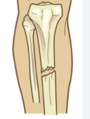
Closed Fracture
Fracture without communication with the external environment caused by blunt trauma, has a lower infection risk than an open fracture.
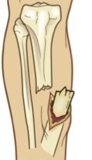
Open Fracture
Fracture communicating with the external environment due to severe trauma causing skin disruption, associated with high risk of contamination, infection, and soft tissue injury, assessed using the Gustilo-Anderson classification.
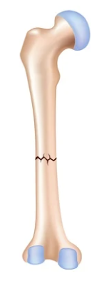
Transverse Fracture
Fracture line runs perpendicular (90°) to the long axis of the bone, usually caused by direct bending force or blunt trauma, considered a simple fracture.
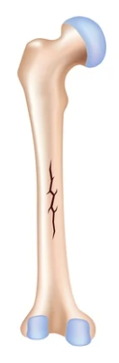
Linear Fracture
Fracture line runs parallel to the long axis of the bone, caused by longitudinal force or compression, often minimally displaced.
Oblique Fracture (Nondisplaced)
Diagonal fracture with fragments remaining aligned, caused by angled force or bending trauma, bone alignment is preserved.
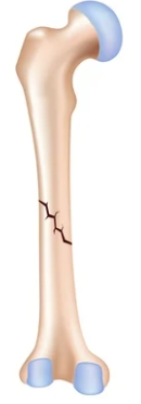
Oblique Fracture (Displaced)
Diagonal fracture with misaligned fragments caused by angled force with displacement, requires reduction to restore alignment.
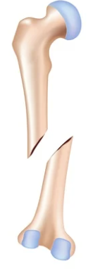
Spiral Fracture
Corkscrew-shaped fracture caused by twisting or rotational forces, hallmark of rotational trauma.
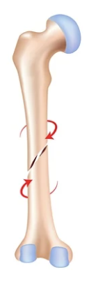
Greenstick Fracture
Incomplete fracture where one cortex breaks while the opposite cortex bends, caused by bending force in immature bone, common in young animals.
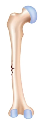
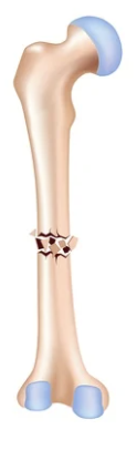
Comminuted Fracture
Bone broken into three or more fragments due to high-impact trauma such as vehicular accidents, severe and often unstable.
Salter-Harris Classification
Classification of growth plate fractures consisting of Types I–V, used only in immature animals with open physes.
Salter-Harris Type I
Fracture through the physis (growth plate) only, occurs only in immature animals with open growth plates.
Salter-Harris Type II
Fracture through the physis and metaphysis, the most common Salter-Harris fracture.
Salter-Harris Type III
Fracture through the physis and epiphysis, involves the articular surface.
Salter-Harris Type IV
Fracture through the metaphysis, physis, and epiphysis, crosses both the growth plate and joint surface.
Salter-Harris Type V
Crush injury to the physis caused by severe compressive trauma, has the worst prognosis with high risk of growth disturbance.
Gustilo-Anderson Classification
Classification system for open fractures based on soft tissue damage and injury mechanism, used to standardize treatment and prognosis, reflects severity of soft tissue injury.
Bone Neoplasia (Bone Tumors)
Abnormal growth of bone tissue that may be benign or malignant, caused by genetic mutations and uncontrolled cellular proliferation, includes primary and secondary (metastatic) tumors.
Osteosarcoma
Most common primary malignant bone tumor in animals arising from osteoid-producing cells, highly aggressive, commonly metastasizes to the lungs, staging is required before treatment because microscopic metastasis may exist despite no gross lesions.
Primary Bone Tumors
Tumors originating directly from bone tissue, often malignant, invasive, and possess high metastatic potential especially to the lungs.
Secondary (Metastatic) Bone Tumors
Bone tumors resulting from metastasis of tumors elsewhere in the body, indicate systemic disease and require identification of the primary tumor.
Bone Tumor Staging
Assessment of tumor extent and metastasis before treatment, essential for prognosis and treatment planning, microscopic metastases may be present despite absence of gross lesions.
Osteomyelitis
Inflammation and infection of bone and bone marrow caused by bacterial or fungal infections, commonly following penetrating wounds, hematogenous spread, fractures, or orthopedic surgery, clinical signs include pain, swelling, lameness, fever, and draining tracts in chronic cases.
Congenital Bone Disorders
Bone abnormalities present at birth due to abnormal embryonic or fetal development, develop before birth, example is persistent ulna or fibula in foals.
Persistent Ulna or Fibula
Failure of normal regression or fusion of the ulna or fibula during development due to a congenital abnormality, reported in foals, may impair limb development and function.
Hereditary Bone Disorders
Inherited diseases affecting bone growth and skeletal development, often complex and transmitted through genetic inheritance.
Nutritional Bone Disorders
Bone abnormalities caused by deficiencies, excesses, or imbalances of calcium, phosphorus, copper, zinc, or magnesium, proper Ca:P ratio is essential for bone homeostasis, excess protein intake may contribute to disorders in growing animals.
Osteomalacia
Defective bone mineralization causing soft weak bones due to calcium and phosphorus deficiency or imbalance, classic nutritional bone disorder with increased fracture risk.
Aseptic Physitis
Inflammation of the growth plate without infection caused by copper deficiency or zinc toxicosis, affects growing animals and is associated with abnormal physeal development.
Osteochondrosis
Developmental disorder involving abnormal cartilage and bone formation associated with nutritional imbalance, rapid growth, and growing animals, may lead to joint lesions.
Vitamin A Imbalance
Nutritional disturbance caused by vitamin A deficiency or excess, impairs normal bone growth and skeletal development.
Vitamin D Imbalance
Nutritional disturbance caused by vitamin D deficiency or excess, alters calcium and phosphorus metabolism, affecting bone mineralization and skeletal development.
Ligament Degeneration
Progressive weakening and deterioration of ligaments caused by chronic degeneration, repetitive trauma, or acute excessive stress, results in ligament rupture, joint instability, and abnormal joint movement, clinical signs include lameness, pain, and joint instability.
Cranial Cruciate Ligament (CCL) Rupture
Tear or complete failure of the cranial cruciate ligament causing knee joint instability, caused by chronic degenerative changes in dogs and chronic degeneration, repetitive trauma, or acute excessive stress in horses, most common ligament injury in dogs causing lameness, pain, and joint instability.
Osteoarthritis (OA)
Chronic progressive degenerative joint disease characterized by cartilage breakdown and joint remodeling, caused by aging, trauma, joint instability, excessive mechanical stress, poor conformation, endocrine disorders, genetics, or sepsis, most common joint disorder in animals causing pain, reduced mobility, and decreased performance, creates a vicious cycle of cartilage damage and increasing joint instability.
Osteoarthritis Hallmark Lesions
Characteristic lesions of osteoarthritis include joint effusion, synovial membrane thickening, decreased synovial fluid viscosity, and articular cartilage degeneration.
Articular Cartilage Degeneration
Progressive breakdown of articular cartilage caused by loss of proteoglycans and increased cartilage water content, resulting in decreased cartilage stiffness, progressive cartilage breakdown, and chronic joint remodeling.
Chronic Joint Remodeling
Permanent structural changes of bone and joints caused by persistent cartilage damage and mechanical stress, leading to decreased joint function.
Inflammatory Joint Disease
Joint inflammation causing pain, joint effusion, and lameness, caused by immune-mediated disorders or infectious agents, severe cases may produce cartilage and subchondral bone erosion, classified as immune-mediated (sterile) or septic (infectious).
Inflammatory Joint Disease Classification
Includes Immune-Mediated Polyarthritis (IMPA) and Septic Arthritis.
Immune-Mediated Polyarthritis (IMPA)
Noninfectious inflammatory arthritis affecting multiple joints, usually idiopathic, may occur secondary to GI infections, non-GI infections, neoplasia, or drug reactions, most common nonerosive inflammatory joint disease in dogs, predisposed breeds include Chinese Shar-Pei, Akita, and Doberman Pinscher, causes pain, joint effusion, and lameness.
Drug-Induced Immune-Mediated Polyarthritis
IMPA triggered by sulfonamides, cephalosporins, or penicillin derivatives, caused by Type IV hypersensitivity, often resolves after discontinuation of the drug.
Erosive Polyarthritis
Immune-mediated inflammatory joint disease causing progressive cartilage and bone destruction, less common than nonerosive forms and may result in permanent joint damage.
Rheumatoid Arthritis (RA)
Chronic autoimmune erosive inflammatory joint disease in which the immune system attacks the synovial membrane, rare in domestic animals but reported in dogs, causes joint swelling, pain, cartilage destruction, and bone erosion.
Feline Progressive Polyarthritis
Immune-mediated erosive polyarthritis affecting young male cats, often associated with Feline Leukemia Virus (FeLV), commonly affects the carpal and tarsal joints, clinical signs include fever, joint pain, swelling, and lameness.
Greyhound Polyarthritis
Immune-mediated erosive polyarthritis affecting Greyhounds, causes severe cartilage destruction in limb joints, clinical signs include shifting-leg lameness, stiff gait, joint swelling, fever, lethargy, and weight loss.
Septic Arthritis
Infectious inflammation of a synovial joint, usually involving a single joint, commonly caused by Staphylococcus spp., clinical signs include joint pain, swelling, effusion, lameness, and decreased range of motion, prompt treatment is required to prevent joint destruction.
Hip Dysplasia
Developmental disorder of the hip joint characterized by joint laxity and instability, caused by polygenic genetics, nutrition, growth rate, exercise, and body weight, common in dogs, frequently progresses to secondary osteoarthritis.
Septic Physitis Extending into Adjacent Joints
Infection and inflammation of the growth plate spreading into adjacent joints due to infectious physitis, may cause joint disease and developmental abnormalities.
Angular Limb Deformities
Developmental abnormalities causing abnormal limb alignment and uneven weight-bearing, resulting in chronic joint stress, degeneration, and osteoarthritis.
Chronic Abnormal Joint Loading
Persistent mechanical stress caused by abnormal weight-bearing or joint mechanics, leading to progressive joint degeneration and secondary osteoarthritis.
Tendon Injuries
Damage to tendons involving tearing or disruption of tendon fibers caused by trauma, excessive mechanical stress, or repetitive strain, heal slowly because of poor blood supply, repair occurs mainly through fibrous scar tissue, healed tendons have reduced tensile strength and increased risk of reinjury.