B3.3: Muscle and Motility | IB Biology (AHL) (copy)
1/26
Earn XP
Description and Tags
Name | Mastery | Learn | Test | Matching | Spaced |
|---|
No study sessions yet.
27 Terms
State that movement can occur within a body or as locomotion from one place to another.
B3.3.1 (AHL): Adaptations for movement as a universal feature of living organisms.
Movement is a universal feature of living organisms.
Movement can occur within a body (sessile) or as locomotion from one place to another (motile)
Compare movement in motile and sessile species.
B3.3.1 (AHL): Adaptations for movement as a universal feature of living organisms.
Both motile and sessile species can move their bodies.
Motile species have adaptations allowing movement (locomotion) from one place to another in their habitat.
Sessile species cannot move from place to place, but they can alter (move) their body form in response to environmental stimuli.
List an example of a motile and a sessile species.
B3.3.1 (AHL): Adaptations for movement as a universal feature of living organisms.
Motile species: Brown-throated three-toed sloth (Bradypus variegatus) can physically move
Also includes: mammals, reptiles, birds, algae, some bacteria
Sessile species: Venus flytrap (Dionaea muscipula) cannot move from one location to another, but they can move their trigger hairs.
Also includes: sponges, corals, anemones, plants
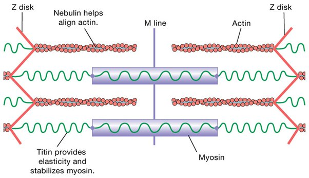
Outline the relationship between muscle fibers, myofibrils and sarcomeres.
B3.3.2 (AHL): Sliding filament model of muscle contraction.
Muscle fibers are thousands of multinucleate cells in an elongated shape, as each fiber is actually several cells that have merged together.
Each muscle fiber contains multiple myofibrils, or protein filaments fiber.
Myofibrils are made of sarcomeres (contracting units) strung out in a single file that make up myofibrils.
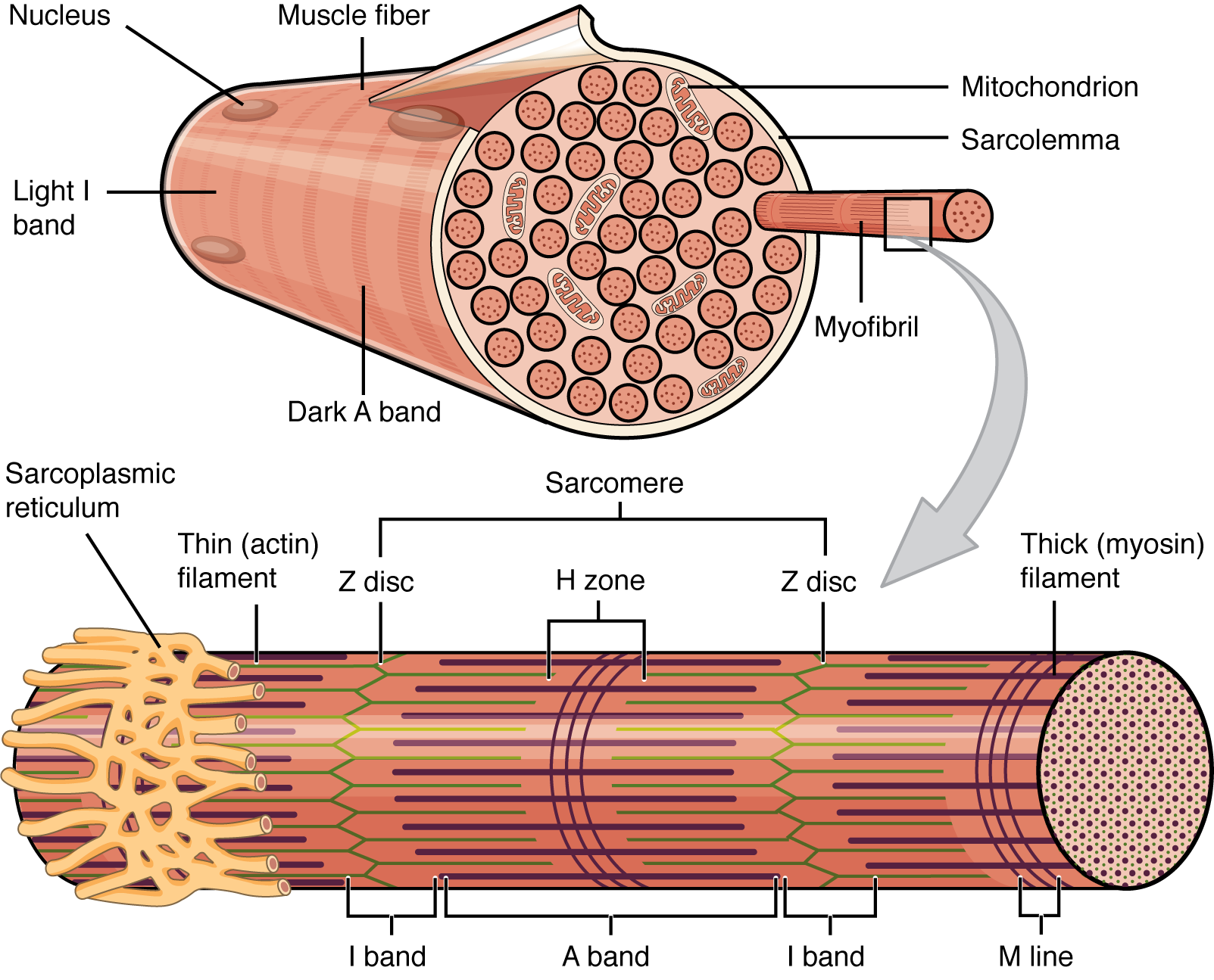
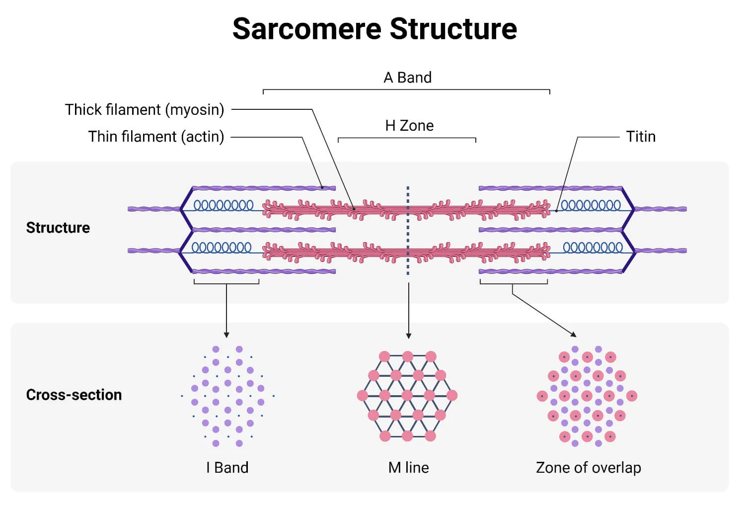
Annotate a diagram of the sarcomere, including the role of the Z line, titin, thin actin filaments, thick myosin filaments, light band and dark band.
B3.3.2 (AHL): Sliding filament model of muscle contraction.
The Z-line are the lines that define the boundary of a sarcomere such that there is one to its right and one to its left.
Titin is a spring that provides stability to the sarcomeres.
Thin actin filaments interact with myosin to produce force, regulate force generation in response to Ca2+ concentration, and transmit the force to the ends of the cell.
The light band (I bands) contain only thin filaments - they only have either actin or myosin, but not both. It shortens during muscle contraction
The dark bands (A bands) contain thick and thin filaments — both actin and myosin. It maintains its width during muscle contraction.

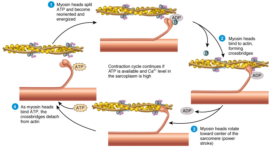
Explain the sliding-filament mechanism of muscle contraction, including the role of actin, myosin heads, cross bridges, ATP, and the power stroke.
B3.3.2 (AHL): Sliding filament model of muscle contraction.
In the sliding-filament mechanism of muscle contraction, actin filaments slide over the myosin fibers.
First, myosin heads are activated by splitting ATP, which causes a change in position of the heads.
Next, myosin heads are attracted to and attach to exposed binding sites of actin to form cross-bridges. Inorganic phosphate is released.
As myosin forms cross-bridges, ADP is released, and myosin bends due to a loss of energy. This bends toward the center of the sarcomere, and the actin moves inward (known as the power stroke — the myosin heads move the actin closer to the center)
Myosin binds to ATP, which allows to detachment of myosin heads from attachment sites.
The contraction cycle continues if ATP is available and Ca2+ levels in the sarcoplasm are high enough.
State the only time a muscle can exert force.
B3.3.3 (AHL): Role of the protein titin and antagonistic muscles in muscle relaxation.
A muscle can only exert force when it contracts.
State what happens when a muscle when relaxes.
B3.3.3 (AHL): Role of the protein titin and antagonistic muscles in muscle relaxation.
Lengthening of a muscle happens when it relaxes.
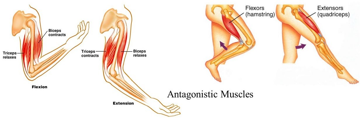
Define antagonistic pairs in relation to muscle movement.
B3.3.3 (AHL): Role of the protein titin and antagonistic muscles in muscle relaxation.
Antagonistic pairs are pairs of muscles that accomplish opposite movements. One of pair exerts a force while the other relaxes and vice versa. They have opposite movements so that there can be a force again.
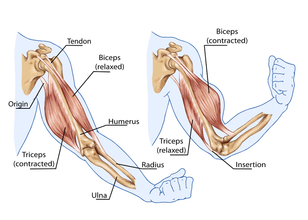
State an example of an antagonistic pair of muscles.
B3.3.3 (AHL): Role of the protein titin and antagonistic muscles in muscle relaxation.
The bicep and tricep is an example of an antagonistic pair of muscles.
Outline the role of the titin protein in muscle stretching.
B3.3.3 (AHL): Role of the protein titin and antagonistic muscles in muscle relaxation.
Titin helps muscles use a force to relax, hold myosin fibers in place in the sarcomere, and prevents muscle fibers from overstretching.
Outline the mechanism by which the protein titin stores potential energy.
B3.3.3 (AHL): Role of the protein titin and antagonistic muscles in muscle relaxation.
During a contraction, sarcomeres shorten, and the 2 sides of the sarcomeres move towards the center. As a result, spring-like tension (potential energy) builds in titin.
Then, when the muscles relax, tension is released, and the sarcomere can undergo a contraction again.
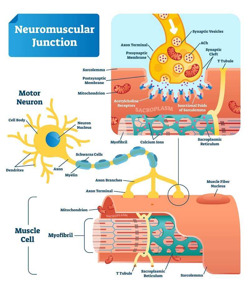
Annotate a diagram of a neuromuscular junction, including the function of the motor neuron, acetylcholine, and muscle fiber.
B3.3.4 (AHL): Structure and function of motor units in skeletal muscle.
The neuromuscular junction is the synapse where a chemical message is sent into the muscle tissue to stimulate a contraction.
Motor neurons are the neurons that carry the signal to stimulate the contraction.
Acetylcholine is the neurotransmitter (the chemical) released at the neuromuscular junction signalling muscle contraction.
Muscle fiber receives the signals to contract.
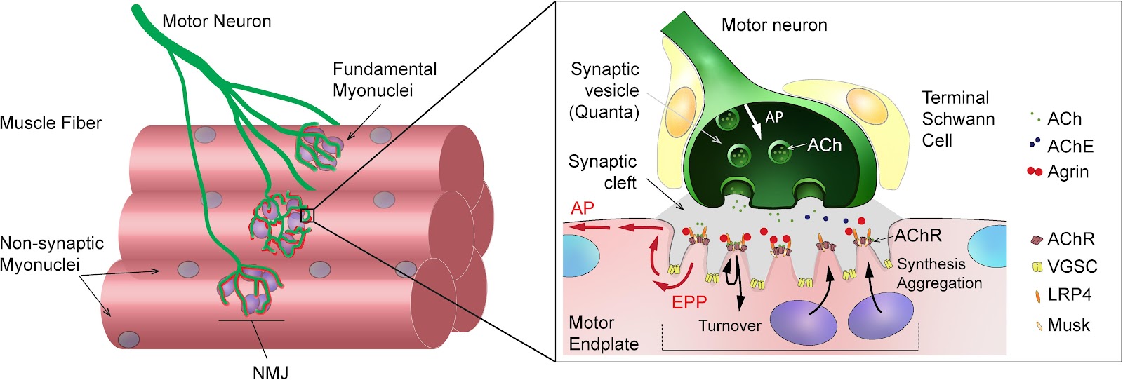
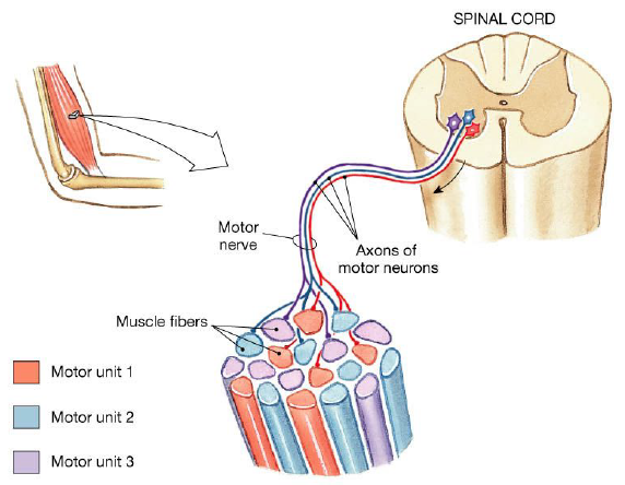
Outline the structure and function of a motor unit.
B3.3.4 (AHL): Structure and function of motor units in skeletal muscle.
A motor unit is a single motor neuron with a set number of muscle fibers that it controls.
It consists of the motor neuron and a certain number of muscle fibers.
It controls which muscle fibers contract.
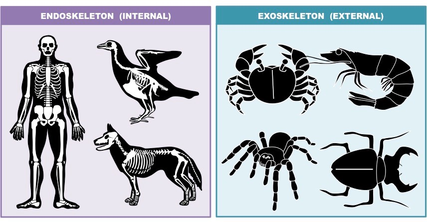
Compare and contrast the structure and function of endoskeletons with exoskeletons.
B3.3.5 (AHL): Roles of skeletons as anchorage for muscles and as levers.
Both endoskeletons and exoskeletons have muscle attachment points. As a result, both endoskeletons and exoskeletons allow bones to act as levers and maximize efficiency for movements.
Endoskeletons are inside the organism’s body, while exoskeletons are outside the body. Therefore, the mucles attach to the outside of bones in endoskeletons, while muscles attach to the inside of the exoskletons. Endoskeletons are made of bones, while exoskeletons are made of chitin.
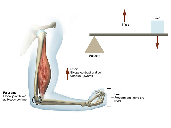
Compare the function of muscles, joints and bones to that of a lever.
B3.3.5 (AHL): Roles of skeletons as anchorage for muscles and as levers.
Many individual bones and segments act as levers to maximize efficiency for movements.
A lever is comprised of a rod that can rotate about the fulcrum. An effort is needed for the lever to achieve this. Similarly, the bone is like the rod and the joint is like the fulcrum. The muscles provide the effort.
The purpose of levers is to lower the force necessary to accomplish work. Similarly, the lever action of bones and joints allow muscles to exert a lower force in order to accomplish a movement.
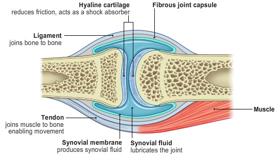
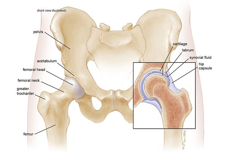
Define synovial joint.
B3.3.6 (AHL): Movement at a synovial joint.
A synovial joint is a type of joint found between 2 bones that move against each other.
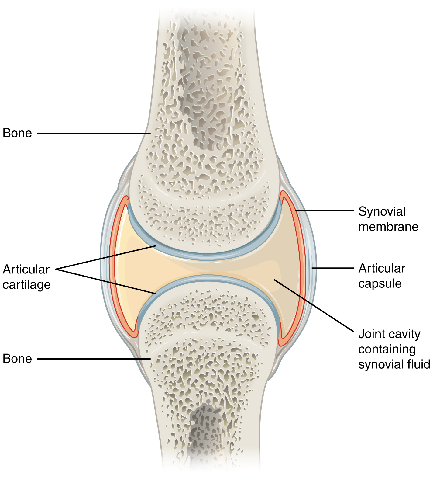
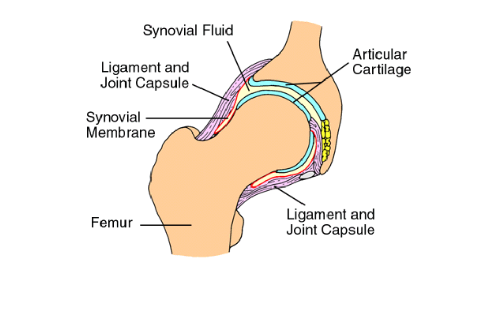
State the function of structures found in a synovial joint, including the bones, cartilage, synovial fluid, ligaments, muscles and tendons.
B3.3.6 (AHL): Movement at a synovial joint.
The bones form the synovial joint (sometimes ball-and-socket).
The cartilage is a smooth protective connective tissue that prevents bone-on-bone contact
Synovial fluid is lubricating fluid (encasing the entire joint) that reduces friction.
Ligaments are tough and fibrous connective tissue encircling the joint that holds the bones in place and allows for a range of motion.
The muscles contract and relax, which allows for motion.
The tendons are connective tissue that connects each muscle to its appropriate bone, which helps controls the movement.
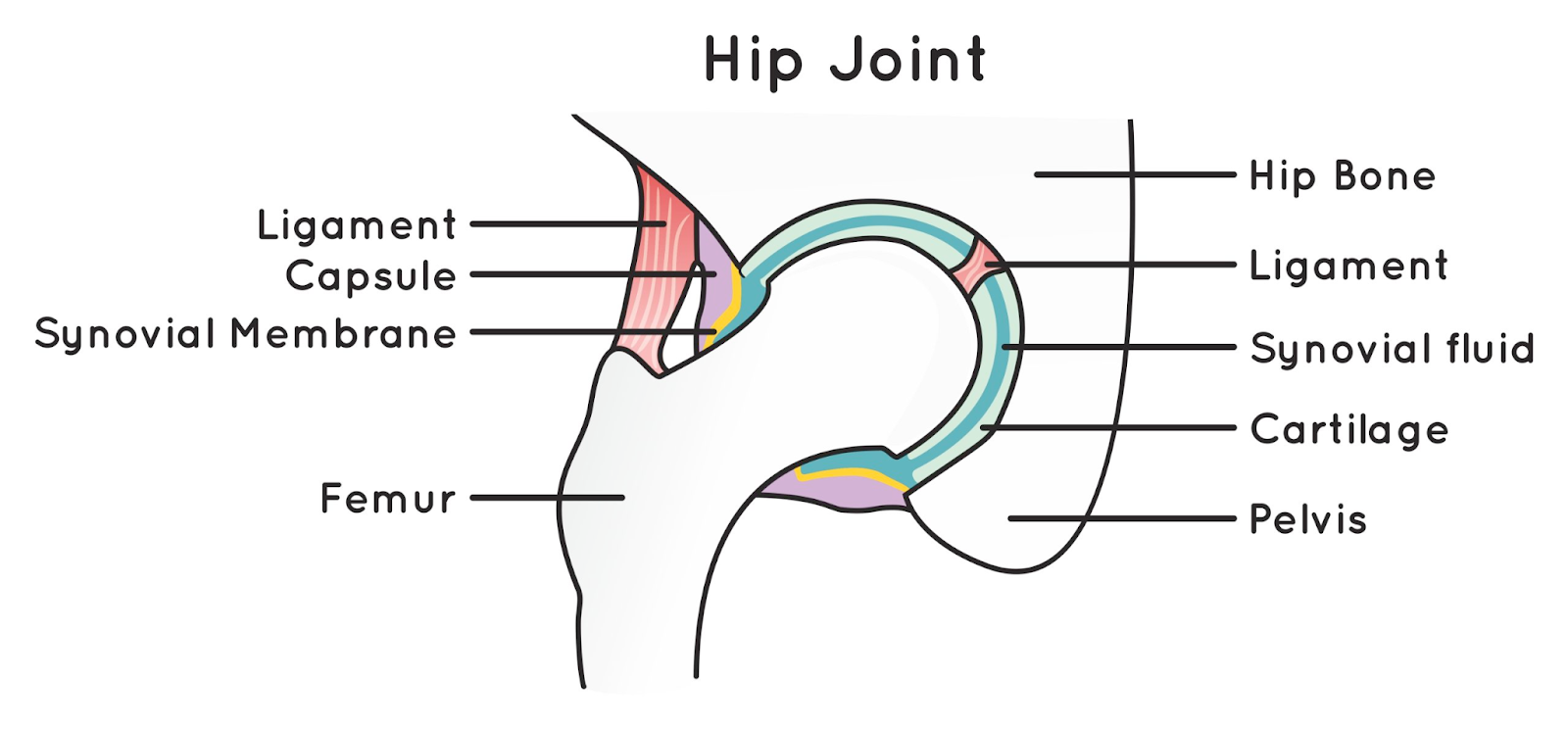
Label a diagram of the human hip joint inclusive of: femur, pelvis, joint capsule, synovial fluid, muscles, ligament and cartilage.
B3.3.6 (AHL): Movement at a synovial joint.
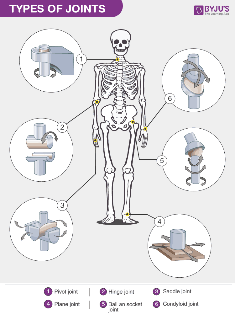
Compare the articulation of hinge joints with that of a ball and socket joint.
B3.3.7 (AHL): Range of motion of a joint.
Hinge joints are more stable than ball-and-socket joints.
Ball-and-socket joints allow a greater range of movement along more than one plane, while hinge joints have less range of movement (only along one plane).
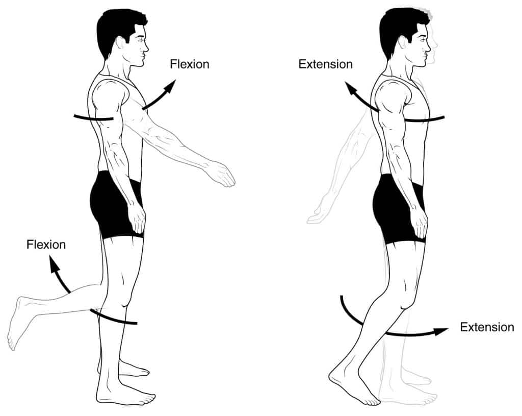
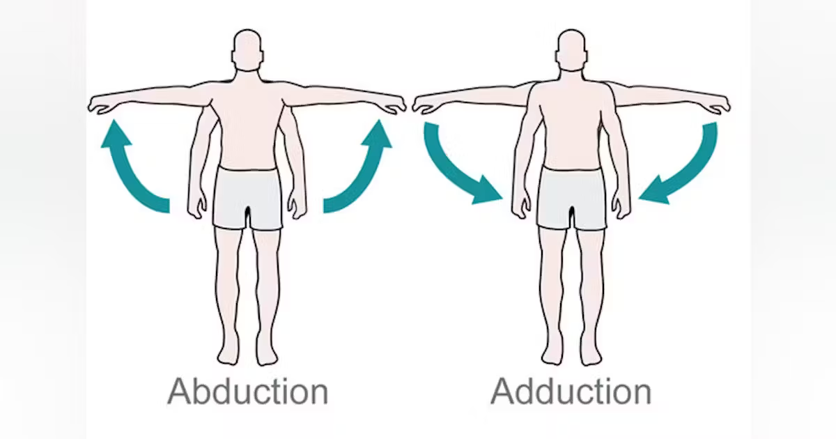
Define range of motion, flexion, extension, rotation, abduction and adduction.
B3.3.7 (AHL): Range of motion of a joint.
Range of motion is the distance and direction that a joint can move (usually in degrees)
Flexion is movement that decreases the angle between 2 body parts along a sagittal (vertical) plane.
Extension is movement that increases the angle between 2 body parts along a sagittal (vertical) plane.
Rotation is movement of the limbs along their long axis. Medial rotation is rotational movement towards the midline (internal rotation), while lateral rotation is rotational movement away from the midline (outward rotation).
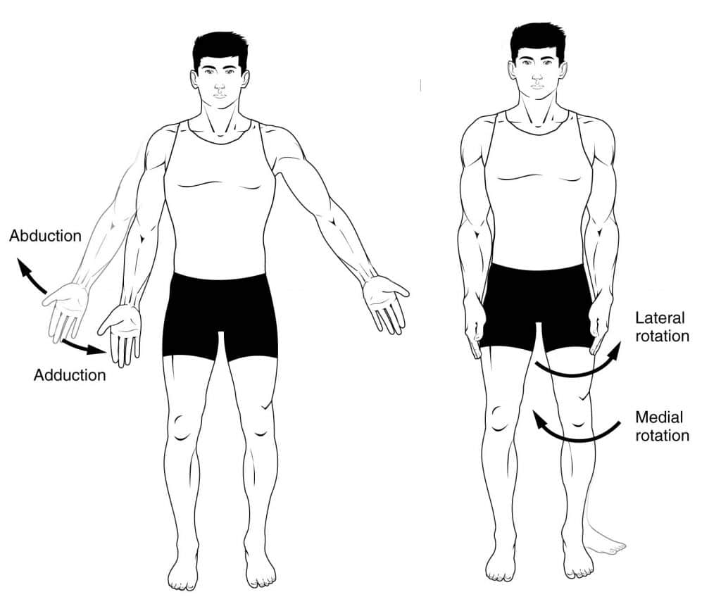
Abduction is movement away from the midline.
Adduction is movement towards the midline.
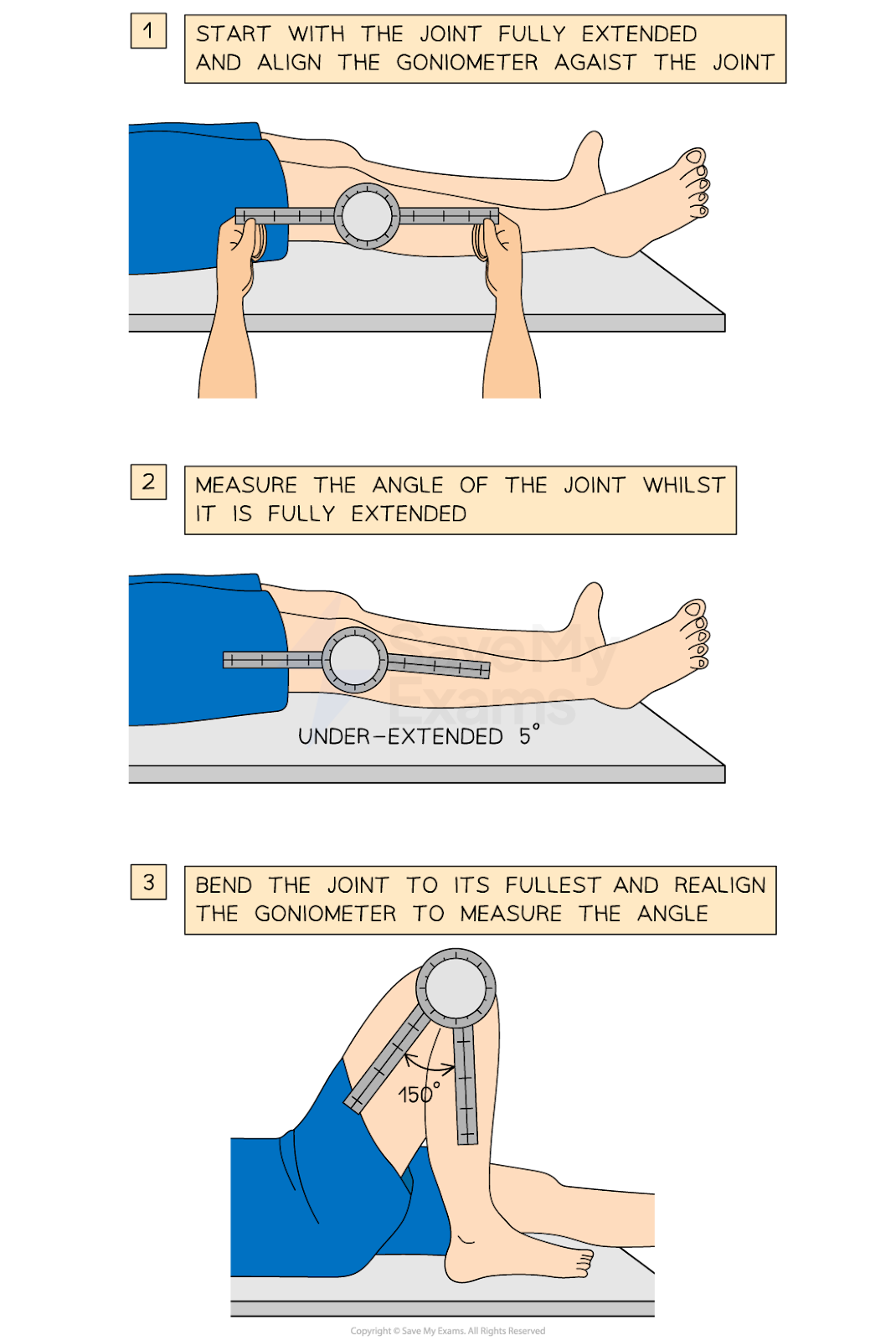
State the purpose of a goniometer.
B3.3.7 (AHL): Range of motion of a joint.
A goniometer is often used to measure the range of motion of a joint.
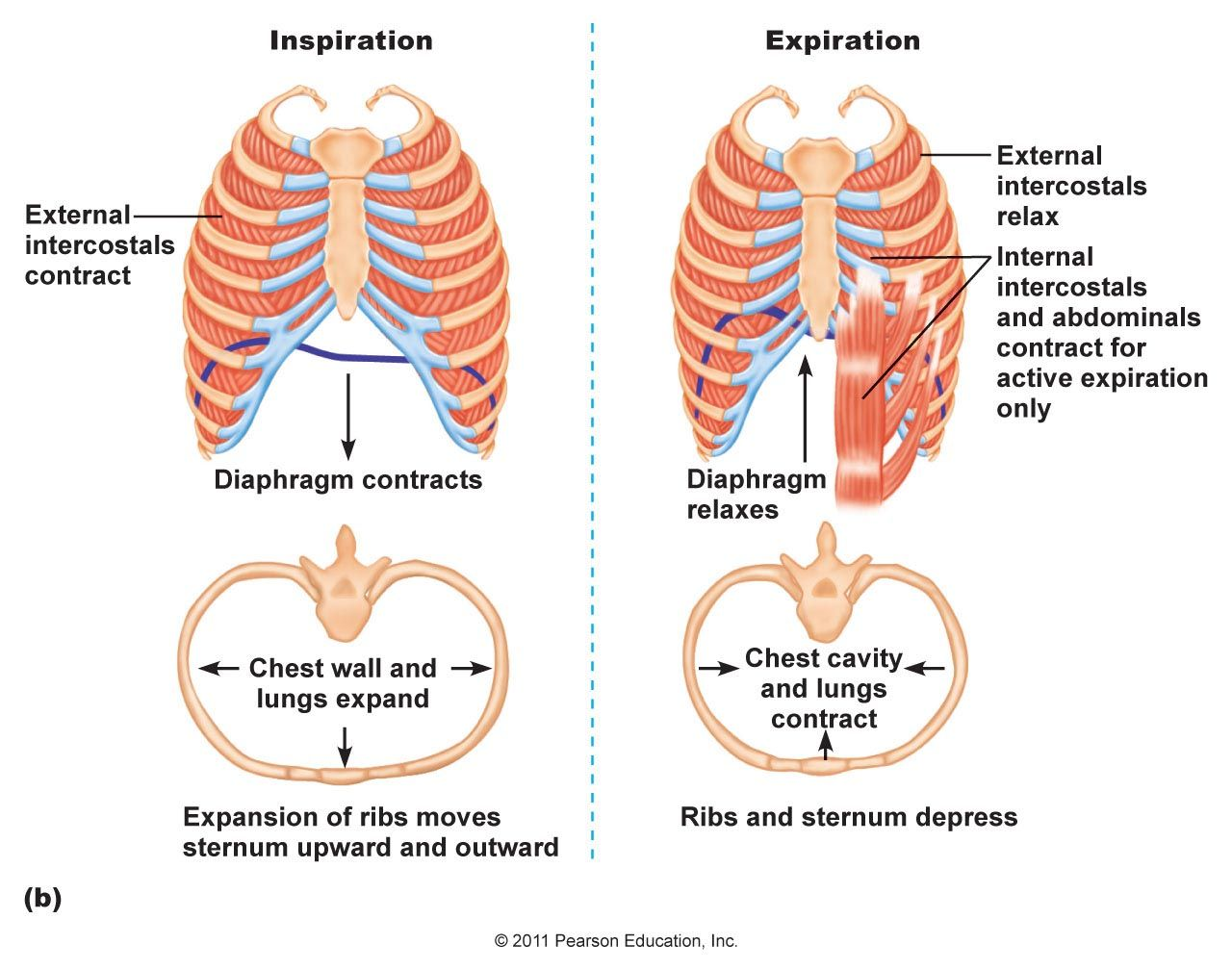
State the location of the internal and external intercostal muscles.
B3.3.8 (AHL): Internal and external intercostal muscles as an example of antagonistic muscle action to facilitate internal body movements.
The intercostal muscles are between each pair of ribs, using ribs as the origin and insertion points.
The internal intercostal muscles are beneat the ribs, while the external intercostal muscles are outside of the ribcage.
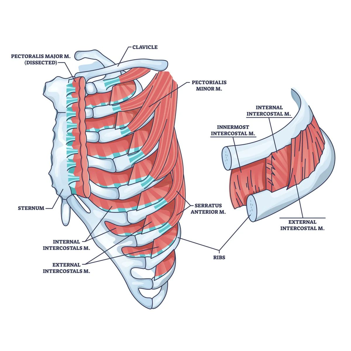
Describe the state of contraction of the internal and external intercostal muscles during inspiration and expiration.
B3.3.8 (AHL): Internal and external intercostal muscles as an example of antagonistic muscle action to facilitate internal body movements.
The different orientations and angles of muscle fibers in the internal and extenral intercostal muscles mean that they move the ribcage in opposite directions.
During inspiration, the external intercostal muscles contract, which pulls the rib cage up and out. The internal intercostal muscles to stretch.
During expiration, the internal intercostal muscles contract, which pulls the rib cage down and inwards. The external intercostal muscles are stretched.
Outline the mechanism by potential energy is stored during contraction of antagonistic muscles.
B3.3.8 (AHL): Internal and external intercostal muscles as an example of antagonistic muscle action to facilitate internal body movements.
The internal and external intercostal muscles are a pair of antagonistic muscles.
When the external intercostal muscles contract (for inspiration), the internal intercostal muscles are stretched, and the titin fibers in the sarcomere are also stretched. As a result, potential energy is stored in the sarcomere protein titin and is used for the next contraction of internal intercostal muscles.
Likewise, the contraction of internal intercostal muscles stores potential energy in the external intercostal muscles’ titin, which also stores potential energy for the next external intercostal muscle contraction.
List reasons for animal locomotion, with one example of each.
B3.3.9 (AHL): Reasons for locomotion.
Foraging for food
E.g. Honey bees flying to collect nectar and pollen
Escaping from danger
E.g. Flying fish escaping predators by swimming fast & gliding over the water
Searching for a mate
E.g. Loggerhead sea turtles swimming back to the beach where they were hatched to mate and lay eggs
Migration
E.g. Arctic terns migrating from Arctic breeding grounds to the Antarctic region and back each year (to take advantage of available food)
Dispersal
E.g. Hoary bat
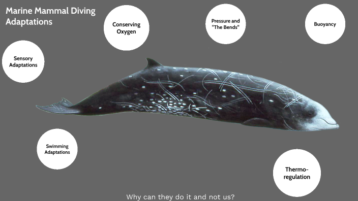
Outline the adaptations for streamlining, locomotion and ventilation of lungs in marine mammals.
B3.3.10 (AHL): Adaptations for swimming in marine mammals
Streamlining
Marine mammals have streamlined bodies, so they can move through viscous water with relative ease and at great speeds.
Marine mammals have almost no body hair, so there is reduced drag through the water.
Locomotion
Marine mammals have a tail adapted to form a fluke, which produces an up-and-down motion for propulsion.
Because they have movement provided by fluke, they lost their rear legs
The front limbs became flippers to help steer.
Ventilation of the lungs
Marine mammals have a blowhole as an airway at the dorsal (top) surface of the head, so they can exchange air at periodic intervals by having the body leave the water.
Marine mammals can seal the blowhole tightly between breaths, so water does not enter the airway.
Marine mammals can stay underwater for several minutes without breathing, so they can make deep dives