Cell Biology Exam 1 Material
 Call Kai
 Learn
Learn Practice Test
 Spaced Repetition
 Match
Match Flashcards
 Knowt Play
1/199

There's no tags or description
Looks like no tags are added yet.
Name | Mastery | Learn | Test | Matching | Spaced | Call with Kai | Chat |
|---|
No analytics yet
Send a link to your students to track their progress
200 Terms
Functions of the plasma membrane
Cell integrity, permeability, adhesion, communication, and membrane potential
Three main components of plasma membrane
phospholipids, proteins, cholesterol
two main types of proteins in plasma membrane
integral and peripheral
phospholipids self assemble into (2)
mycelles, bylayers
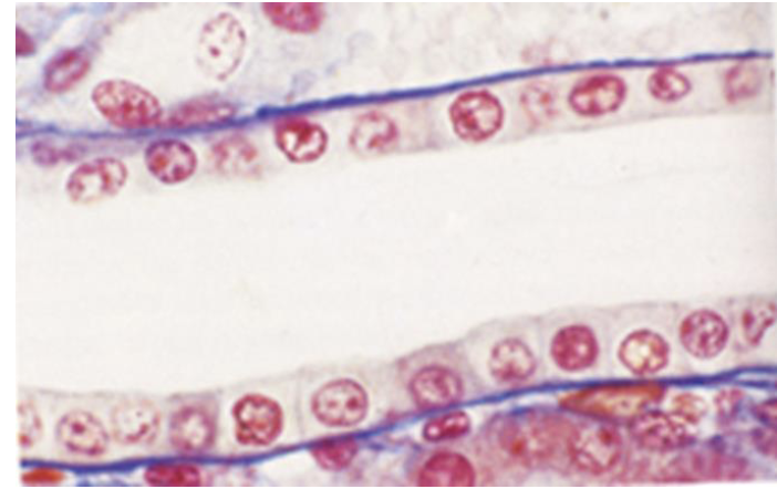
what is this image of?
Epithelium
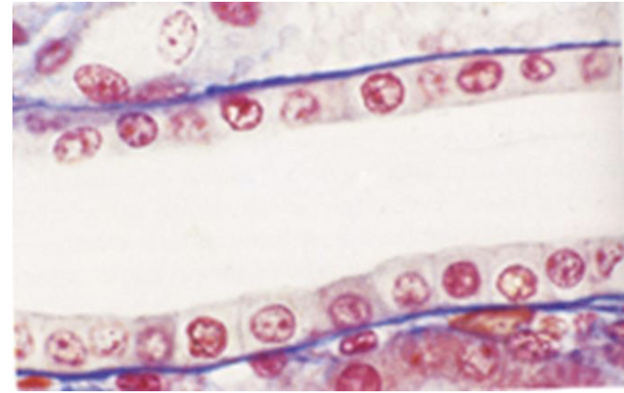
what does the blue line represent
plasma membrane of the basal surface
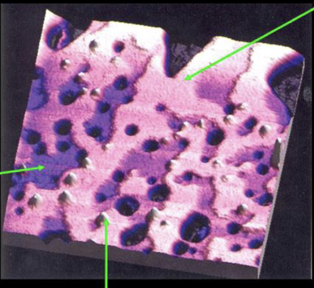
what does the pink area of this image represent (top left arrow)
Lipid raft
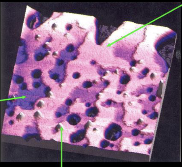
what does the dark purple area represent (left arrow)
non-raft area
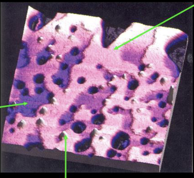
what does the white represent (bottom arrow)
bacteral protein
as cholesterol is added to the gaps formed by unsaturated fatty acids the fluidity of the plasma membrane decreases
decrease
Move ions/molecules across the membrane with energy
pupps
Move ions/molecules across the membrane without energy
channels
Bind molecules in extracellular space
Receptors
Cell attachment to extracellular matrix (ECM)
Linker proteins
Attach cells to one another
Structural proteins
what side does the p face on (intercellular/extracellular)
intercellular
what side does the e face on (intercellular/extracellular)
extracellular
what face has most integral proteins
p face
this transport system only moves one molecule at a time
uniport
this transport system moves two different molecules the same direction
symport
this transport system moves two different molecules opposite direction
antiport
what molecules can move freely through the plasma membrane?
uncharged gasses, lipids, and lipid soluble molecules
name of channel that helps bring water into cell
aquaporins
facilitated diffusion is (faster/slower) than simple diffusion
faster
name of protein that is mutated in cystic fibrosis
CFTR
what is the purpose of the CFTR protein (cystic fibrosis) and what does it impact
Cl ion transport; water movement
two mechanisms of viscular transport
Endocytosis and Exocytosis
three methods of endocytosis
pinocytosis, phagocytosis, and receptor mediated endocytosis
This is the non specific uptake of fluid and small molecules
pinocytosis
helps with pinocytosis (3)
caveolin, flotillin, or actin cytoskeleton
This type of endocytosis is for the entry of specific molecules into the cell
receptor mediated endocytosis
molecule that helps with receptor mediated endocytosis
clathrin
name of protein that pinches off vesicle in receptor mediated endocytosis
dynamin
this is a disease caused by mutation in low density lipoprotein receptor
familial hypercholesterolemia
what is mutated in familial hypercholesterolemia
low density lipoprotein receptor
what does familial hypercholesterolemia lead to, what does it result in
excess cholesterol in blood stream, CAD or xanthomas
this is caused a mutation in the CFTR protein, what is a major symptom
cycstic fibrosis, accumulation of mucus in the lungs
this form of endocytosis is the engulfment of large particles
phagocytosis
what molecule helps phagocytosis
actin
inhibition of endocytosis also inhibits (and vice versa)
exocytosis
this is a receptor that can bind to a variety of different proteins and polysaccharides to identify many ‘large particles‘ for phagocytosis
C3b
most common fate of receptor and ligand after use
receptor recycled and ligand degraded
fate of LDL receptor
receptor recycled and ligand degraded
fate of transferrin receptor
both recycled
fate of EGF receptor
both degraded
fate of igA secretions
transcytosis
what is transcytosis
movement from one side of cell to the other
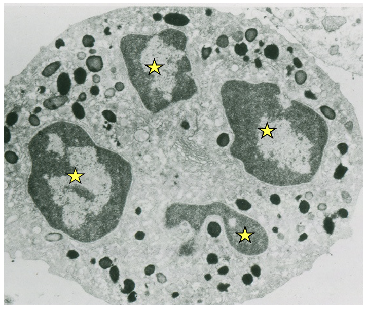
this image has only one nucleus how is this possible
multiple lobes
this cell has no nucleus
RBC
this cell has multiple nuclei
muscle cells
what are histones
small basic proteins that allow for tight packing of DNA
charge of histones
positive
heterochromatin
highly condensed inactive DNA
euchromatin
dispersed active DNA
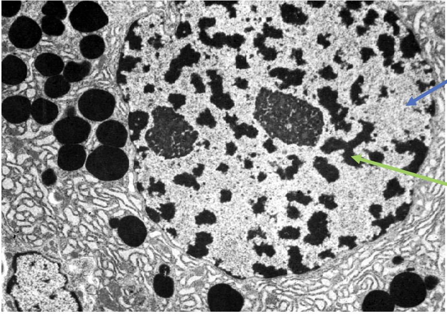
what type of DNA is the green arrow pointing to
heterochromatin
what type of DNA is the blue arrow pointing to
euchromatin
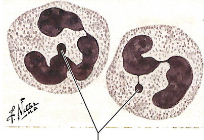
what is this and what is its purpose
barr body; inactivation of the second X in females
the sorting and pairing of individual chromosomes based on size shape and color pattern
karyotype
how many histones are in one nucleosome
8
what is a nucleosome
DNA wound around histones
name all the proteins in a nucleosome (4×2)
H2A, H2B, H3, and H4
when are chromosomes visible
mitosis and miosis
this is important for rRNA processing and regulation of p53 expression
nucleostemin
the nucleolus contains mostly this
rRNA
spot for rRNA synthesis and ribosome assembly
nucleolus
space between inner and outer membrane of nuclear envelope
perinuclear space
the perinuclear space / cisterna is continuous with this structure
rough ER
provides structural support for nuclear envelope
nuclear lamina
purpose of nuclear lamina
sturcutral support and attachment site for chromatin
what filaments form the lattice like network for the nuclear lamina
lamins A and C
what intermediate filaments bind receptors in envelope
lamin B
what happens to the nuclear lamina during mitosis
disassembles then reforms
what can lamin mutations effect
striated and cardiac muscle, adipose tissue, nervous system, and premature aging
lamin mutation affecting adipose tissue can end up causing
lipodystrphy syndromes
lamin mutations affecting the nervous system can end up causing
peripheral neuropathy
what proteins make up nuclear pores (~100)
nucleoporins
small water soluble molecules can move via this through the nucelar pores
passive diffusion
Primary energy source for transport through the nuclear pore complex?
GTP
this sequence is recognized by importins in the nucleus
nuclear localization signal
this sequence is recognized by exportin in the nucleus
Nuclear Export Sequence
nuclear localization signal (NLS) is a sequence that is recognized by this to enter the nucleus
inportin
Nuclear Export Sequence (NES) is a sequence that is recognized by this to exit the nucleus
exportin
three types of cell cycle populations
static, stable, and renewing
in these cells no dicision occurs
static
in these cells division is sporadic
stable
in this cells regular division occurs
renewing
examples of static cells
neurons and cardiac myocytes
examples of stable cells
endothelial cells or fibroblasts
examples of slow renewing cells
smooth muscle of hollow organs
examples of rapid renewing cells
epithelial cells of GI tract
longest phase that is the period of cell growth (collect nutrients and synthesize proteins )
G1
phase where DNA synthesis occurs
S
phase with RNA and protein synthesis in preparation for mitosis
G2
stage where cells are no longer dividing
G0
protein expressed during g1 through mitosis (immunofluorescence)
ki67
protein expressed during S and G2 (immunofluorescence)
PCNA
protein expressed during S (immunofluorescence)
MCM2
given that the specimen is alive DNA in the S phase can be labeled with this for immunofluorescence
BrdU
in this phase there is a nuclear envelope and nucleolus, and chromatin is disbursed
interphase
in this phase DNA is condensed and the nuclear envelope and nucleolus is lost
prophase