lecture 2 - neurophysiology pt 2
 Call Kai
 Learn
Learn Practice Test
 Spaced Repetition
 Match
Match Flashcards
 Knowt Play
1/68

There's no tags or description
Looks like no tags are added yet.
Name | Mastery | Learn | Test | Matching | Spaced | Call with Kai | Chat |
|---|
No analytics yet
Send a link to your students to track their progress
69 Terms
Nervous tissue has 2 types of cells:
Neurons
Glial Cells
Neurons
Nerve cells that transmit information
Made of:
Cell body → integrates incoming and outgoing information
Dendrites → information moves towards the cell body
Axon → information moves away from the cell body
Capacity to generate action potential and convey information

Multipolar
mainly in the CNS
Pseudounipolar
mainly in the PNS
Bipolar
mainly in sensory organs
Sensory (afferent)
Carry information from PNS → CNS
Motor (efferent)
Carry information from CNS → muscles and glands
Interneurons (association)
Relay information between neurons within the CNS
Specialized receptors
Transducers = convert stimuli into signals
Glial Cells
Non-neuronal cells
About 10× more abundant than neurons
Types include:
Oligodendrocytes
Astrocytes
Ependymal cells
Microglia
Functions
Provide structural support
Oligodendrocytes → form myelin
Astrocytes → secrete glutamate and can affect neuronal excitation
Microglia → can perform phagocytosis
Contact blood vessels and neurons
Help transport nutrients to neurons
Neurons do not store glucose or O₂, so they need a constant supply
Grey Matter
Mainly contains cell bodies
White Matter
Contains bundles of neuron processes
Appears white because of myelin
Nerves
Bundles of axons
Run to or from the CNS
Ganglia
Clusters of sensory neuron cell bodies
Located outside the CNS
Motor Neurons
Cell bodies are located in specific areas of the CNS
CNS = brain and spinal cord
Myelin
white lipid surrounding nerve fibers
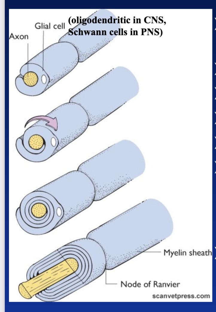
Myelin Sheaths
Myelin = white lipid surrounding nerve fibers
Made when glial cells wrap around an axon
Cytoplasm is lost, leaving layers of lipids
Found in white matter
Not all nerve fibers are myelinated

Function; Myelin Sheaths
Acts as an electrical insulator
Makes action potential transmission faster
Nodes of Ranvier
Gaps in the myelin sheath
Occur about every 1–2 mm
Exposed areas of the axon allow depolarization
Allow transmission of the action potential (AP)

Membrane Potential
Every cell has a membrane potential
Resting Membrane Potential (RMP) = difference in electrical charge across the cell membrane
Inside of the cell is negative relative to outside
RMP varies depending on:
Amount of charges
Ion channels
Membrane thickness
Nerve Cells
Average RMP = about –70 to –90 mV
What Maintains the RMP?
Three main factors:
Selective permeability
Ions passively move through channels by diffusion
Na⁺/K⁺ pump
Pumps 3 Na⁺ out
Brings 2 K⁺ in
Large anions
Negative molecules trapped inside the cell
Selective Permeability
Ions can passively leak through channels according to their concentration gradient
At rest, the membrane is:
Very permeable to K⁺
Barely permeable to Na⁺, Ca²⁺, and Cl⁻
Positive charges therefore tend to accumulate outside
Na⁺/K⁺ Pump
Ion concentrations need to stay relatively constant
The pump compensates for ion leakage
Moves:
3 Na⁺ OUT
2 K⁺ IN
Moves ions against their concentration gradients
Requires a lot of energy
Can use up to 40% of ATP availability
Important
Neurons do not store glucose or O₂ → they need a constant supply.
Excitable Cells
Cells that can generate electrical impulses (action potentials)
Must first be stimulated
Stimuli can be:
Chemical
Electrical
Physical
A stimulus changes the membrane potential.
If the membrane reaches threshold:
→ voltage-gated ion channels open
→ action potential occurs
Depolarization
Na⁺ channels open
Na⁺ rushes into the cell
Membrane becomes less negative
Can eventually become positive
This is called depolarization
In some nerve endings and smooth/cardiac muscle cells, Ca²⁺ can be involved instead.
Repolarization
K⁺ channels open
K⁺ flows out of the cell
Membrane potential returns toward the RMP
This is called repolarization
Generation of an Action Potential
cont
1. Initial Depolarization
A stimulus causes depolarization
Must reach threshold
2. Na⁺ Channels Open
Voltage-gated Na⁺ channels open
Na⁺ rushes into the cell
Causes rapid depolarization
3. Na⁺ Channels Close
After about 0.5 ms, Na⁺ channels rapidly close
4. K⁺ Channels Open
K⁺ voltage-gated channels open
K⁺ flows out
Causes repolarization
5. Hyperpolarization
K⁺ channels close gradually
K⁺ continues leaving after reaching the RMP
Membrane becomes more negative than the RMP
This is hyperpolarization
6. Return to RMP
Gated channels close
Ions return to their proper compartments through:
Diffusion
Na⁺/K⁺ pumps
Refractory Period
Neuron cannot be re-stimulated until the RMP is restored.
Ion-Gated Channels
Several types of gated ion channels:
Voltage-gated channels
Open in response to changes in membrane potential
Ligand-gated channels
Have binding sites for neurotransmitters
Open when a neurotransmitter binds
Channel Structure
Each channel is made of several subunits
Channels have different levels of specificity
All-or-None Rule
Nerve cells follow the all-or-none rule
If threshold is reached → an action potential (AP) is generated
If threshold is not reached → no AP
The amplitude of the AP is fixed for that neuron
Important
The intensity of a signal is coded by the frequency of APs, NOT their amplitude.
Stronger stimulus → more frequent APs
Conduction of Action Potential
Depolarization and repolarization propagate along the cell membrane
The change in membrane potential must reach threshold in the nearby area
This triggers the opening of gated channels
The AP then continues along the axon
Nerve Velocity Depends On:
Thickness of myelin
Diameter of the fiber
Thicker fiber → faster conduction
Unmyelinated Axons
AP occurs continuously along the cell membrane
Myelinated Axons
AP occurs only at the Nodes of Ranvier
Myelin prevents ion leakage
Electrical current jumps from node to node
This is called saltatory conduction
Advantages of Saltatory Conduction
Faster transmission
Less membrane is affected
Requires less energy to transport ions
Conduction Speeds
Can range from about 0.5–100 m/sec
About 250–2500 impulses/sec
Synaptic Transmission
Allows a signal to pass:
From one neuron → another neuron
From a neuron → target cell, such as a skeletal muscle
Synaptic Gap
Cell membranes are made of phospholipids
Act as an electrical insulator
There is a gap between the two cell membranes
This is called the:
Synaptic gap
Synaptic cleft
Electrical Synapses
Sometimes there is direct continuity of the electrical impulse through gap junctions
Found in:
Cardiac muscle
Some smooth muscle
Chemical Synapses
In vertebrates, neuronal synapses are mostly chemical
Neurotransmitters
Molecules that transmit information from a neuron
Convert an electrical signal (AP) → chemical signal
Process
Released by the presynaptic neuron
Enter the synaptic gap
Bind to specific receptors on the postsynaptic membrane
Cause a response
Types; Neurotransmitters
cont
Small molecules
Made in the nerve terminals
Made using specific enzymes
Include:
Amino acid derivatives
Biogenic amines
Neuropeptides
Made of 3–40 amino acids
Synthesized in the cell body
Packaged into secretory vesicles
Transported to the site of release
Neuromuscular Synapse
Acetylcholine (ACh) = neurotransmitter at the neuromuscular synapse
Postsynaptic membrane has folds
Increases surface area
Common at neuromuscular synapses
Not found in interneurons
Steps
1. Action potential (AP)
↓
2. AP opens voltage-gated Ca²⁺ channels
↓
3. Ca²⁺ enters the cell
↓
4. Ca²⁺ triggers exocytosis of neurotransmitter
↓
5. Neurotransmitter diffuses across the cleft
↓
6. Neurotransmitter binds to specific receptors
↓
7. Ion channels open on postsynaptic membrane → depolarization
↓
8. Neurotransmitter is inactivated → signal ends
Small Molecules
Can be terminated by:
Being taken back up by the presynaptic neuron
Recycled for future use
Being broken down in the cleft by enzymes
Example: Acetylcholinesterase (AChE)
Neuropeptides
Can be terminated by:
Being taken into the postsynaptic cell through endocytosis
Being broken down by cellular enzymes
Being broken down by extracellular peptidases in the gap
The receptor can become desensitized
Integration of Multiple Synapses
cont
Neuromuscular Synapse
1 neuron → AP → muscle cell depolarization
Neuron-Neuron Synapse
One neuron can receive signals from many other neurons
Synapses can be:
Excitatory
Inhibitory
One impulse does not always cause a response
The neuron must reach threshold to generate an AP
Excitatory Synapse
Causes depolarization
Usually involves Na⁺ entering
Inhibitory Synapse
Causes hyperpolarization
Can involve:
Cl⁻ entering
K⁺ leaving