Ultrasound Display Modes and Imaging Systems
1/41
Earn XP
Description and Tags
Vocabulary flashcards covering ultrasound display modes (A, B, M, and 2-D), scanning techniques, resolution principles, recording equipment, and digital communication systems like DICOM and PACS.
Name | Mastery | Learn | Test | Matching | Spaced | Call with Kai |
|---|
No analytics yet
Send a link to your students to track their progress
42 Terms
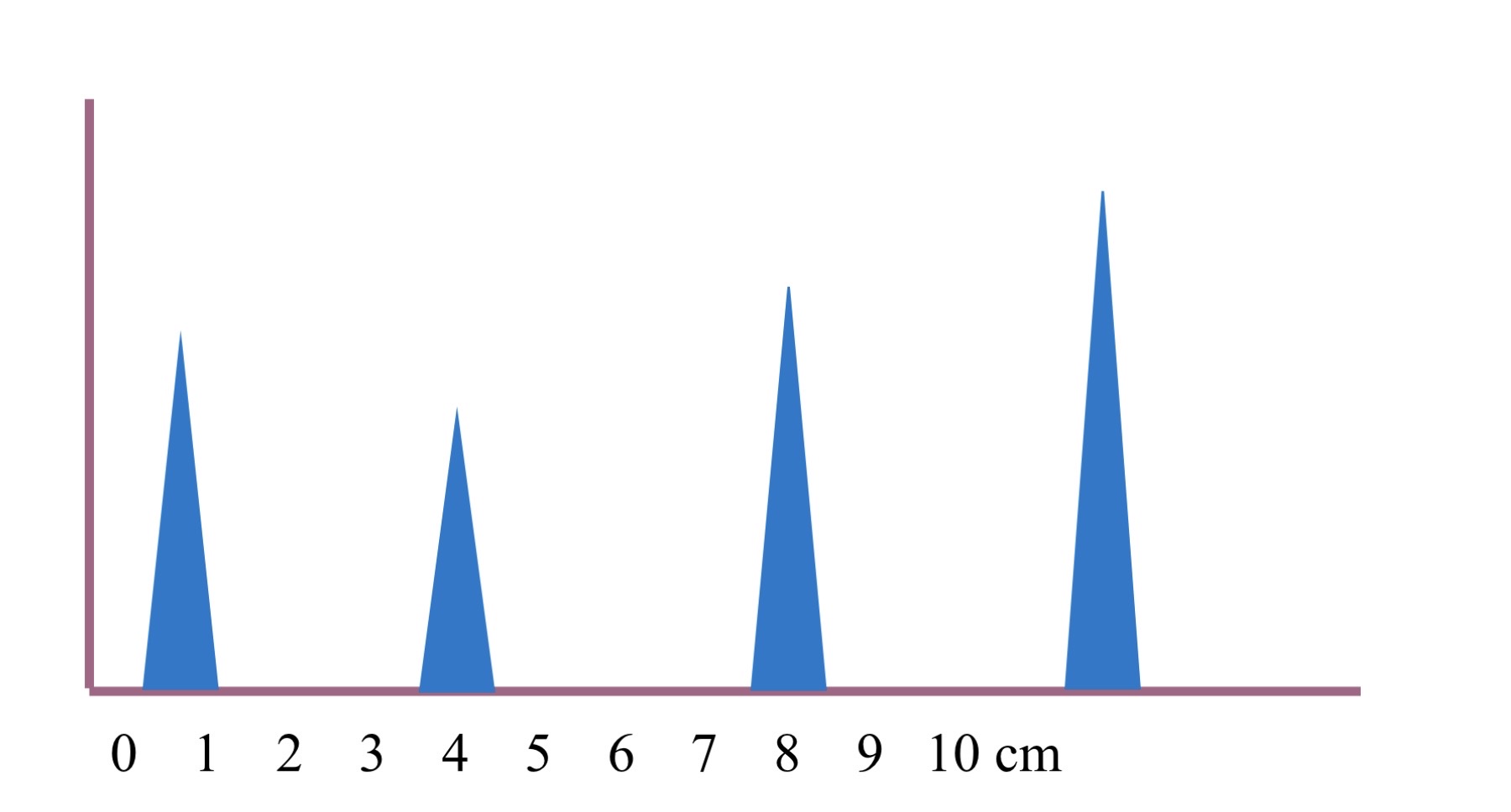
A Mode is also known as?
Also known as amplitude mode.
A mode was original display mode for ?
US
In A mode each spike represents a
reflector or Interface in the body.
In A mode the higher the spike ?
Higher the amplitude the stronger the reflector and show depth image
A mode still used in
Ophthalmic studies also really archaic
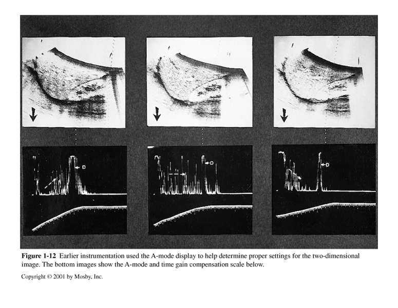
What does these image describes?
A mode
B Mode also know ?
Also known as brightness mode.
B Mode is a series of ?
Series of dot
In B mode each dot represents a
Reflector or interface in the body.
In B mode the brighter the dot. Also represent ?
The stronger the reflector. Also still represents depth along the horizontal axis
M Mode also known as ?
Also known as motion mode.
In M mode straight lines indicates
No reflector motion
In motion mode moving lines indicates
Motion
M mode used most commonly with
Heart imaging
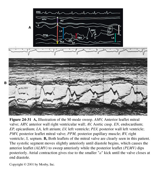
This is image is example of ?
M mode/ motion mode

This is image is example of ?
M mode
What is B Scan?
A method where B mode is combined with scanning motion.
B mode dots are image
In a vertical lines
In scan line many vertical lines
Place side by side
In B scan dots represent ?
Depth and location or position within the body and forms a a 2 dimensional image
What are the two types of B scan
Static scanning
Real time of dynamic
Static Scanning
Also called compound or contact scanning, it uses a transducer mounted on an articulated arm that a Sonographer physically moves to 'paint' an image on the screen; it cannot display motion.
Real-Time Scanning
Also called dynamic scanning, the sound beam is swept mechanically or electronically to produce a series of frames in rapid sequence, creating a motion picture effect.
Frame Rate
The number of images produced per second, measured in units of per second or Hz; the average is 30 frames per second.
Flicker Threshold
The human eye can perceive flickering up to 15 images per second; above this rate, the image appears dynamic or real-time.
Line Density
The number of scan lines used to create a single frame; greater density improves detail resolution but requires more time to create the frame.
Multi-focus Scanning
A process where multiple scan lines are created for each position prior to moving; for example, 3 focal zones require 3 scan lines.
PRF Penetration Equation
The relationship used to avoid range ambiguity, defined as penetration (cm)×PRF (kHz)<77.
Temporal Resolution
The ability of a display to distinguish closely spaced events in time; it improves with an increased frame rate.
Coded Excitation
A technique utilizing ensembles of pulses to generate a scan line with a decoder that interprets the code, resulting in higher intensity driving pulses and a more sensitive receiving system.
Harmonics
A type of filtering that removes the fundamental frequency to improve images by narrowing the primary beam, eliminating grating lobes, and reducing artifacts.
Pulse Inversion
A method using two pulses per scan line (one being the inverse of the first) to improve detail resolution, though it compromises temporal resolution.
Elastography
A technique that estimates tissue stiffness, often called 'imaging' palpation, used to evaluate masses for malignancy and assess myocardium viability.
Fusion Imaging
The combined presentation of an ultrasound image with another imaging modality.
Tissue Doppler
A mode used specifically to measure the movement of muscle.
Laser Film
A recording medium providing superior image quality with 256 to 4096 shades of gray, typically used in 14×17 size.
Color Video Printer
A device that heat-processes images onto paper using cyan, magenta, and yellow ribbons; all three colors together make black.
VCR (Video Cassette Recorder)
A recording device where standard VHS tapes can record 240 lines and Super VHS tapes can record 425 lines.
DICOM (Digital Imaging and Communication in Medicine)
A standardized file format and communication protocol for patient data and images, allowing transmission over networks between multiple modalities and PACS.
PACS (Picture Archiving and Communication Systems)
A system consisting of digital acquisition, display workstations, and storage devices interconnected through a Local Area Network (LAN) to handle digital information.
Teleradiology
A PACS application involving the electronic transfer of images from one location to another, allowing physicians to review exams remotely.