ZOOL 463 Midterm 2
1/242
Earn XP
Description and Tags
ZOOL 463 UofC
Name | Mastery | Learn | Test | Matching | Spaced | Call with Kai |
|---|
No analytics yet
Send a link to your students to track their progress
243 Terms
MODULE 6: OSMOTIC & IONIC REGULATION
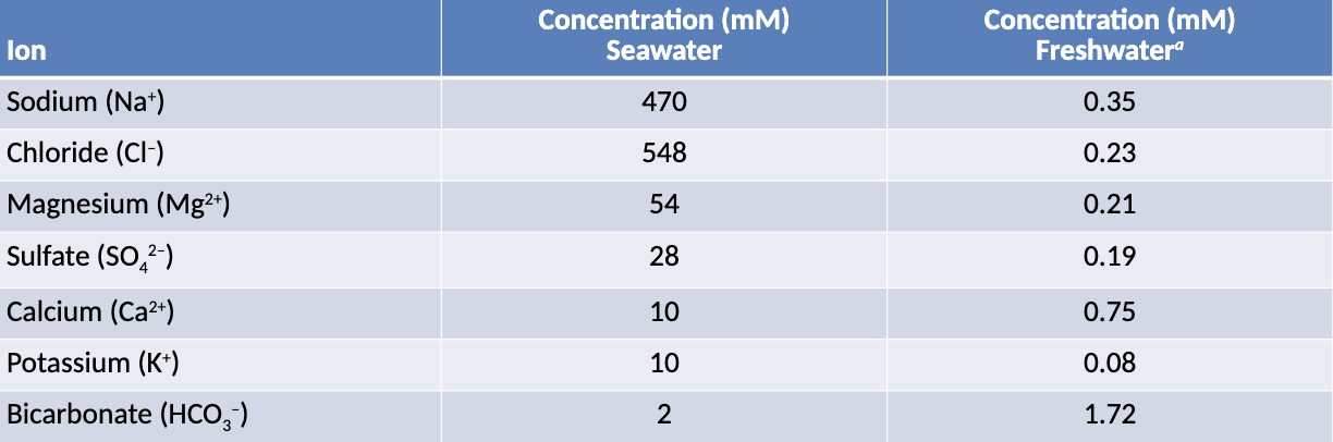
Aquatic Environments: Salinity (2)
Aquatic environments can vary in their salinity gradients (E.g. Chesapeake Bay where freshwater rivers dilute saltwater from the ocean)
Saltwater has a greater concentration of ions relative to freshwater
2 Strategies to Function in Varied Environments
Regulators: Maintain tight control of internal environment (regardless of external environment)
Conformers: Maintain normal cellular function while matching external environment (match amount of dissolved solutes to internal environment)
3 Methods of Regulatory Control
Osmotic Regulation: Controlling total amount of dissolved solutes in internal environment to maintain water balance
Ionic Regulation: Regulation of specific ions, independent of total solute concentration
Volume Regulation: Organisms that are changing their physical size through precise regulation of water and salt balance
*Volume Regulation: Example
Some crustaceans temporarily increase water uptake to swell and break out of their rigid carapace during molting, allowing them to grow before the new exoskeleton hardens
Graphing Osmotic Regulators vs. Conformers - Similarities (3)
On the y-axis, blood osmotic pressure (total dissolved solutes in the blood) in mOsm is plotted
On the x-axis, ambient osmotic pressure (solute concentration of the environment) in mOsm is plotted
The dotted diagonal line represents the isoosmotic line, where blood osmotic pressure equals environmental osmotic pressure
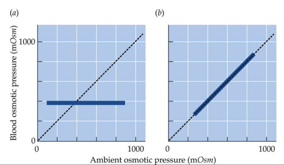
Graphing Osmotic Regulators vs. Conformers - Differences (2)
In the left graph, an osmotic regulator maintains a relatively constant blood osmotic pressure regardless of changes in the environment, reflected by a horizontal line (does not follow isoosmotic line)
In the right graph, an osmotic conformer, whose internal osmotic pressure increases proportionally with environmental osmotic pressure, closely follows the isoosmotic line

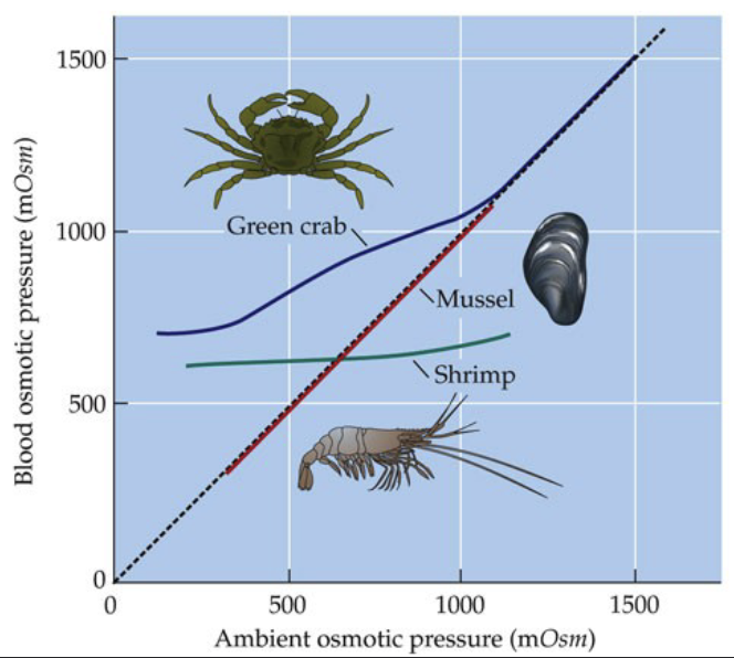
*Graphing Osmotic Regulators vs. Conformers - Examples
Shrimp is a regulator, mussel is a conformer, and the green crab is unique in which it tends to be a regulator until a high osmotic pressure (at which they become conformers)
Osmosis (2)
Osmosis is the diffusion of water across a semipermeable membrane, where water moves from areas of low solute concentrations to high solute concentrations (areas of higher free water to less free water)
This occurs because dissolved solutes bind surrounding water molecules, forming hydration shells that leave less “free water available
Osmolarity
Refers to the total concentration of dissolved solutes in a solution and determines its osmotic pressure, which drives the movement of water during osmosis
Osmoticity: Osmotic Regulators (2)
In freshwater, the external environment is hypoosmotic relative to body fluids, meaning it has a lower osmotic pressure (lower solute concentration)
In saltwater, the external environment is hyperosmotic relative to body fluids, meaning it has a higher osmotic pressure (higher solute concentration)
If the external and internal osmotic pressures are equal, the condition is described as isoosmotic
Osmoticity & Osmotic Regulators: Challenges to Freshwater Regulators (2)
External environment is hypoosmotic, so water is constantly moving into the organism
Constantly losing ions
Osmoticity & Osmotic Regulators: Challenges to Marine Regulators (2)
External environment is hyperosmotic, so water is constantly moving out of organism
Constantly loading ions
Organs of Salt/Water Balance: Gills (3)
Gills are highly permeable structures with a large surface area and thin epithelium, adaptations that maximize gas exchange
The extensive surface area increases osmotic water movement, and the high permeability allows ions to diffuse across the gill surface
As a result, animals with high O2 demands require larger gill surface areas, but this also increases water and ion exchange (osmoregulatory challenge)
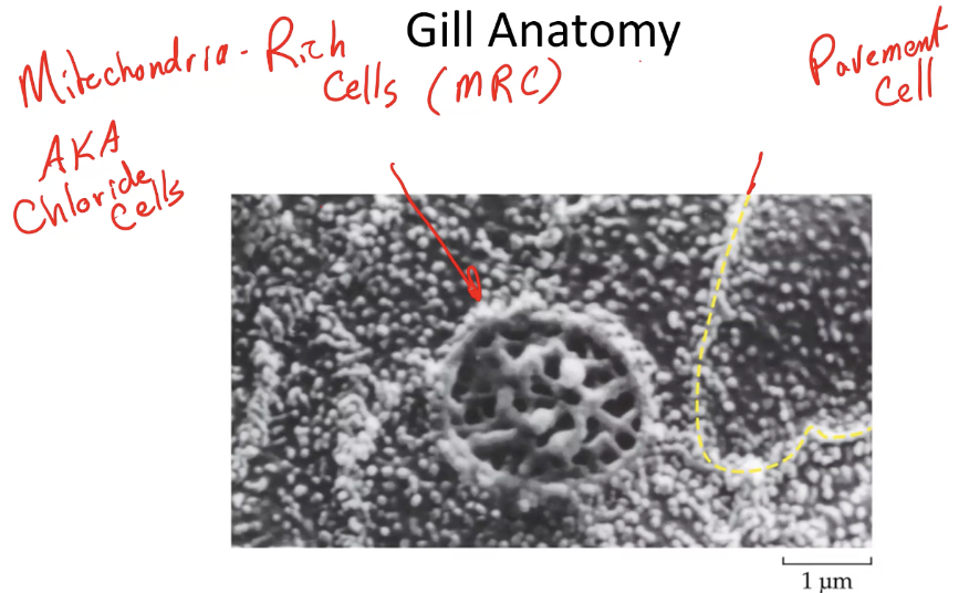
Gill Anatomy: Two Types of Cells
Pavement Cells
Make up most of the gills, and majority of the oxygen transport
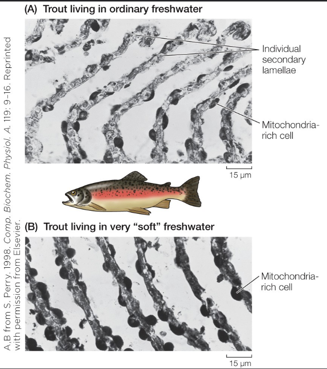
Mitochondria-Rich Cells (MRCs - aka Chloride Cells)
Specialized for ion regulation rather than gas exchange
Gill Cell Types: Mitochondria-Rich Cells (2)
Uptake of Cl-, Na+, and Ca2+ in freshwater (compensate ion loss), but excretion of Cl- and Na+ in saltwater (counteract ion gain)
The density and functional type of MRCs can change depending on environmental conditions, and are partially under hormonal control
Solution to Osmoticity Challenges: Active Transport (2)
In freshwater fish, MRCs actively transport Na+ and Cl- into the body to replace losses, while excess water is eliminated through production of large amounts of dilute urine
In saltwater fishes, MRCs actively excrete excess ions, and water loss is compensated by drinking seawater
Ion Transport by Fish Gills: Anatomy
The membrane facing the external environment is the apical membrane, while the membrane facing the blood supply is the basolateral membrane
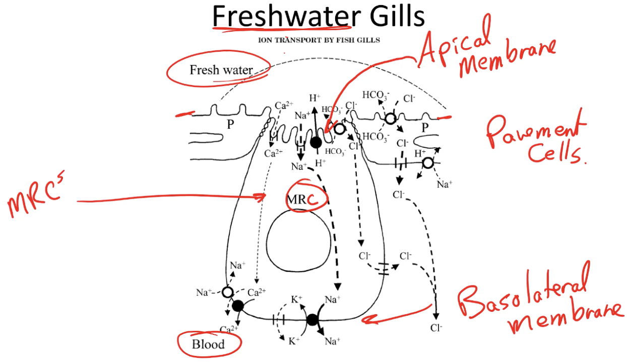
Ion Transport by Fish Gills: Freshwater Gills - Apical Membrane (3)
Ion uptake begins with a V-type (vacuolar) H+-ATPase in the apical membrane which actively pumps protons out of the cell into the surrounding water to create a negative electrical potential inside the cell
This attracts positive ions such as Na+ & Ca2+ to enter passively through sodium channels & calcium channels respectively in the apical membrane
Chloride uptake occurs through an electroneutral anion exchanger in the apical membrane, which exchanges external Cl- for intracellular HCO3-

Ion Transport by Fish Gills: Freshwater Gills - Basolateral Membrane (3)
The Na+/K+-ATPase actively pumps Na+ out of the cell into the blood while bringing K+ into the cell; the K+ that enters the cell leaks back into the blood, helping preserve the negative membrane potential
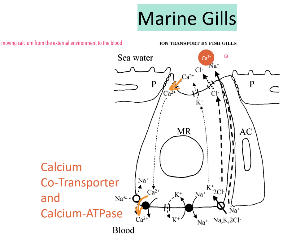
Calcium can be transported across the basolateral membrane either by Ca2+-ATPase or by a Na+/Ca2+ exchanger (NCX) (uses inward Na+ gradient as driving force - created by Na+/K+-ATPase)
As Cl- accumulates in the cell, it can move across the basolateral membrane through channels such as the Cystic Fibrosis Transmembrane Regulator (CFTR) which is a passive channel

The Three Active Transporters During Ion Transport by Freshwater Fish Gills Are ___
V-type pump, sodium-potassium pump, calcium-ATPase
Cystic Fibrosis (3)
Caused by a genetic mutation in the CFTR gene, reducing Cl- clearance from the cell, making the intracellular environment more negative than normal
This reduces the movement of positively charged ions such as Na+ out of the cell, and the buildup of ions increases osmotic water movement into cells, contributing to thickened secretions and inflammation
In epithelial tissues in the lungs and GI tract, this leads to mucosal buildup (and its associated complications)
Saltwater Fishes: Drinking Seawater (3)
When seawater is consumed, the fluid in the gut becomes hyperosmotic relative to the blood plasma; as a result, water moves by osmosis out of the blood plasma into the gut lumen
At the same time, Na+ and Cl- diffuse from the gut into the bloodstream down their concentration gradients; later parts of the intestine continue to actively transport Na+ and Cl- out of the gut and into the blood (allows animal to retain water despite ingesting a hyperosmotic fluid)
Net result is concentrated (hyperosmotic) blood plasma which is why mammals cannot drink seawater to rehydrate (lack specialized transport mechanisms)
Saltwater Fishes: Drinking Seawater - Percentages (2)
As Na+ and Cl- are absorbed, water follows by osmosis and 50-85% of the ingested water can be absorbed into the blood
For this process to be effective, 97% of Na+ and Cl- must be transported into the blood plasma to maintain the gradient for water absorption
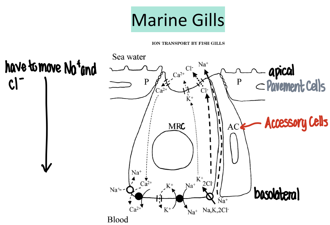
Ion Transport by Fish Gills: Marine Gills - Basolateral & Apical Membrane (4)
The Na+/K+-ATPase at basolateral membrane actively pumps Na+ out of the cell and K+ into the cell (K+ leaks back into blood), maintaining a low intracellular Na+ and negative membrane potential
This sodium gradient drives the NKCC cotransporter which brings 1 Na+, 1 K+, and 2 Cl- into the cell from the blood with each cycle
The accumulation of Cl- rises above that of seawater, allowing it to passively diffuse out across the apical membrane into the external environment
Sodium follows paracellulary due to the electrical gradient, allowing marine fish to effectively excrete excess salts absorbed from seawater
Ion Transport by Fish Gills: Marine Gills - Calcium Transport
*Calcium transport in marine gills is same as freshwater gills
Terrestrial Organisms: Marine Birds/Reptiles (2)
Live in close association with seawater and are hypoosmotic relative to seawater
The two major challenges they face are water loss and salt loading
Terrestrial Organisms: Marine Birds/Reptiles - Challenges (2)
Water tends to leave their bodies by osmosis, while salts enter through ingestion of seawater and consumption of marine prey
Have less permeable integuments which helps reduce water loss across the body surface, but water loss still occurs through respiration
Marine Birds - Strategies for Salt Balance (2)
Rely on specialized nasal salt glands that actively remove excess NaCl from the blood by producing a highly concentrated salt solution that is excreted through the nostrils
This secretion is often more concentrated than seawater, allowing them to eliminate excess solutes while conserving water
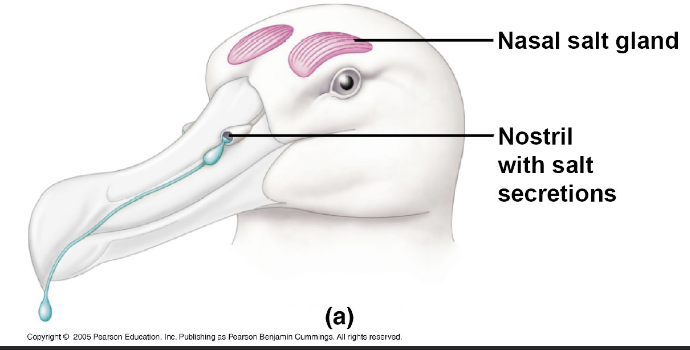
*4 Marine Birds That Utilize Nasal Salt Glands
Gulls (Charadriiformes)
Penguins (Sphenisciformes)
Albatross (Procellariiformes)
Pelicans (Pelecaniformes)
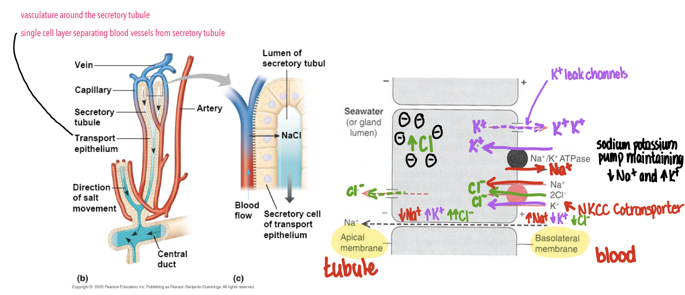
Marine Birds: Physiology of Nasal Salt Glands (4)
The nasal salt gland consists of highly vascularized secretory tubules surrounded by capillaries, separated by a single epithelial cell layer that allows efficient ion transport from blood to gland lumen
On the basolateral membrane, the Na+/K+-ATPase pumps Na+ out of the cell and K+ into the cell (out through K+ leak channels to stabilize membrane potential), maintaining low intracellular Na+ for negative membrane potential
This sodium gradient drives the NKCC cotransporter (same function as before), resulting in a highly concentrated NaCl solution in the tubule lumen
The concentrated NaCl is then transported through a central duct and excreted through the nostrils
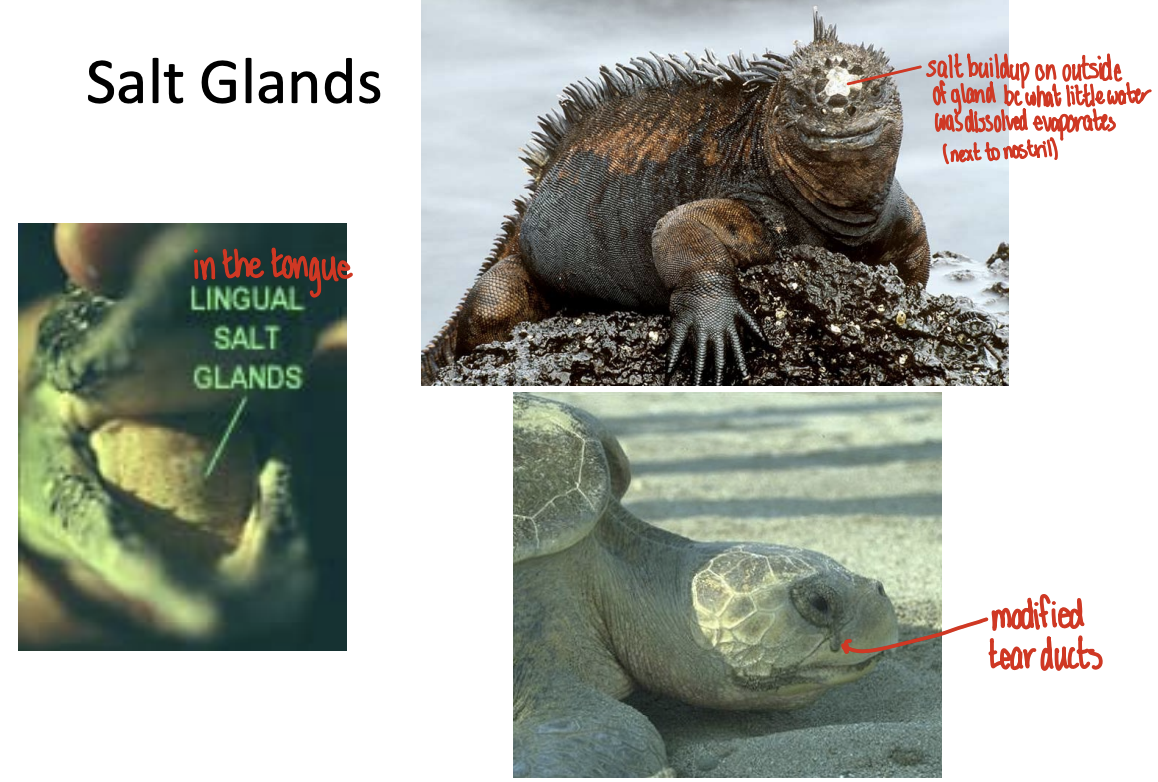
Marine Reptiles: Salt Glands
Function similarly to that of the marine birds, but differ in anatomical location (e.g. lingual (tongue) salt gland, modified tear ducts, etc.,)
Marine Fish: Special Cases to Osmotic Balance - Elasmobranchii (3)
Elasmobranchii (sharks, rays, skates) are marine osmoconformers/ionic regulators that take a unique approach to osmotic balance
Instead of actively secreting large amounts of salt, they produce high concentrations of organic solutes in their blood, primarily urea and trimethylamine oxide (TMAO)
The accumulation of organic osmolytes raises osmotic pressure of blood plasma to be nearly isosmotic with seawater, reducing osmotic water loss
Marine Fish: Special Cases to Osmotic Balance - Salmon
Salmon are born in freshwater, migrate to seawater to grow, and return to freshwater to reproduce
Salmon: Behavioural Strategies (2)
Salmon often spend time in brackish water (estuaries) that serve as a transitional environment between freshwater and full-strength seawater (reduces osmotic stress)
Additionally, salmon can adjust drinking behaviour to reduce water intake when transitioning between environments
Salmon: Physiological Strategies (2)
Kidney function changes as in freshwater salmon produce a high volume of dilute urine to eliminate excess water, whereas in seawater urine volume decreases
Gill function also shifts through changes in MRCs, where in freshwater they specialize in ion uptake, while in seawater they switch to ion excretion
MODULE 7: TERRESTRIAL OSMOREGULATION & THE KIDNEY
The greatest challenge to terrestrial organisms is ___
Water loss
3 Sources of Water Loss
Respiratory Water Loss: Water vapour lost during breathing
Evaporative Water Loss (EWL): Water loss across the skin/body surface
Excretory Water Loss: Water loss through urine and feces
Excretory Water Loss (3)
EWL is necessary because organisms must remove metabolic waste products, particularly nitrogenous wastes produced from breakdown of proteins
Nitrogen is initially generated as ammonia (toxic), and many terrestrial vertebrates convert it into urea for safer transport and excretion
Eliminating these wastes requires water, so to prevent excessive dehydration organisms regulate the composition, concentration, and volume of urine
Urine Concentration Modification (2)
During times of drought, organisms retain as much water as possible by producing highly concentrated urine
During times of water loading, excess water is eliminated through production of large volumes of dilute urine
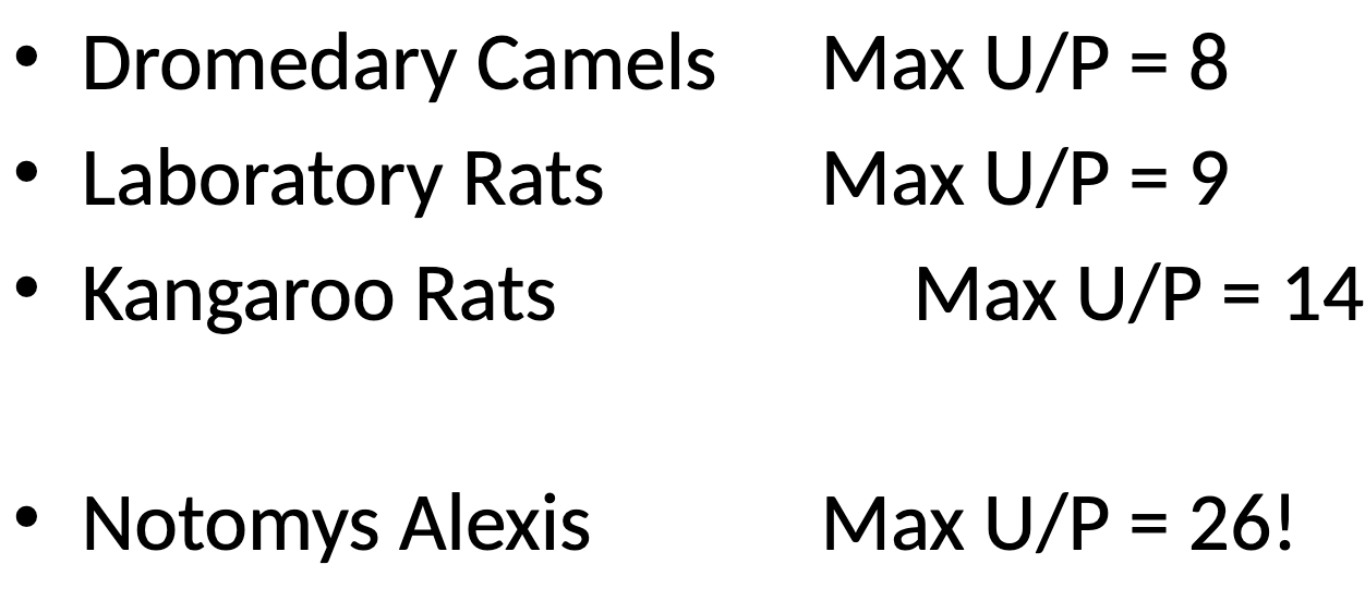
The degree of urine concentration is expressed as ___
U/P ratio; concentration of substance in urine divided by its concentration in plasma (primarily reflected by adjustments in urine concentration)
U/P Ratio (3)
U/P = 1; urine is isoosmotic to plasma
U/P < 1; urine is hypoosmotic (dilute) relative to plasma (water excretion)
U/P > 1; urine is hyperosmotic (concentrated) relative to plasma (water loading)
The U/P ratio in humans ranges from ___
0.1-4
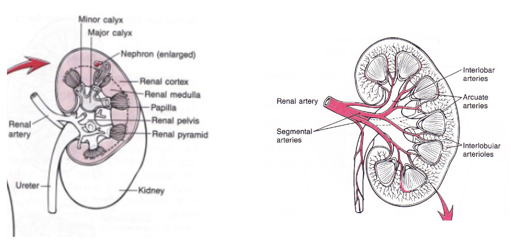
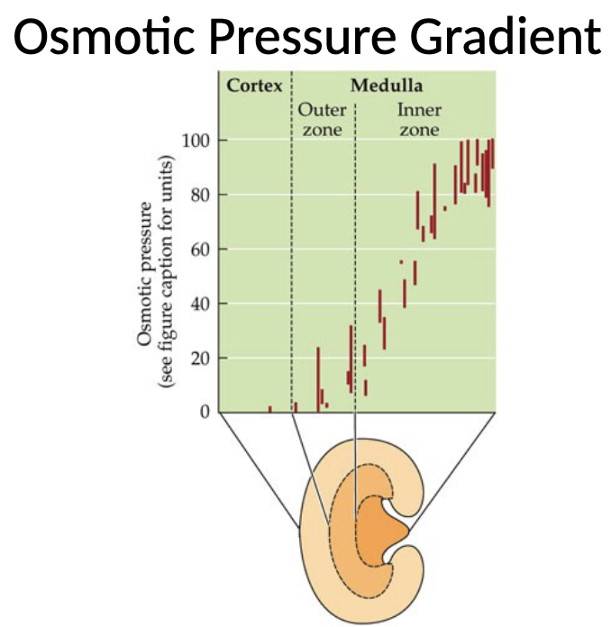
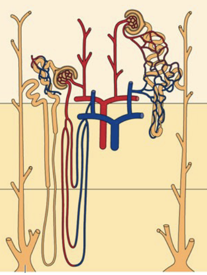
The Mammalian Kidney
Its concentrating ability regulates urine production, and thus H2O and solute concentration
*Dotted line on left diagram is border between renal cortex and renal medulla
*Renal pyramid is the structure that carries a collection of nephrons (functional unit of water and salt balance for the kidneys - 1 million nephrons per kidney)
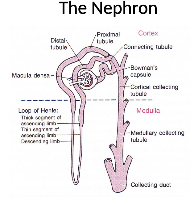
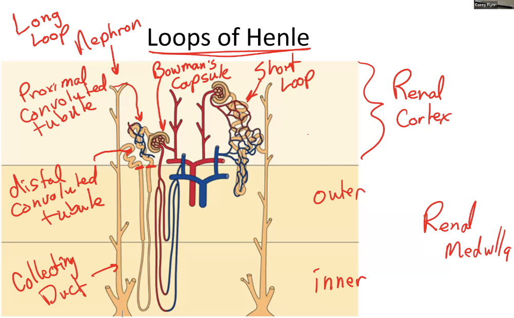
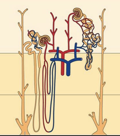
The Nephron (5)
The nephron is the functional unit of the mammalian kidney and is responsible for filtering blood and forming urine
It begins in the renal cortex with Bowman’s capsule, which surrounds the glomerulus and is where filtration occurs
The filtrate then passes through the proximal tubule, where most reabsorption of water and solutes takes place
It continues into the loop of Henle which extends into the medulla and consists of descending and ascending limbs that establish the osmotic gradient necessary for urine concentration
The filtrate then moves into the distal tubule, and finally into the collecting duct (which runs through the renal medulla)
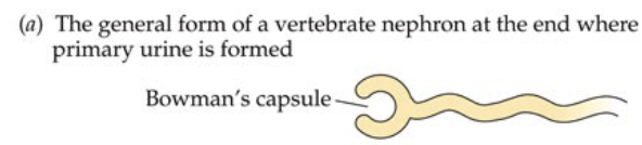
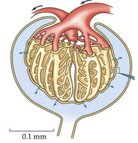
Bowman’s Capsule (3)
Located in the cortex and surrounds the glomerulus, a network of capillaries that provides direct access to the blood
This close association allows filtration of blood plasma under pressure, forming the initial filtrate (primary urine)
Small solutes and water pass from the blood into Bowman’s capsule, while large molecules (proteins and cells) are retained in the blood
Glomerulus (2)
Blood enters the glomerulus through the afferent arteriole and exits through the efferent arteriole
Exit of blood in the capillary bed through an arteriole rather than a venule allows tight regulation of contraction through systole pressure (heart contraction)
Together, Bowman’s capsule and the glomerulus form the ___
Renal corpuscle
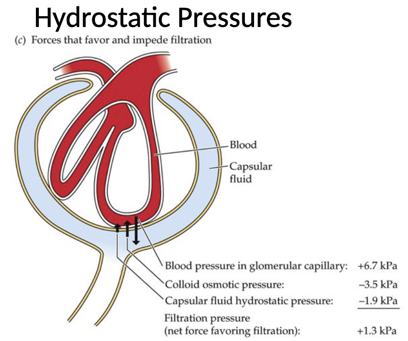
Filtration at Glomerulus: Hydrostatic & Osmotic Forces (3)
The primary force favouring filtration is the hydrostatic blood pressure within the glomerular capillaries which push fluid into the capsule
Opposing this movement are two forces: colloid osmotic pressure of blood (generated by plasma proteins that remain in capillaries), and hydrostatic pressure of the capsular fluid within Bowman’s capsule
The osmotic (oncotic) pressure of the blood is slightly higher than that of the filtrate, which pulls water back towards the capillaries
Glomerular Ultrafiltration (2)
Ultrafiltration occurs at the glomerulus when hydrostatic pressure in the glomerular capillaries is greater than pressure within the tubule lumen of Bowman’s capsule
This pressure difference (generated by cardiac systole) forces water and small solutes out of the blood and into the filtrate, but not large solutes
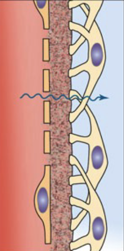
Glomerular Ultrafilter: 3 Components
Glomerular Capillaries
Composed of a single endothelial layer containing fenestrations that allow water and small solutes to pass only
Basement Membrane
On the outside of blood vessel; collagen-rich layer that acts as a selective barrier based on size and charge (restricting large proteins)
Podocytes
Have a central cell body with extending foot processes that interlock to form openings known as slit diaphragms
Primary Urine (2)
Primary urine consists of water and small solutes that first enter the nephron after filtration into the Bowman’s capsule
Primary urine has a U/P ratio close to 1 (blood slightly higher osmotic pressure)
Definitive Urine (2)
Is the final excreted product after reabsorption and secretion along the nephron
Can have a much higher or lower osmotic concentration than plasma depending on whether the body is conserving water, or eliminating excess water
Glomerular Filtration Rate (GFR) (2)
GFR is the rate of production of primary urine (approximately 120mL per minute)
The kidneys filter a volume equivalent to the entire blood volume roughly every 30 minutes
Loop of Henle (3)
The ability to change the concentration of urine starts with the Loop of Henle
Nephrons are classified as long-loop (juxtamedullary) or short-loop (cortical) nephrons
Long-loop nephrons have Loops of Henle that extend deep into the inner renal medulla; short-loop nephrons remain mostly in the cortex and outer renal medulla and do not contribute to urine concentration
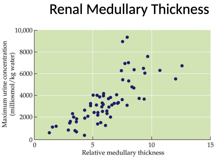
Renal Medullary Thickness (2)
Renal medullary thickness can be measured as the thickness of the inner renal medulla relative to the overall thickness of the kidney
There is a positive relationship between relative medullary thickness (longer Loops of Henle) and maximum urine concentration
Primary Urine Flow: Loops of Henle (3)
After filtration in Bowman’s capsule, primary urine flows into the proximal convoluted tubule and then into the Loop of Henle
The filtrate first travels down into the inner renal medulla through the thin segment of the descending limb
When the loop turns and ascends back toward the renal cortex, it enters the ascending limb (which is divided into a thick and thin segment)

Loops of Henle: Descending Thin Segments - Permeability
The descending thin segments are highly permeable to water, and moderately permeable to solutes
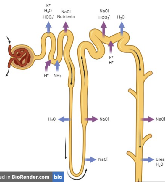
Loops of Henle: Ascending Thin Segments - Permeability
The ascending thin segment is impermeable to water, but moderately permeable to solutes

Loops of Henle: Ascending Thick Segments - Permeability
The ascending thick segment is impermeable to water, and is involved in active transport of NaCl

___ can leave the descending limb, but not the ascending limb
Water
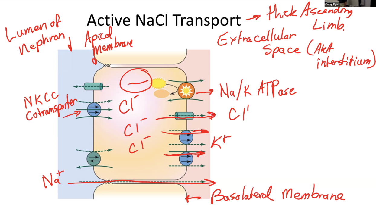
Loops of Henle: Ascending Thick Segments - Active NaCl Transport (4)
The Na+/K+-ATPase located on the basolateral membrane actively pumps Na+ out of the cell (into interstitium), while most of the K+ leaks back into the interstitium through leak channels
This low Na+ level provides the driving force for the NKCC cotransporter on the apical membrane, which brings 1Na+, 1K+, and 2Cl- ions into the cell from lumen of nephron
The net effect is accumulation of Cl- within the cell, which then diffuses passively through chloride channels into the interstitium
Additionally, the movement of ions creates an electrical gradient that drives Na+ movement from lumen to interstitium through paracellular pathways
Two Osmotic Pressures in Loop of Henle
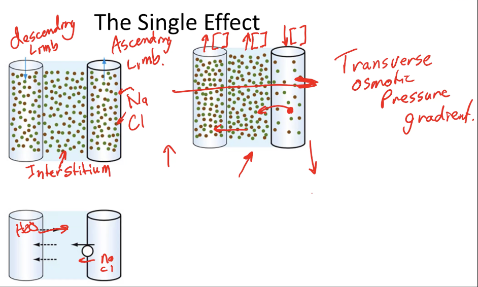
Single Effect
Refers to the initial local change in concentration produced by active NaCl transport
Countercurrent Multiplication
Occurs because fluid in the descending and ascending limbs flow in opposite directions, causing the single effect to be repeatedly applied along its length as new filtrate enters and moves through the loop
Single Effect: Scenario (4)
Imagine starting with equal solute concentrations in the ascending limb, descending limb, and surrounding interstitium
Active transport pumps NaCl out of the ascending limb into the interstitium, decreasing NaCl concentration in the ascending limb while increasing it in the interstitium
As the interstitium becomes more concentrated, NaCl diffuses into the descending limb, and water moves out of the descending limb into the interstitium
This single active transport process therefore creates concentration differences across the two limbs and the interstitium to establish a transverse (horizontal) osmotic pressure gradient between the limbs
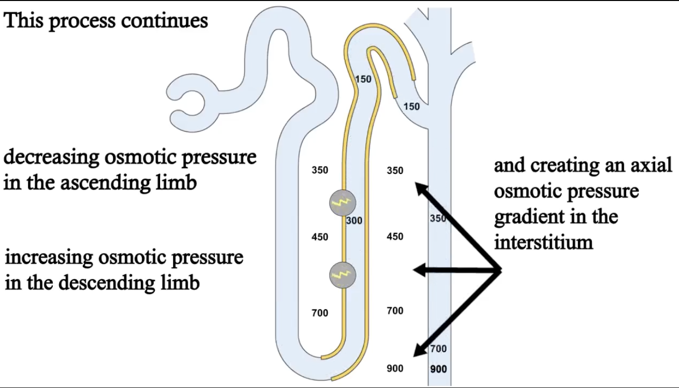
Countercurrent Multiplication (2)
Countercurrent multiplication transforms the transverse gradient created by the single effect into an axial osmotic pressure gradient within the renal medulla
Osmotic pressure increases as you move deeper into the renal medulla, with solute concentrations becoming highest in the inner medulla
Collecting Ducts (2)
The loop of Henle creates the osmotic gradient in the renal medulla, and is continuously removing NaCl making the tubular fluid dilute
The concentration of urine primarily occurs in the collecting ducts
As fluid moves down the collecting duct toward the inner renal medulla, ___
it encounters an interstitium with higher osmotic concentration
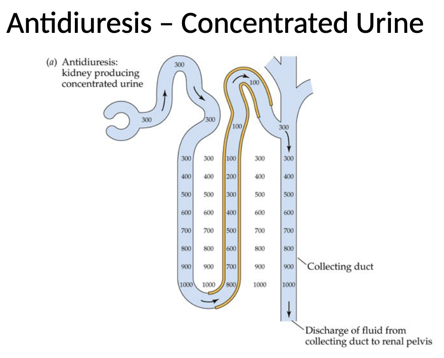
Antidiuresis: Concentrated Urine - Drought (2)
During antidiuresis, the collecting duct is permeable to water, so kidney produces concentrated urine because water is reabsorbed from the collecting duct into the interstitium
Most solutes remain in the duct, so fluid inside becomes increasingly concentrated as it descends
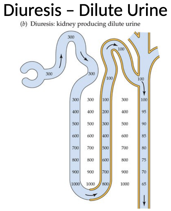
Diuresis: Dilute Urine - Water Loading (2)
During diuresis, the collecting duct becomes impermeable to water
As a result, the tubular fluid becomes progressively more dilute, and is excreted as large volumes of dilute urine
Water retention of the collecting duct is governed by ___
hormonal control
Hormonal Control of Urine Concentration (3)
Hormonal control of urine concentration is primarily regulated by antidiuretic hormone (ADH), aka vasopressin or arginine vasopressin
ADH modulates the permeability of the collecting duct to water
When ADH is present, it stimulates the insertion of aquaporin-2 water channels into the membrane of collecting duct cells, allowing water to move into the interstitium (antidiuresis)
Antidiuretic Hormone: Detection
ADH release is upregulated by low levels of blood plasma, meaning the body needs to conserve water
Antidiuretic Hormone: Stimulation of Release - Baroreceptors (2)
Low blood volume is detected by baroreceptors which are stretch-sensitive receptors located in the pulmonary venous system, cardiac atria, aortic arch, and carotid sinus
When these receptors sense reduced stretch (indicating lower blood volume), they signal for increased ADH release
Antidiuretic Hormone: Stimulation of Release - Osmoreceptors (2)
Osmoreceptors in the hypothalamus detect increases in blood osmolarity (when blood becomes more concentrated due to water loss)
These receptors stimulate neurons in the hypothalamus that trigger the posterior pituitary to release ADH into the bloodstream
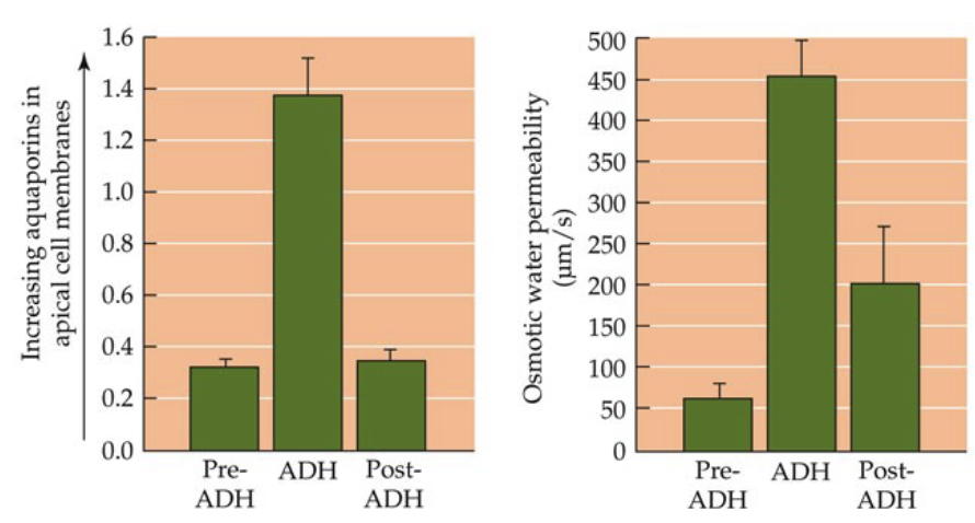
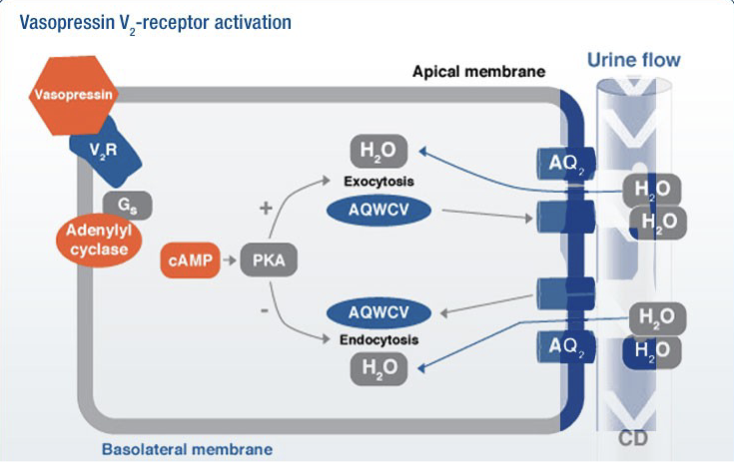
Aquaporin-2: Second-Messenger Signalling Pathway (4)
ADH binds to V2 receptors on the basolateral membrane of collecting duct cells which activates a G-protein (Gs)
This activates adenylyl cyclase which converts ATP into cAMP, which then activates protein kinase A (PKA)
PKA triggers exocytosis of aquaporin-containing vesicles (AQWCV), inserting aquaporin-2 channels into the apical membrane of the collecting duct (concentrated urine)
When ADH is absent, the opposite occurs where aquaporin-2 channels are removed from the membrane by endocytosis (dilute urine)
Urea (2)
Ammonia (NH3) is a nitrogenous waste product formed during protein metabolism (toxic); the liver converts ammonia into urea, a less toxic compound that is transported to the kidneys for excretion
Urea is freely filtered at the glomerulus as the concentration of urea in Bowman’s capsule is nearly equal to that of blood plasma
Urea Has Low Permeability In: (3)
Distal convoluted tubule
Thick ascending segment of Loop of Henle
Cortical and outer renal medulla
Urea Has High Permeability In: (2)
Collecting duct
Inner renal medulla
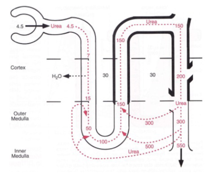
Urea Permeability: Urea Recycling (3)
As fluid moves down the descending limb of the loop of Henle, the concentration of urea increases as water leaves into the interstitium, but then stabilizes as it enters the ascending limb and upper portion of the collecting duct
In the lower portion of the collecting duct in the inner renal medulla, the epithelial cells contain high levels of UT-A1 and UT-A3, which are transporters that allow urea to diffuse out of the collecting duct into the interstitial fluid
Once in the interstitium, some of the urea diffuses back into the thin segments of the loop of Henle (increasing urea concentration)
Urea transporter proteins are upregulated by ___
ADH
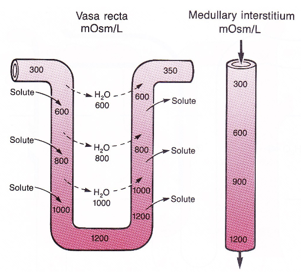
Renal Medulla Blood Supply: Vasa Recta (3)
The capillaries that supply the medulla (vasa recta) are highly permeable, allowing water and solutes to move easily between the blood and surrounding interstitial fluid
As blood flows from the renal cortex down into the medulla, it encounters an environment with progressively higher osmotic pressure
As a result, water diffuses out of the plasma into the interstitium, while the blood takes up NaCl and urea
Vasa Recta (2)
The vasa recta supplies oxygen and nutrients to the medulla without destroying osmotic pressure gradients because of countercurrent exchange (minimizes washout of solutes)
Only 1-2% of total renal blood flow passes through the vasa recta (also minimizes washout)
Vasa Recta: Countercurrent Exchange (3)
As blood enters the descending limb of the vasa recta, it encounters increasingly concentrated interstitium, and water diffuses out of the blood vessel while solutes (NaCl and urea) diffuse into the blood
When the blood turns and ascends back toward the cortex, the situation reverses as the blood inside the vessel is now more concentrated, so water moves back into the blood and solutes diffuse into the interstitium
The blood leaves the vasa recta and returns to the venous circulation has a composition similar to the blood that entered, and this mechanism prevents the osmotic gradient of the renal medulla from being washed out
MODULE 8: CARDIOVASCULAR 1
Cadiovascular System
Refers to the system involving the heart (cardio) and blood vessels (vascular) that circulate blood throughout the body
Lumen
The hollow internal space within a blood vessel or heart chamber through which blood flows
Circulation (3)
Refers to the pressure-driven bulk flow of blood through vessels and passages
Mechanistically, it is the movement of blood caused by pressure differences generated by the heart
Functionally, circulation transports commodities (oxygen, nutrients, wastes, hormones, etc.,) while also providing hydraulic pressure that supports organ function
Perfusion
Refers to the forced flow of blood through blood vessels supplying tissues, ensuring that organs receive oxygen and nutrients
Convective Transport
Refers to the movement of substances by bulk flow of fluid, meaning materials are carried along with flowing blood rather than diffusion
The circulatory system includes the ___, ___, ___, and ___
Heart (blood pump)
Blood vessels
Circulatory passages
Blood
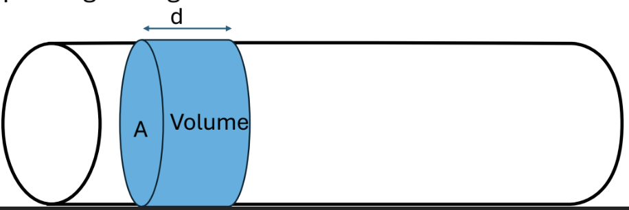
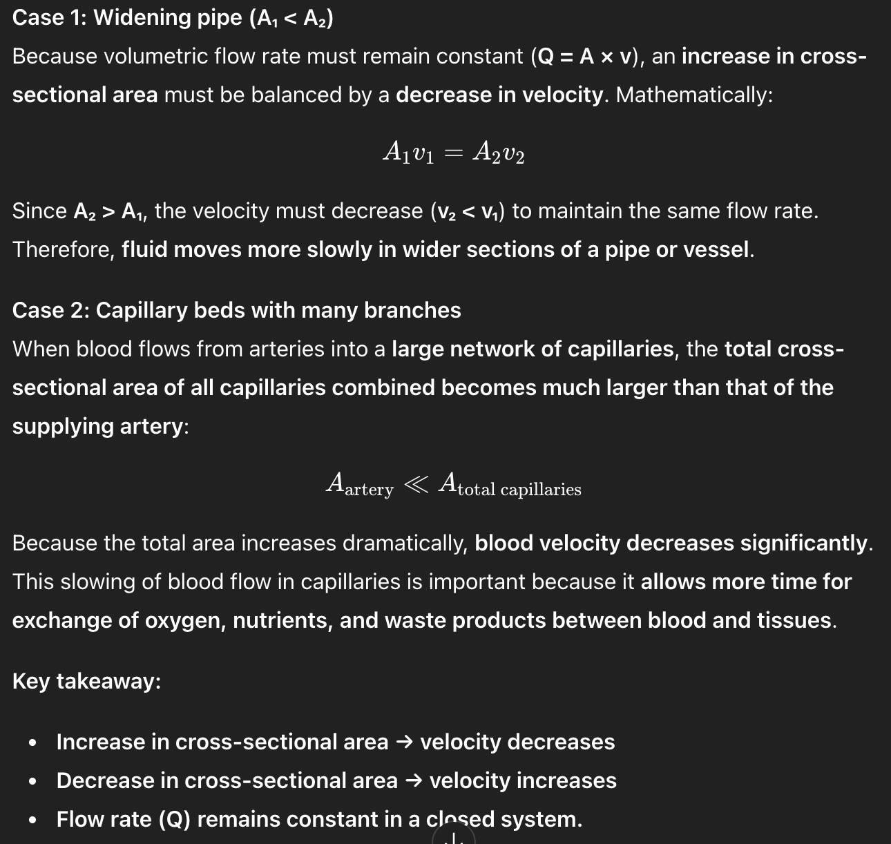
Volumetric Flow Rate (Q) (2)
Volumetric flow rate (Q) describes the volume of fluid that passes through a cross-section of a vessel per unit time (L/min or m2/min)
It is calculated using the equation: Q = A x v where A is the cross-sectional area of the vessel (m2) and v is the velocity of the fluid (m/s)
Volumetric Flow Rate (Q): Continuity Principle (2)
In a closed system, Q is conserved, meaning the same volume of fluid must pass through each section of the system per unit time
If the radius of a vessel decreases, the cross-sectional area decreases; since flow must remain constant, the velocity of fluid must increase to compensate
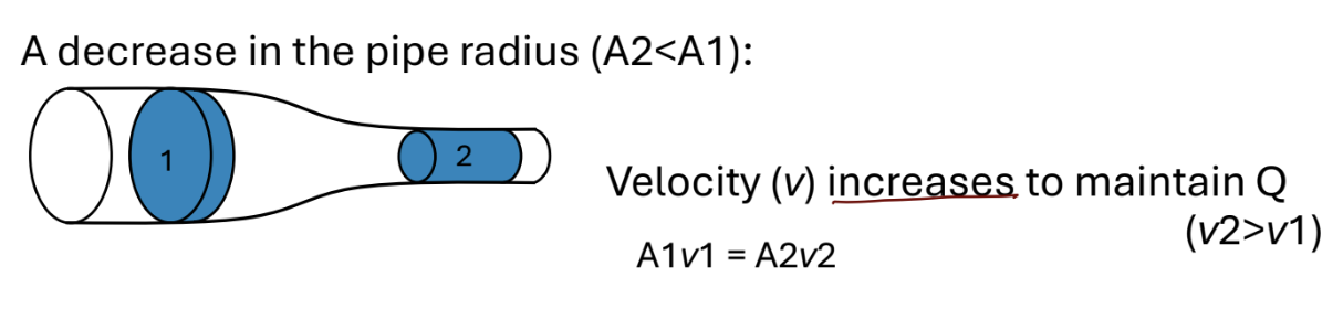
*Volumetric Flow Rate (Q): Continuity Principle - Mathematical Approach (2)
Because Q is constant: A1V1 = A2V2
If A2 < A1, then V2 > V1; meaning fluid moves faster in narrow vessels
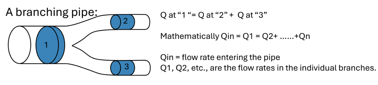
Volumetric Flow Rate (Q): Branching Vessels (2)
When a vessel branches, the flow entering the branch must equal the sum of the flows in all downstream branches (Qin = Q1 = Q2 + Q3 + Qn)
This means the total flow entering a branching point equals the total flow leaving it
Volumetric flow rate explains ___ and ___
Why blood velocity increases in narrower vessels
Why flow from larger arteries is distributed among multiple smaller branches while maintaining conservation of flow
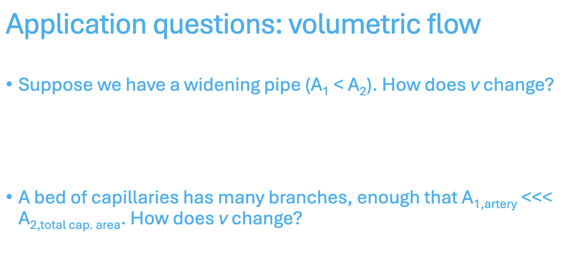
*Application Question: Volumetric Flow
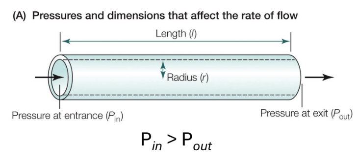
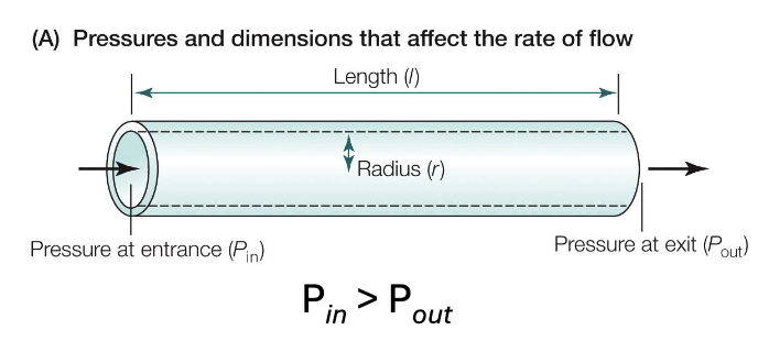
5 Factors Affecting Q
Pressure Difference
ΔP between the entrance and exit of the vessel is proportional to flow; larger pressure gradient produces greater flow (blood flows from high to low - Pin > Pout)
Vessel Radius (r)
Larger radius increases flow; resistance to flow decreases
Vessel Length (l)
Longer vessels increase resistance (reducing flow)
Viscosity of Fluid (v)
Higher viscosity (thicker blood) increases resistance and reduces flow
Elasticity & Compliance
Blood vessels that are more elastic and compliant can expand in response to pressure
*VELPR
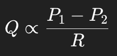
Hagen-Poiseuille Law (3)
Describes how flow (Q) through a cylindrical vessel depends on the pressure difference and resistance
Where P1-P2 is the pressure difference between the entrance and exit of the vessel, and R is the resistance to flow
This means that greater pressure differences increase flow, while greater resistance decreases flow
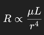
Resistance in a Vessel (3)
Resistance depends on viscosity of the fluid (µ) length of the vessel (L), and radius of the vessel (r)
Resistance increases with greater viscosity and longer vessel length, and decreases dramatically as radius increases (strongest effect)
Viscosity is influenced by the number of red blood cells (hematocrit) and plasma proteins (affect how thick blood is)