Review test flashcards
 Call Kai
 Learn
Learn Practice Test
 Spaced Repetition
 Match
Match Flashcards
 Knowt Play
1/87

There's no tags or description
Looks like no tags are added yet.
Name | Mastery | Learn | Test | Matching | Spaced | Call with Kai | Chat |
|---|
No analytics yet
Send a link to your students to track their progress
88 Terms
What is primary cell structure and what organelles can be seen using light microscopy?
They are structures that can be seen using a light microscope, organelles that can be seen are the nucleus, cytoplasm, cell membrane, cell wall, chloroplast.
What is Cell Ultrastructure and what organelles can be seen using electron microscopy
Structures that can be seen using an electron microscope, visible organelles are the mitochondria, ribosomes, Endoplasmic Reticulum, Golgi, Nucleolus, chromatin.
Nucleus
Cell division, protein synthesis
Mitochondrion
Respiration pathways
Chloroplast
Photosynthetic pathways
Lysosome
Digestion, recycling and isolation
Golgi Apparatus
Secretion, reprocessing, lysosome synthesis
Endoplasmic Reticulum (ER)
Support, Golgi apparatus synthesis
Ribosome
Protein synthesis
Label the nucleus


Nucleus Diagram
Nucleus Function
Main site of DNA in eukaryotic cells
Preservation, replication and expression of genetic information
It makes RNA for proteins synthesis
It copies DNA for cell division
Label the Mitochondria


Mitochondria Diagram
Mitochondrion Function
The inner membrane contains the enzyme necessary for the synthesis of Adenosine Triphosphate (ATP)
The mitochondria are closely associated with the pathways of respiration
These metabolic pathways are divided up and supported by the membranes

Label the Chloroplast

Chloroplast Diagram
Chloroplast Function
Photosynthesis
The metabolic pathways are closely associated with the membranes as in the case of the mitochondrion.
Organelles and Evolution
Both chloroplasts and mitochondria are double membrane bound
They involved in energy reactions
They constrain extra nuclear DNA and characteristic small ribosomes of their own
This has led biologists to believe that there may be some similarity in their origins in the cells of eukaryotes
The endosymbiotic theory
Lysosome function and structure
Structurally simple, spherical, single membrane bound
Lysosomes contain a large number of CATABOLIC enzymes. Catabolic enzymes digest materials by hydrolysis
It’s function is the digestion of compounds taken in by the cell by endocytosis And the recycling of material within the cell
Golgi Apparatus Diagram


Label the Golgi Apparatus
Golgi Apparatus Function
Processing and packaging
Synthesizing lysosomes to contain the potentially dangerous catabolic enzymes
Producing secretory vesicles - mucus
Making more plasma membrane

Endoplasmic Reticulum diagram

Label the Endoplasmic Reticulum
ER functions
The ER starts the biosynthetic pathways form many protein and lipid molecules in the cell
These continue in the Golgi apparatus
Rough ER has ribosomes attached to it as opposed to Smooth ER
The proteins are made on Rough ER will eventually be secreted outside the cell.

Ribosome diagram

Label the ribosome
Ribosome Function
NOT membrane bound
Found both in pro and eukaryotes
The subunits are synthesized separately in the nucleolus of the nucleus of eukaryotes.
Ribosomes in the cytoplasm
Single free-floating
Attached to rough ER
linked together as a poly ribosome or polysome
Their function is protein synthesis
Explain the structure of Carbon
Carbon has 4 available binding sites
Carbon can form a single, double, or triple covalent bond
Carbon is able to bond covalently with a large number of elements
Carbon can form both long chains and rings of atoms
Tetrahedral structure when bound allows for isomerism
Organic Compounds
Compounds containing carbon found in living organisms
Not including carbonates, hydrogen carbonates, CO2 or CO
Often based upon the skeleton of carbon
Evolution has chosen a few for use in living organisms
There are four principle groups: sugars, fatty acids, amino acids, and nucleotides
Carbohydrates
Organic molecules commonly referred to as sugars
Made up of Carbon, hydrogen and oxygen. The general formula is C(H2O)n
There are three types of carbohydrates: mono, Di, and poly saccharides
Monosaccharides
Monosaccharides are the simplest form of carbohydrates
Contain a carbonyl group and at least two hydroxyl groups
Empirical formula is CH2O
6 carbon monosaccharides include glucose, fructose and galactose (heroes sugars).
5 carbon monosaccharides include ribose and deoxyribose (pentose sugars)
All hexose sugars have the molecular formula C6H12)6, there are also isomers.

Straight Chain and Cyclic Structure
The previous slide contained the straight chain structure of three monosaccharides.
The carbons are numbered starting at the carbonyl group. However, in aqueous solution monosaccharides adopt a ring structure.
This occurs as a result of the intermolecular reaction between the aldehyde group on C1 and the OH group on C5
Ring Structure Isomers
The intermolecular reaction that causes the ring structure to form in aqueous solution produces an asymmetric carbon at C1
Therefore, there are two ring structure isomers of glucose called a-glucose and b-glucose
The difference has an effect on the properties of their polymer.

Condensation reactions
Monosaccharides undergo condensation reactions in order to form disaccharides and polysaccharides.
Hydroxide groups on adjacent monosaccharides react to form a bond, known as a glycosidic link, and a molecule of water.
Two molecules of a-glucose condense to form a 1-4 glycosidic link which produces the disaccharide maltose.
Disaccharides
Disaccharides are composed of two monosaccharides joined together.
Combining different monosaccharides produces different disaccharides.
Disaccharides are soluble molecules that can be broken down into their component monosaccharides through acid hydrolysis or by enzyme catalysed reactions (digestion)
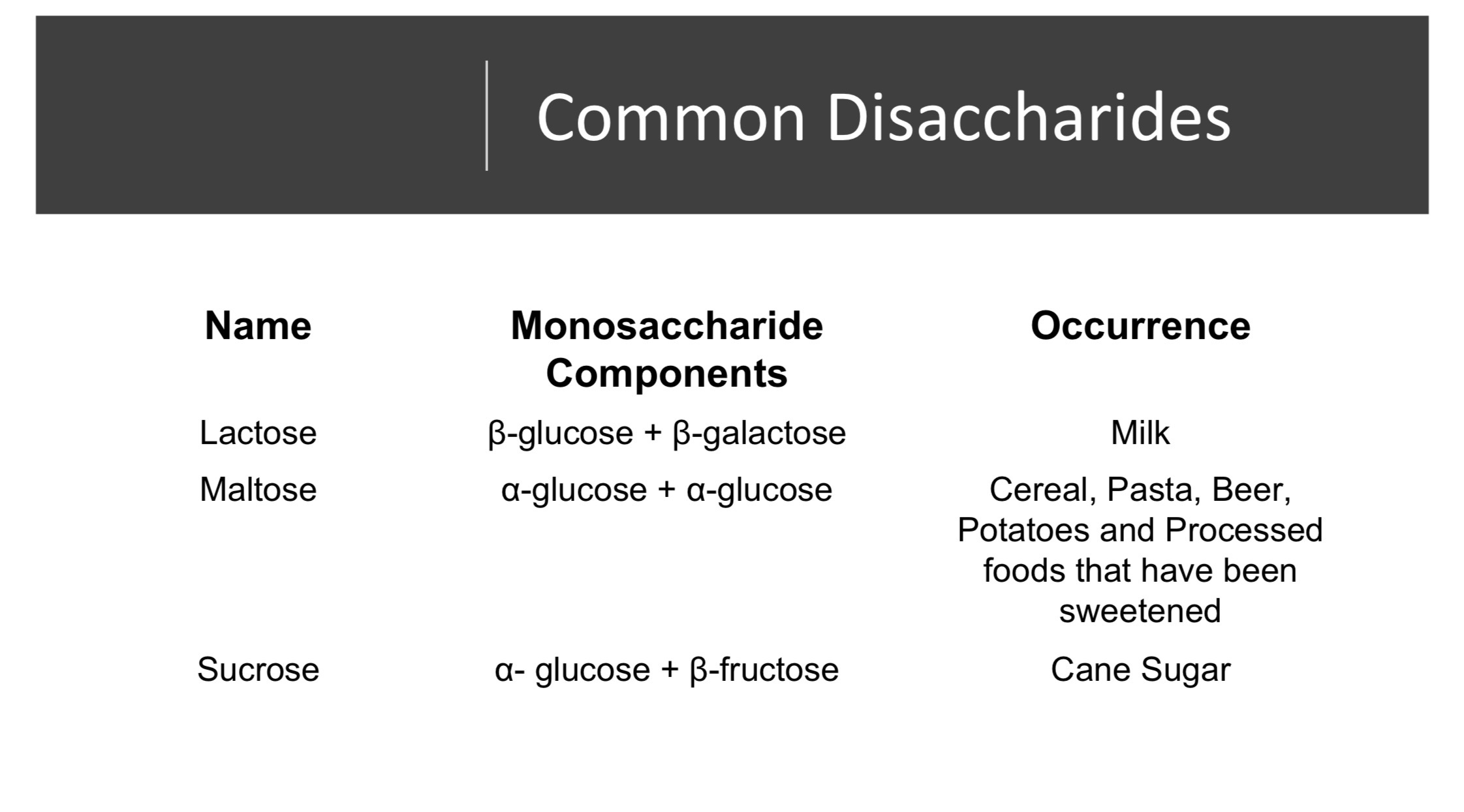
Common Disaccharides
Polysaccharides
Polysaccharides are long chains of monosaccharides held together by glycosidic bonds.
Have large molar mass, are not sweet, are insoluble or slightly soluble in water and are non-reducing.
Polysaccharides differ in the nature of their recurring monosaccharide, their bonds, the length of their chains and the degree of branching.
Glucose Based Polysaccharides
Starch
Glycogen
Cellulose
Starch
Starch is a polymer of a-glucose and id the main form of carbohydrate storage in plants.
Starch can form a compact spiral structure and is stored in the starch granules of plants.
There are two forms of starch;
Amylose- a straight chain polymer of a-glucose with 1-4 glycosidic bonds.
Amylopectin- a branched polymer of a-glucose with both 1-4 and 1-6 glycosidic bonds.
Glycogen
A polymer of a-glucose
Main form of carbohydrate storage in animals, found in the liver and muscle tissue
Similar to amylopectin but with more 1-6 glycosidic branches.
Cellulose
A polymer of B-glucose
It is the structural material in the cell walls of plants
A linear polymer with 1-4 B-glycosidic linkages
This forms an uncoiled structure with alternate glucose molecules upside down
This allows cellulose to form cables known as micro fibrils that give a rigid structure.
Digestion
Polysaccharides are broken down into their monosaccharide units during the digestive process.
The human body has enzymes that digest starch and glycogen through a series of enzyme controlled reactions (hydrolysis) during which the glycosidic links are broken
The human body does not produce enzymes that work on cellulose.
Carbohydrate Functions
Sugars (mono and disaccharides ) small molecules and soluble in water:
Maintainence of osmotic balance ( salts in blood plasma, plant cell turgidity)
Transport of energy reserves (glucose in blood or sucrose in sap)
Energy substrate (respiration and photosynthesis)
Energy store (sugar cane)
Flavouring (fruits) reward (nectar)
Precursors (building blocks) of polysaccharides, nucleotides and amino acids
Polysaccharides: Large molecules insoluble in water
Osmotically inactive carbohydrate storage (seeds, roots, chloroplasts)
Structural (cellulose in Plants)
Lipids
Lipids contain carbon, hydrogen, and oxygen
More hydrogen (more reduced) than carbohydrates
Lipids are insoluble in water but they are soluble in polar solvents (organic solvents such as alcohols, acetone, chloroform)
The most common lipids are fats, oils, steroids, and phospholipids.,
Fats and Oils
The major component of fats and oils are triglycerides.
Glycerides are composed of glycerol and fatty acids
Glycerol is a molecule of three car bonds, each of which contains an alcohol group
Fatty acids are long chain carboxylic acids
An esterification reaction takes place between the carbonyl group of a fatty acids and each of the hydroxide groups of the glycerol
This forms an ester link
A glycerol condenses with three fatty acids to form a triglyceride.
The three fatty acids that form a triglyceride do not have to be the same
Fatty Acids
Fatty acids differ in the length if the carbon chain (14-22) and the position and number of double bonds between car bonds.
Fatty acids with no double bonds are called saturated
Fatty acids with one double bond are called monounsaturated
Fatty acids with more than one double bonds are called polyunsaturated
Saturated Fatty Acids
Saturated fatty acids have high melting points, are solid at room temperature.
These are known as as fats and are derived from animals
These properties are the result of the carbon-carbon single bonds angles being tetrahedral (109.5) which allows them to pack closely together which leads to significant intermolecular forces between molecules.
Unsaturated Fatty acids
Unsaturated fatty acids have lower melting points and are liquid at room temperature
These are known as oil and are derived from plants
The properties are caused by the carbon-carbon double bonds angles being having a bond angle of 120 which creates kinks in the chain than make packing closely together together difficult which leads to lower intermolecular forces.

Summary table for fatty acids
Essential fatty acids
The human body can synthesize all of the fatty acids it requires except two:
Linoleum acid (omega 6 fatty acid)
Linolenic acid (omega 3 fatty acid)
The terms omega 6 and omega 3 indicate the position of the first double bond in the molecule relative to the terminal CH3 group to represent its distance from the carbonyl group
These fatty acids must be obtained in the diet
They are found in plants and fish
These essential fatty acids play a role in prostaglandin production and help lower LDL cholesterol
Digestions of Fats and Oils
Since fats and oils are insoluble they must be broken down into fatty acids and glycerol in order to be transported
This is done through the process of hydrolysis which is controlled by a group of enzymes known as lipases.
These are the slowest molecules to break down in the digestive system as lipases act sequentially in different parts of the digestive system.
Phospholipids
Similar to triglycerides but they only have two fatty acids condensed onto the glycerol
The remaining hydroxide group condenses with a phosphate group
Phospholipids vary in the fatty acids attached and the group that binds with the phosphate
The most common phospholipid is lecithin.
Phospholipids have a hydrophilic head region and a hydrophilic tail region
This causes them to spontaneously form phospholipid belayers that maximize the interaction of the polar region with water while the interior is non-polar
The bilayer is the basis of membrane structure
Other lipids
Steroids: multiple ring structures (cholesterol)
Functions: cell membrane structure, digestions (help to emulsify fats), hormones (testosterone), vitamins, poisons
Waxes: long chain alcohol + fatty acids
Water proof coating to leaves, fur, feathers, insect exoskeletons
Used by bees to construct their honey combs
General lipid function
Structural: biological membranes (phospholipids, steroids, glycolipids) cushioning (fat deposits around kidneys)
Electrical Insulation: myelin sheath around axons
Thermal insulation: subcutaneous fat deposits
Water proofing: waxes and oils
Energy store and substrate: very condensed for of energy used by animals and seeds
Hormones: steroids
Buoyancy: oil droplets in plankton

Lipids in the diet
The food industry used addition reactions to chemical modify the fats we eat
This decreases the degree of unsaturation across the fat
This creates a fat with a higher melting point which allows it to be solid at room temperature for easy transport and storage
This also gives the fat a longer shelf life
Hydrogenation requires the fat to be exposed to heat and pressure which affects the remaining double bonds by altering the position of the groups
The groups are altered from a cis position to a trans position
These are known as trans fats
Trans fats are present in many processed foods and are always present if the food is labelled partially hydrogenated
Consuming trans fats raises the level of ldl cholesterol which is a risk factor for heart disease and reduces HDL cholesterol which protects against heart disease
Effects of excess lipids
Atherosclerosis is caused by lipid deposits on the walls of major blood vessels
The low solubility of lipids allows this deposit to occur. The deposits lead to high blood pressure and heart disease
Obesity of caused by the storage of excess lipids in adipose tissue
Linked to many health issues such as diabetes, cancer, and heart disease

Proteins
These are organic compounds that contain N, C, H, and O.
Proteins are responsible for most of the chemical functions that take place in the cells of the body.
Proteins are polymers of amino acids
Amino acids contain an amine functional group at one end and a carboxylic acid functional group at the other end
Amino acids bond together in long chains to form proteins
There are 20 naturally occurring amino acids
Types of amino acids
Amino end and carboxylic end can be ionized NH3+ and COO- to give acidic and basic characteristic
Most amino acids are neutral as they contain one acid group and one alkaline group
The residues are side chains which give the individual properties to the amino acids contain (acidic, basic, neutral and nonpolar)
Some amino acids contain side chains (R groups) that contain -NH2 groups which makes them basic amino acids; while others contain -COOH in their side chains which makes them acidic amino acids
Functions of Amino acids
Protein synthesis, energy reserve, hormones (thyroxin)
20 different amino acids used in protein synthesis though others do occur in nature
Essential amino acids canton be synthesized by the organism and must form part of their diet
The formation of proteins
Amino acids undergo condensation reactions to form substituted amides in the presence of enzymes
Ex. Glycine and alanine can combine to form two possible dipeptides.
A dipeptide is a substituted amide made up of two amino acids joined by a peptide bond or a peptide linkage
Water is formed in this enzyme controlled reaction
If a compound contains many peptide bonds it is considered a polypeptide, and after some folding, a protein
Protein Conformation
A functional protein is not just a polypeptide chain but one or more polypeptide chains twisted, folded and coiled into a molecule of unique shape. This three-dimensional shape is known as the protein conformation.
Protein conformation determines the function of the protein
Four levels of protein structure
When a ribosome synthesizes polypeptides the chain folds spontaneously to assume the functional conformation of that protein.
This is reinforces by a variety of chemical bonds between parts of the chain
In this, three levels of structure have been identified, primary, secondary and tertiary
A fourth levels of structure known as quaternary occurs when a protein contains two or more polypeptide chains
Primary Structure
The primary structure of a protein is its unique sequence of amino acids acids
Even a slight change in this primary structure can affect its conformation and ability to function
Ex. Sickle cell disease is an inherited blood disorder in which one amino acid is substituted for another in a single position in the primary sequence of hemoglobin

Secondary structure
Most proteins have segments of their polypeptide chain repeatedly coiled or folded in patterns that contribute to the overall conformation of the protein
This is the result of the hydrogen bonds at regular intervals along the polypeptide backbone
Alpha (a) helix:
A type of secondary structure
A coil held together by a hydrogen bonds held every fourth peptide bond
Pleated (B) sheet:
A type of secondary structure
The polypeptide chain folds back and fourth or where two regions are parallel to each other
Hydrogen bonds between the parallel regions hold this structure together
Tertiary Structure
This is a series of irregular contortions caused by bonding between the Sid chains (R-groups) of the various amino acids.
Two major factors contribute to tertiary structure
Hydrophobic interactions
Disulfide bridges
Hydrophobic Interactions
As the polypeptide folds into its conformation, the amino acids with nonpolar (hydrophobic) side chains usually move towards the core of the protein, out of contact with water.
This keeps the hydrophobic side chains together in localized clusters
Caused by the behaviours of water molecules as they hydrogen bonds between to one another and hydrophilic side chains.
Disulfide bridges
Strong covalent bonds form where to cysteine monomers (AA with sulfhydryl groups - SH) are brought close together by the folding of the protein.
The sulfur of one cysteine bonds to the sulfur of a second and the Disulfide bridge rivets parts of the protein together
Hydrogen bonds between side chains and ionic bonds (salt bridges) between positively and negatively charged side chains also help stabilize tertiary structure.
Quaternary Structure
When two or more polypeptide chains aggregate to form one functional macromolecule
This is the overall protein structure that results from the aggregation of these polypeptide subunits
Collagen
A fibrous protein that has helical subunits supercoiled into a triple helix
Hemoglobin
A globular protein that consists of two kinds of polypeptide chains, two of each kind for four subunits per molecule
Nucleotide Structure
DNA and RNA are made up of nucleotides
Each nucleotide is made up of 3 parts:
Phosphate group
Nitrogenous base
Pentose sugar
The sugar (DNA and RNA)
In DNA the sugar is called deoxyribose
Deoxyribose is a pentose sugar because it has 5 carbon atoms in its structure
In RNA the sugar is called ribose
Ribose is also a pentose sugar with a 5 carbon ring structure
The nitrogenous Bases
These are bases that contain nitrogen
The nucleotide is names for the nitrogenous base attached
There are 4 nitrogenous bases found in DNA
There are 4 nitrogenous bases found in RNA (thymine is replaced by Uracil)
These fall into two categories:
Purine-Adenine and Guanine
Pyrimidines-Thymine, Cytosine, and Uracil
Purines
The nitrogenous bases that have a double ringed structure
Pyrmidines
The nitrogenous bases that have a single ringed structure
DNA and RNA molecular structure
In DNA nucleotides form long strands which hydrogen bond across two antiparallel strands to form a double stranded molecule in the shape of a double helix.
In RNA nucleotides form long chains in a single stranded molecule

Metabolism
Metabolism is the integrated network of all the biochemical reactions of life.
Metabolic pathways are the chemical processes that occur in all cells that maintain life
Cellular respiration and photosynthesis are two key metabolic pathways.
Metabolic pathways consist of enzyme catalysed chains or cycles of reactions
Simple metabolic pathways involve a series of steps, each controlled by an enzyme, which convert substrates into a final product

Enzymes
Enzymes are proteins that act to speed up chemical reactions by lowering the activation energy required for the reaction to occur
Enzymes are not used up in the reaction
Metabolic reactions that occur in living things must occur at body temperature, which is never high enough for activation energy.
With the use of enzymes, biological reactions release more energy that they require for activation and are said to be exothermic.
In the structure of every enzyme is a specially shaped region called the active site.
Substrates bind to the active site of an enzyme because their shapes are complimentary. This is called the enzyme-substrate complex

The induces-Fit Model
Developed by Daniel Koshland in 1958
The lock and key model of enzyme action cannot account for the binding and simultaneous change that is seen in many reactions
It also does not account for the fact that some enzymes can bind with multiple similarity shaped substrates
This model states that the substrate induces a slight change in the active site so it can fit perfectly
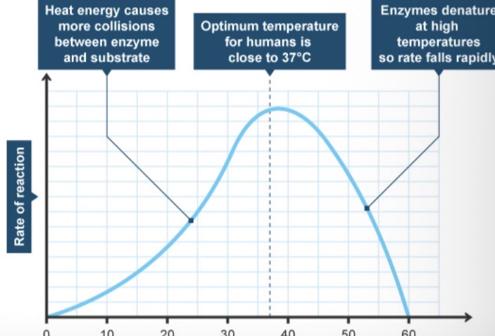
Factors affecting Enzyme Action
Temperature
In the human body the optimal temperature for reactions is 37ºC. Deviations from this affect the reaction rate.
At low temperatures the molecules of enzyme and substrate are moving slowly, therefore, fewer collisions occur per unit time.
At high temperature the atoms within the enzyme molecules are moving more energetically causing a strain on the bonds that hold the atoms together. Eventually they break and the enzyme is denatured and no longer functional.

Ph (enyzymes)
Not all enzymes have the same optimal pH.
Enzymes are affected by pH because the amino acids that make up the molecule contain positive and negative regions around the active site.
Excess H+ or OH- can lead to bonding in the charged areas which could affect the matching process.

Concentration of Substrate
Reactions are a product of collisions
The greater the concentration of the substrate the more collisions per unit time that can occur
This increases the rate of the reaction to a limit determined by the amount of enzyme present. Once this limit is reached increasing substrate concentration has no effect on reaction rate.
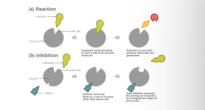
Inhibitors
Enzyme inhibitors are substances that reduce or prevent an enzymes activity.
There are two types of inhibitors:
Competitive
2. Non-competitive

Competitive Inhibitors
Have a structure similar to that of the substrate that would normally bind with the enzyme
They compete with the substrate to occupy the active site of the enzyme and prevent the substrate from binding
The inhibitors do not affect the enzyme and do not form products so they tend to remain in the active site.
The rate of reaction is decreased because substrate cannot enter the active site of the enzyme
At high concentrations of substrate the effects of inhibition are decreased as the substrate can outcompete the inhibitor.

Non-Competitive Inhibition
Also combine with enzymes but not at the active site
They bind to another part of the enzyme where they either
Partly block access to the active site
2. Cause a change in shape of the enzyme so that the substrate cannot enter the active site.
Increasing the concentration of the substrate has no effect on inhibition

Control of metabolic pathways
Metabolic pathways are often controlled by end-product inhibition
This process used the end product of the pathway to inhibit an enzyme in that pathway
May be competitive or non-competitive. However in most cases it is non-competitive
This prevents over production
Overproduction wastes energy or could be toxic
Enzymes that are acted upon by a non-competitive end-product inhibitor are known as allosteric enzymes.
The product is called an allosteric inhibitor and binds to the allosteric site
An example of end product inhibition is found in the pathway of threonine being converted to isoleucine.
Negative feedback
As the end product beings to accumulate the inhibitory effects become more prevalent and production decreases
When the end products starts to be used up, the inhibitory effects are reduced and more product can be created