Lab 15: Eye Muscles, Optic Pathway, Tunic
 Call Kai
 Learn
Learn Practice Test
 Spaced Repetition
 Match
Match Flashcards
 Knowt Play
1/24

There's no tags or description
Looks like no tags are added yet.
Name | Mastery | Learn | Test | Matching | Spaced | Call with Kai | Chat |
|---|
No analytics yet
Send a link to your students to track their progress
25 Terms
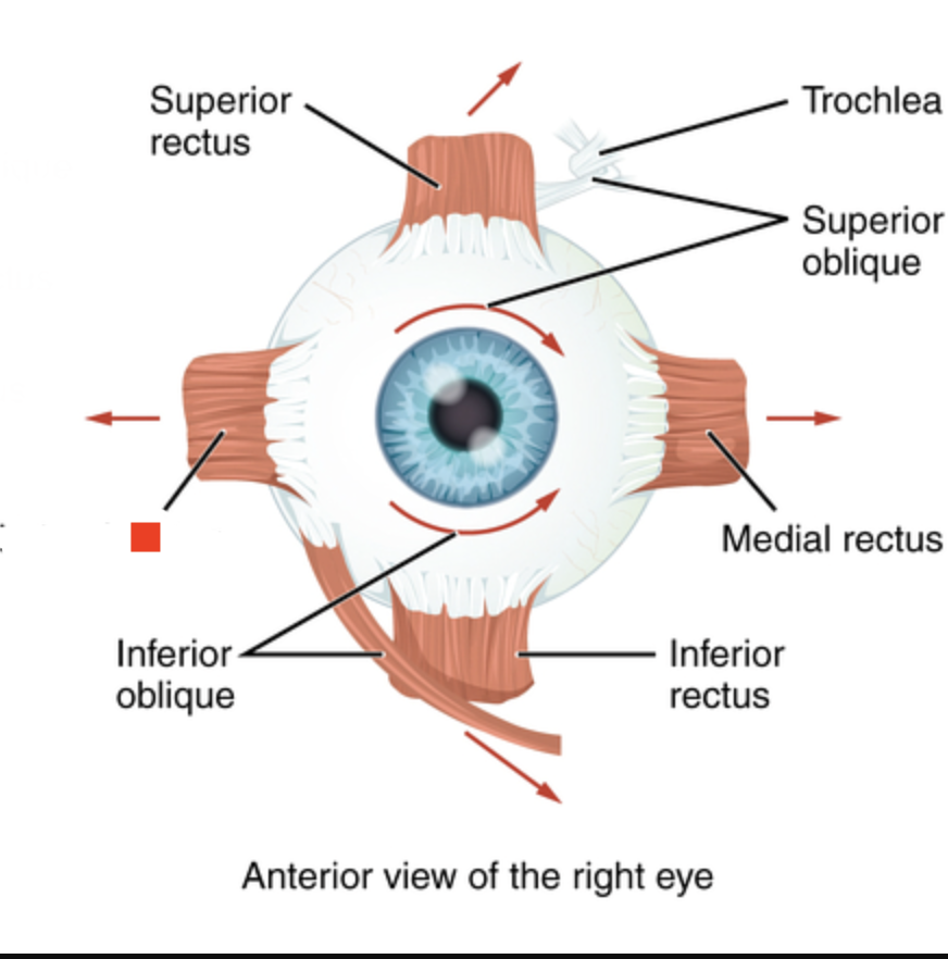
Medial rectus
Moves eye medially (adduction).
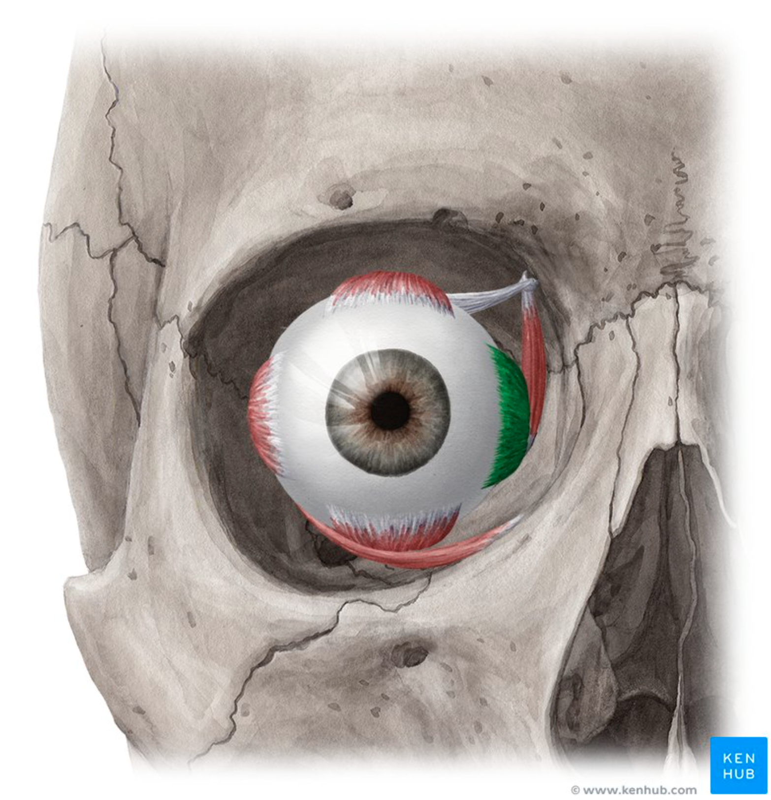
Lateral rectus
Moves eye laterally (abduction).
RED SQUARE.
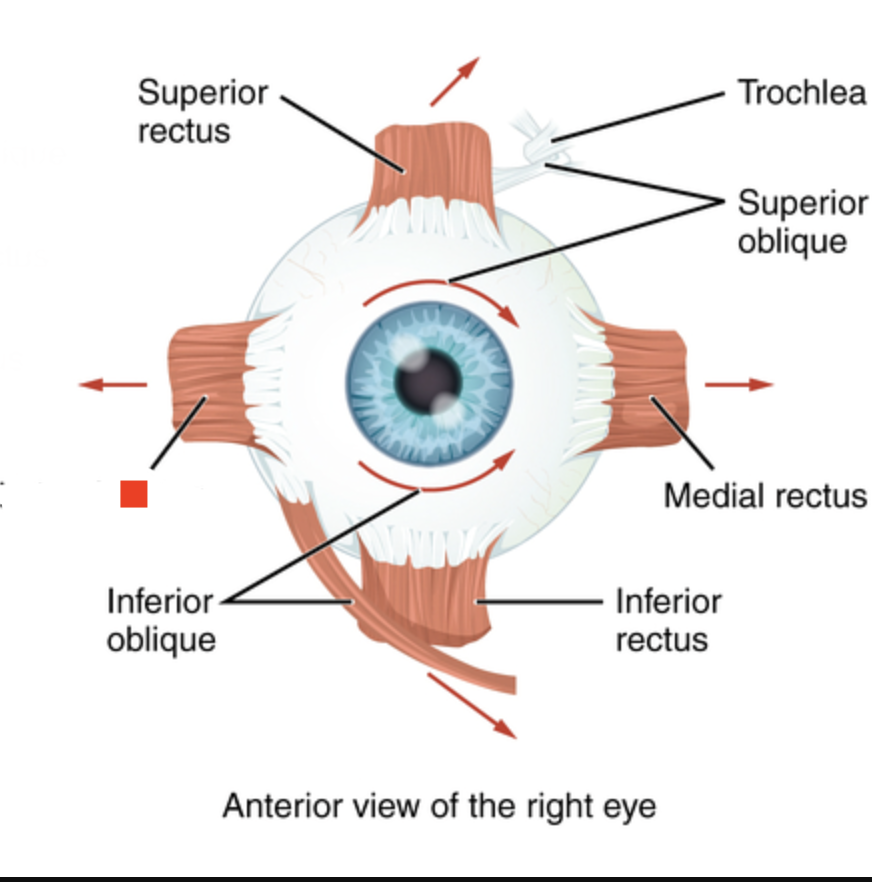
Superior rectus
Moves eye up and medially.
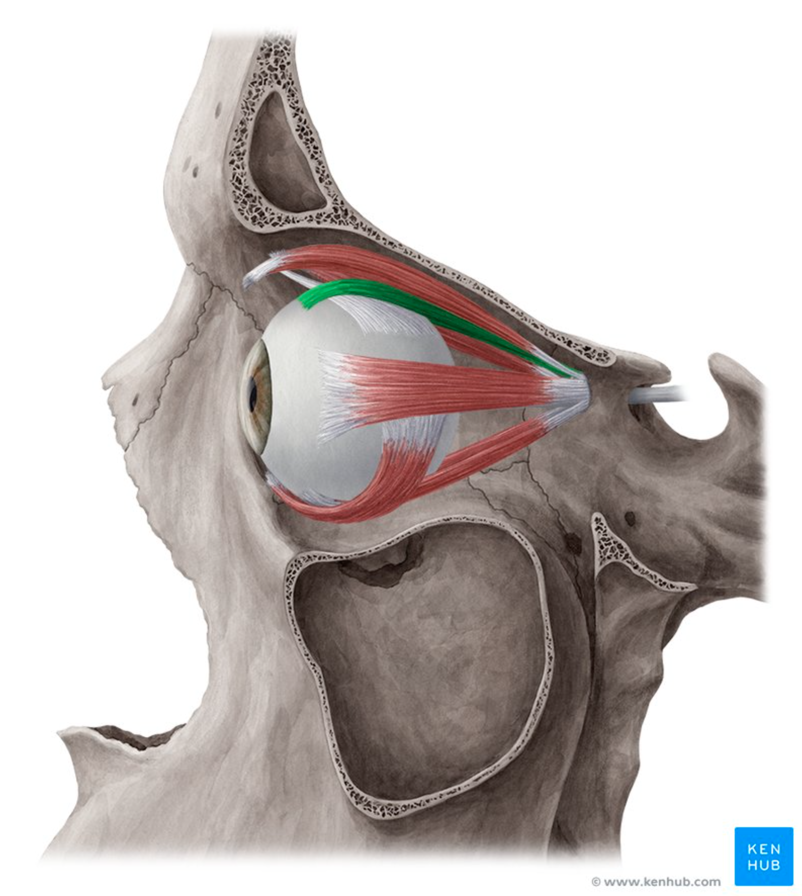
Inferior rectus
Moves eye down and medially.
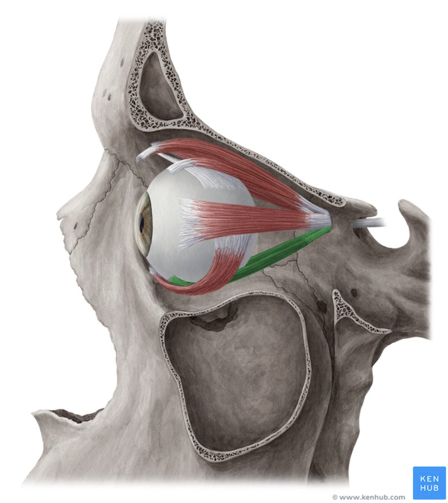
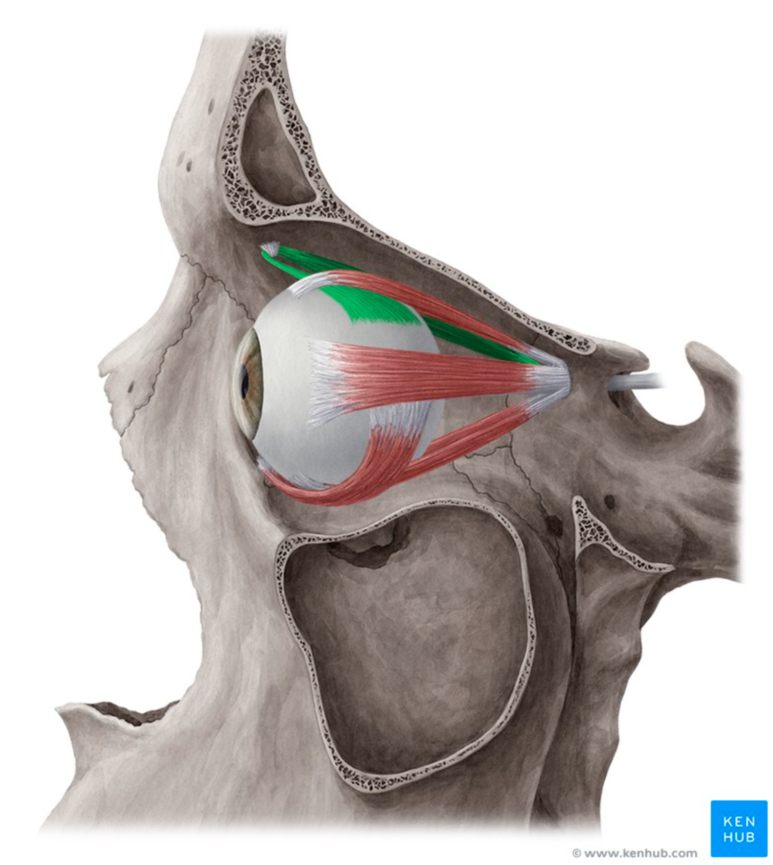
Superior oblique
Moves eye down and laterally.
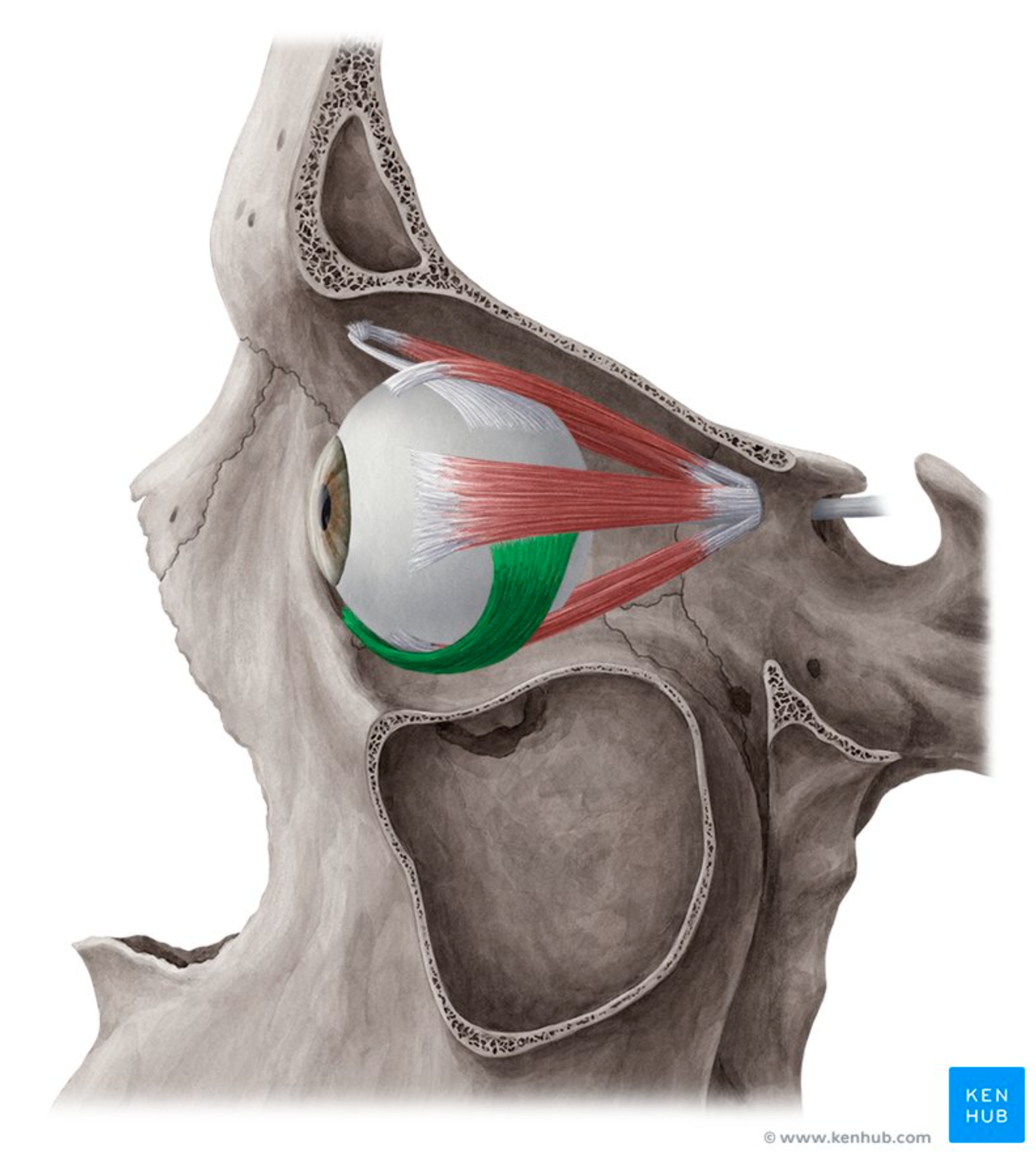
Inferior oblique
Moves eye up and laterally.
Lateral rectus
What muscles is used when LOOKING LEFT?
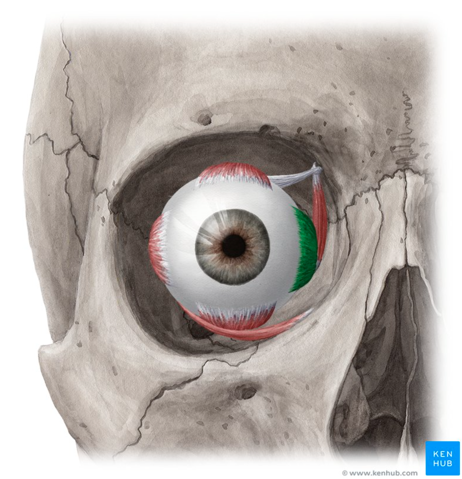
Medial rectus
What muscles is used when LOOKING RIGHT?
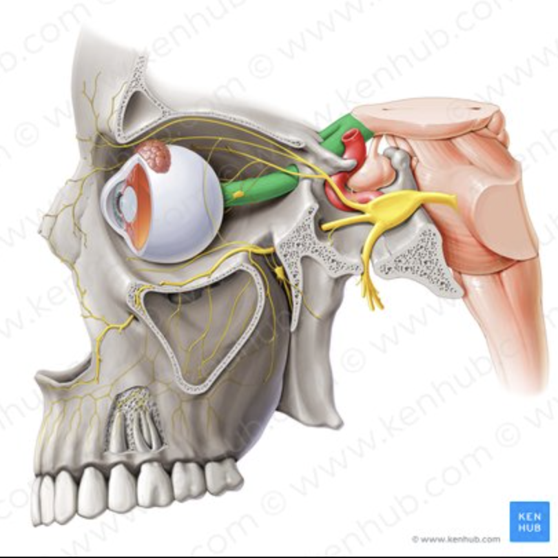
Optic nerve
Carries visual information from each eye.
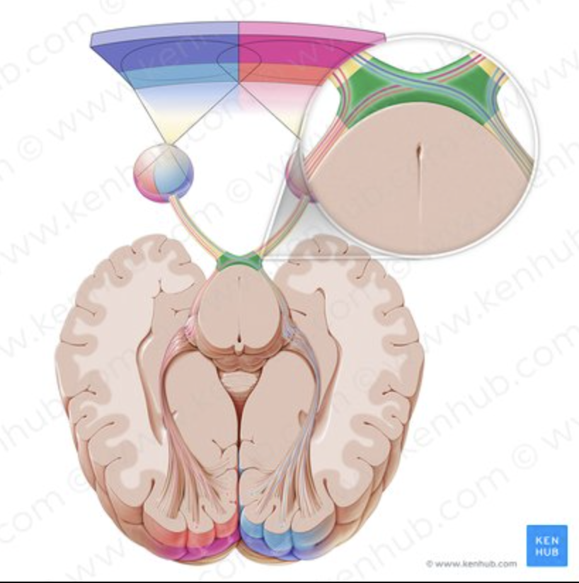
Optic chiasm
Some nerve fibers cross to the opposite side.
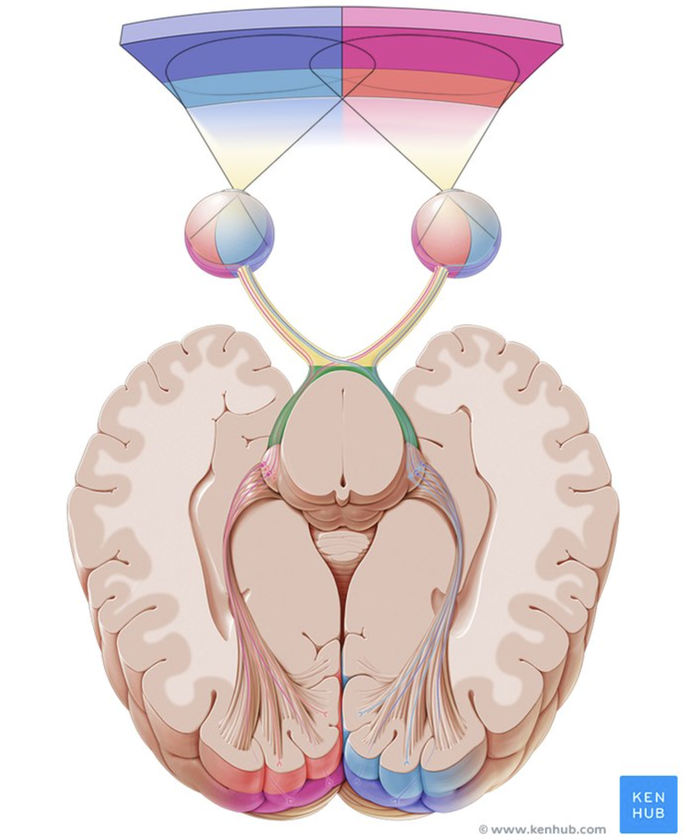
Optic tract
Carries visual information to the brain.
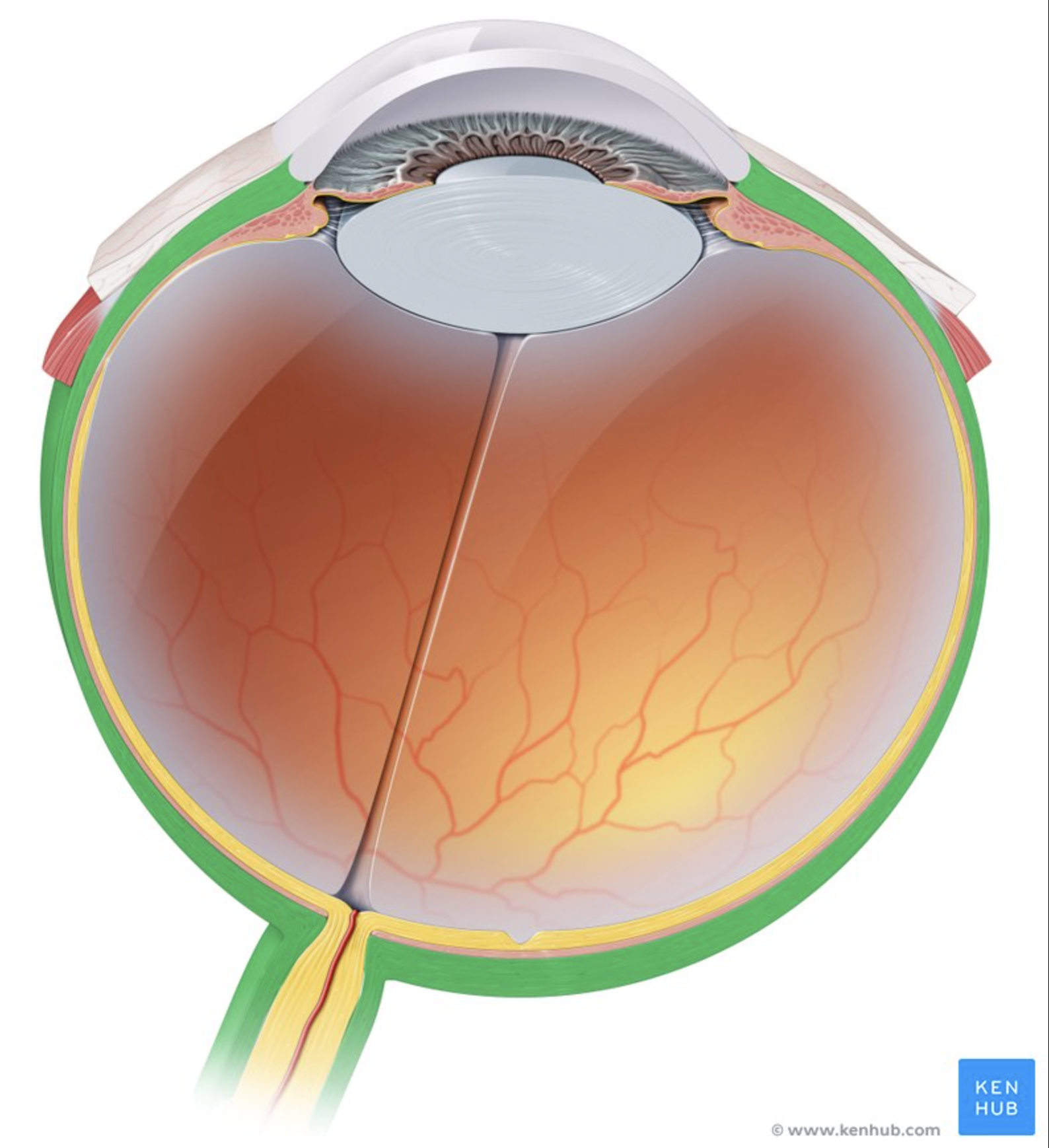
Sclera
Tough white outer layer.
Protects the eye.
(FIBROUS TUNIC)
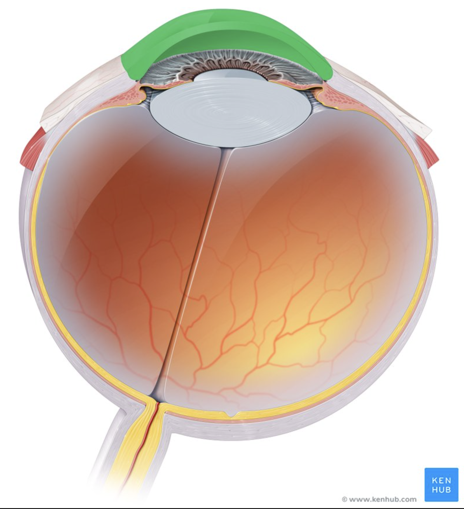
Cornea
Clear front of the eye.
Bends (refracts) light.
(FIBROUS TUNIC)
Choroid
Contains blood vessels.
Nourishes the eye.
(VASCULAR TUNIC)

Ciliary body
Changes lens shape for focusing.
(VASCULAR TUNIC)

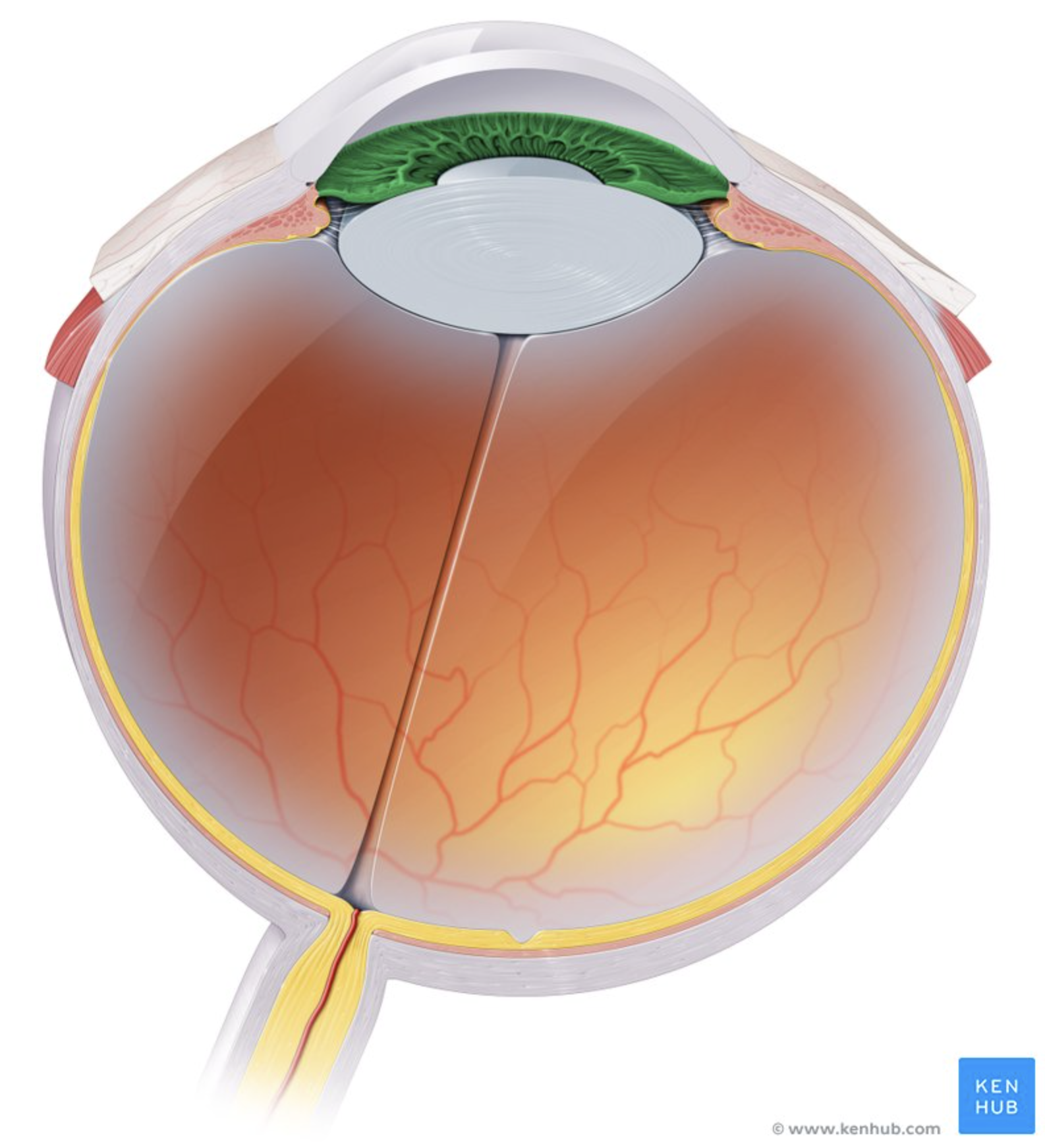
Iris
Colored part of the eye.
Controls pupil size.
(VASCULAR TUNIC)
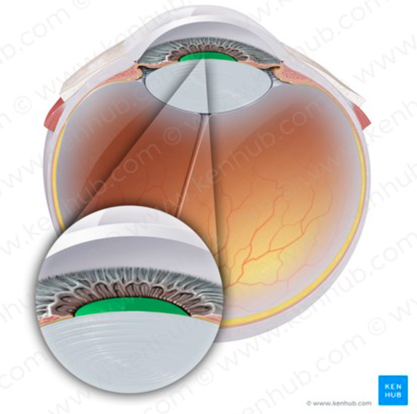
Pupil
Opening that lets light enter.
(VASCULAR TUNIC)
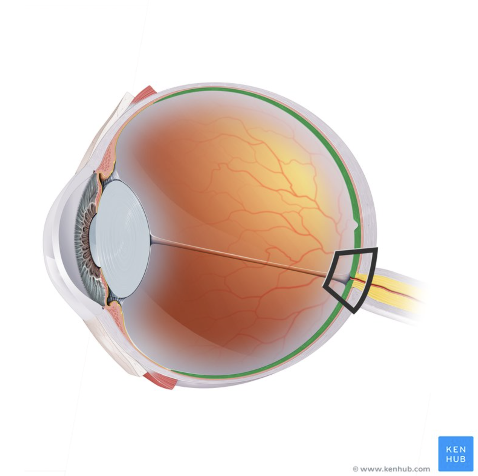
Retina
Contains photoreceptors.
Converts light into nerve signals
(NEURAL TUNIC)
Macula lutea
Area of sharp central vision.
(NEURAL TUNIC)
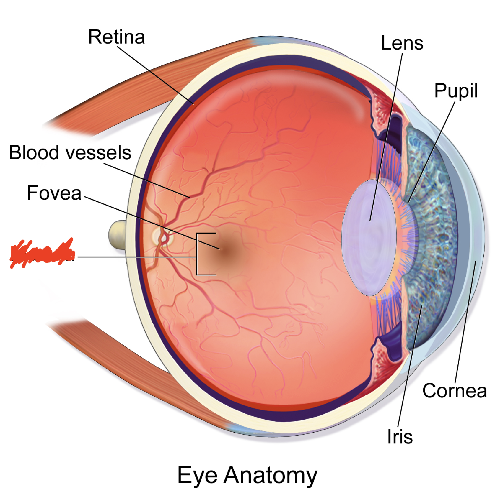
Fovea centralis
Sharpest vision.
Highest cone density
(NEURAL TUNIC)
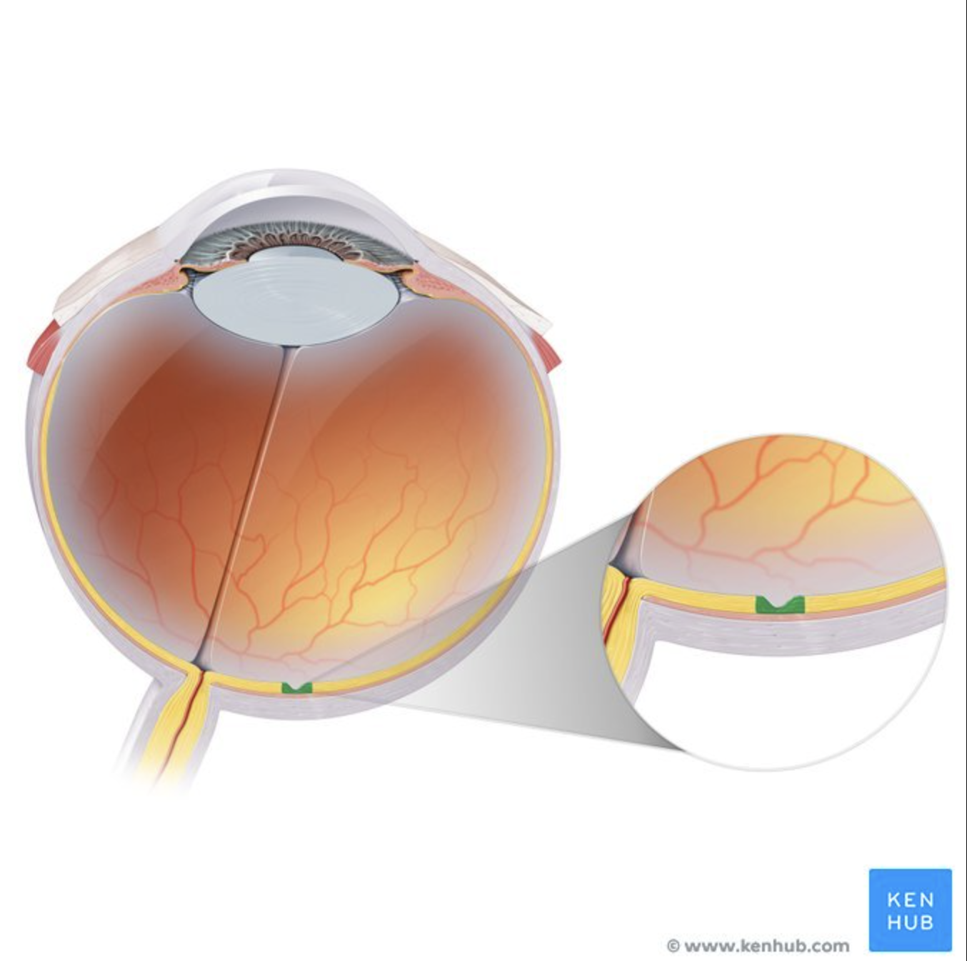
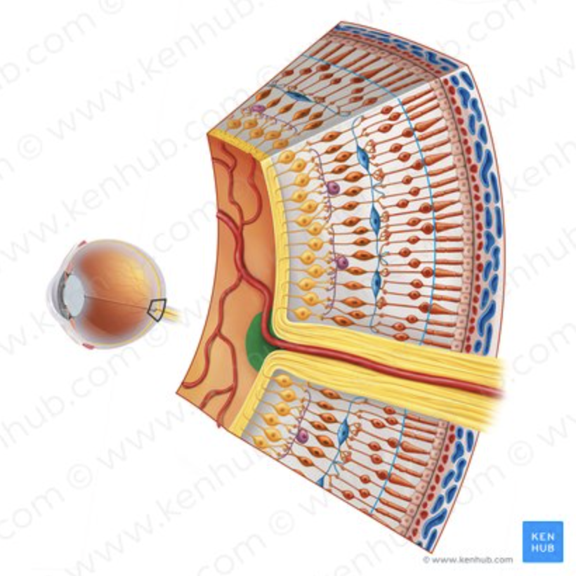
Optic disc
Blind spot where optic nerve exits.
(NEURAL TUNIC)
Fibrous tunic, vascular tunic, neural tunic
What are the three TUNICS?
Sclera, cornea
What is in the FIBROUS TUNIC (OUTER LAYER)?
Choroid, ciliary body, iris, pupil
What is in the VASCULAR TUNIC (MIDDLE LAYER)?
Retina, macula lutea, fovea centralis, optic disc
What is in the NEURAL TUNIC (INNER LAYER)?