MCB 2210 L25: Interphase
 Call Kai
 Learn
Learn Practice Test
 Spaced Repetition
 Match
Match Flashcards
 Knowt Play
1/20

There's no tags or description
Looks like no tags are added yet.
Name | Mastery | Learn | Test | Matching | Spaced | Call with Kai | Chat |
|---|
No analytics yet
Send a link to your students to track their progress
21 Terms
Direction of MT motors
Kinesin → (+) end
Dynein → (-) end
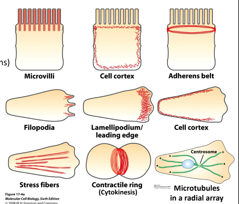
Describe actin + MT + motor functions
Actin
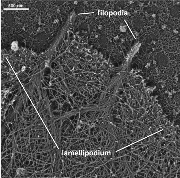
Membrane protrusion (lamellipodia + filopodia)
Cell adhesion (stress fibers + focal adhesions)
Endocytosis + trafficking (Clathrin pits + endosomes)
Cytokinesis
Microtubules
Organelle position (ER + Golgi)
Anterograde transport (ER → Golgi → Plasma Membrane)
Retrograde Transport (Endosome → Golgi → ER)
Mitosis
What cell is commonly used to observe membrane protrusions?
What edge of the cell drives motion?
Which 2 actin-based structures are involved in membrane protrusions?
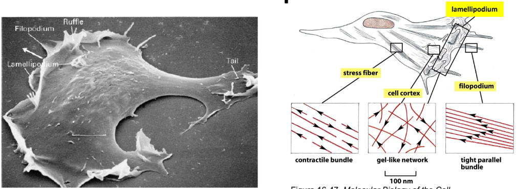
Fibroblast = cell of connective tissue
Leading edge = dynamic, forward-facing protrusive edge of migrating cell that drives forward motion
Lamellipodia = thin sheet-like structures
Filopodia = thin needle-like structures
Push plasma membrane forward by adhering to substrate or sweeping over top of cell as a ruffle
Stress fibers = cell adhesion + contraction
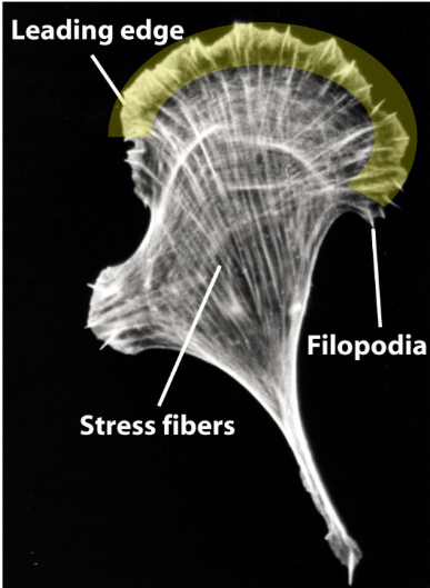
How can the behavior of actin filaments of leading edge be observed?
Microinjecting cell w/ fluorescently labeled actin
Fluorescent actin = incorporated into filaments @ extreme leading edge
Demonstrates that actin polymerization is taking place @ leading edge
What drives membrane protrusion?
What describes the rearward movement of cytoskeleton?
Actin assembly → pushes membrane forward
Energy from ATP hydrolysis by actin → drives process
Retrograde flow = continuous rearward movement of actin cytoskeleton
Filaments pushed back by membrane resistance + myosin-driven contraction
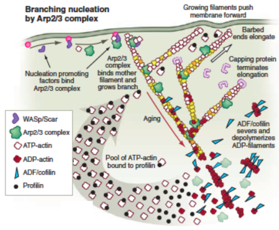
TREADMILLING
Cofilin = actin severing protein → actin depolymerization @ leading edge
Describe what fluorescence speckle microscopy + what phenomenon it demonstrates
Fluorescence speckle microscopy = live-cell imaging technique using very low [fluorescently-labeled subunits] → “speckle” pattern
Visualize retrograde flow
Meshwork pushed backwards as membrane = moving outward
Membrane → resistant
Pushes meshwork inwards
![<p><strong>Fluorescence speckle microscopy</strong> = live-cell imaging technique using very low [fluorescently-labeled subunits] → “speckle” pattern</p><ul><li><p>Visualize retrograde flow </p></li><li><p>Meshwork pushed backwards as membrane = moving outward</p></li><li><p>Membrane → resistant </p></li><li><p>Pushes meshwork inwards </p></li></ul><p></p>](https://assets.knowt.com/user-attachments/b93d7f3e-ad08-4155-bee5-3efc72d12538.png)
Describe how F-actin is organized in protrusive structures
Barbed (+) ends → membrane (direction of protrusion)
Lamellipodia = branched actin network
Nucleated by Arp2/3 complex + WASP protein
Filopodia = tight parallel bundles of straight actin = finger-like protrusions
Describe actin assembly into stress fibers
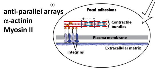
Stress fibers = long bundles of actin filaments below cell surface
Focal adhesions = where stress fibers terminate + attach to underlying substrates
α-actinin = cross-linker proteins
Myosin thick filament = bridge bundles together
Integrins = transmembrane protein that connect actin → extracellular matrix
What triggers dynamic actin specialization?
What are the 3 types relevant to actin specialization?
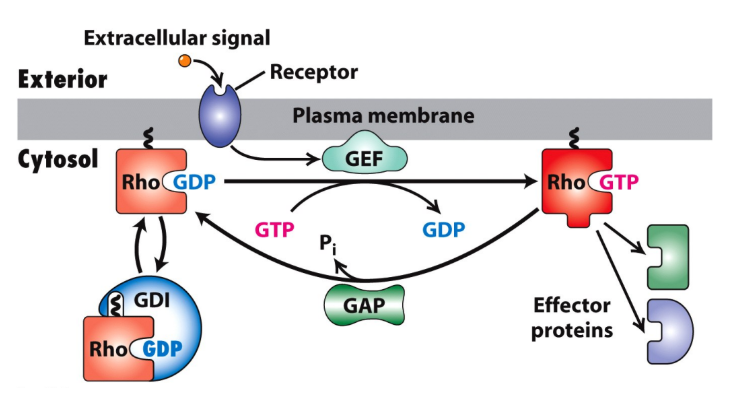
Small G-proteins → Rho family GTPases
Rho family GTPases = members of Ras
Rho, Rac, Cdc42
Act as small molecular switches b/w:
Active form = bound to GTP
Inactive form = bound to GDP
GEFs = Guanine Nucleotide Exchange Factors = GDP → GTP = ACTIVATE
GAPs = GTPase Activating Proteins (GAPs) = GTP → GDP = DEACTIVATE
GDIs = Guanine Nucleotide Dissociation Inhibitors (GDIs) = stabilize GDP
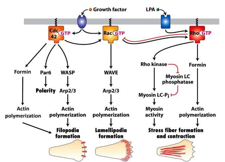
What are the three types of pathways that activate WASPs and/or Formins and drive actin assembly in cells?
Rho → Formin → Stress fiber (straight tightly bundled actin)
Rac → Arp2/3 + WASP → Lamellipodia (branched actin)
Cdc42 → Formin + Arp2/3 + WASP → Filopodia (finger-like protrusions, spikes)
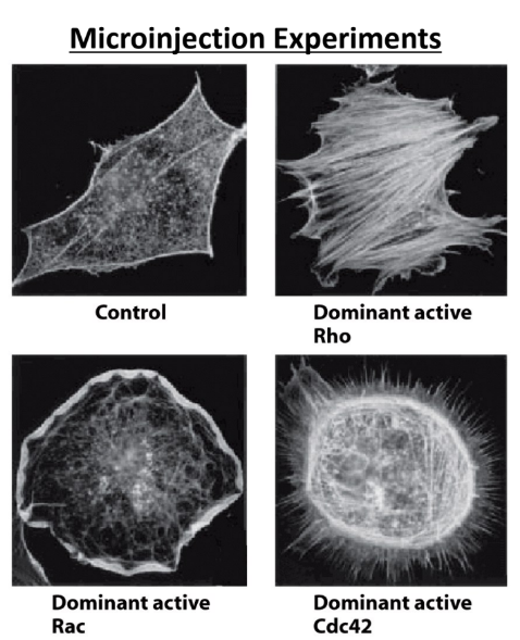
Explain an experimental method that can be used to create the 3 distinct actin structures.
Microinject GTPase of interest in dominant active form (constantly active) + observe actin forming structures
Rho → stress fibers (tightly bundled straight actin)
Rac → lamellipodia (branched actin)
Cdc42 → filopodia (finger-like protrusions, spikes)
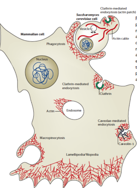
What 2 membrane trafficking processes does actin cytoskeleton direct?
Endocytosis
Myosins → move on actin near cell surface
Switch to MT motor-based movement on MTs
Endosome trafficking
Endosome rocketing = endosomes polymerize actin on one side through cytoplasm by propelling fast actin polymerization
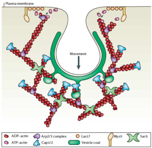
What type of actin assembly facilitates endocytic internalization?
Branched actin assembly → Arp2/3 complex, CapZ, WASP
How is actin assembly involved in membrane sorting in endosomes?
Actin assembly marks regions of membrane that need to be recycled back → different patches of membrane = recycled
Actin = markers to sort membrane into endosomes
What 2 processes of the secretory pathway are controlled by the MT skeleton?
Organelle position
Membrane transport
Ex. Anterograde transport (ER → Golgi → Plasma membrane
Use tracks of MTs + motor proteins → travel through compartments
Why are ER membranes dynamic?
ER membranes = connected to MTs
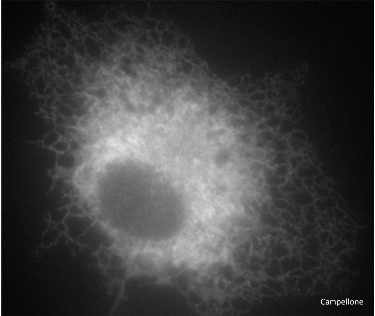
Explain Golgi and ER position in the cell in relation to motor proteins
How do drugs affect the position of the ER and Golgi?
MTs = help position Golgi + ER
Golgi → centrosome
Dyneins help keep Golgi near centrosome
ER → along MTs + cytoplasmic branches
Kinesins help keep ER positioned to cell periphery
Drug added → MT = disrupted → Golgi fragmented + ER collapses
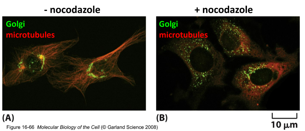
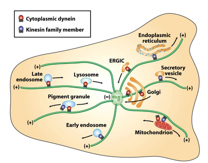
What motor protein controls retrograde transport?
What motor protein control anterograde transport?
Dynein → (-) direction
Golgi @ centrosomes (-) ends
Kinesins → (+) direction
ER @ cell periphery (+) ends
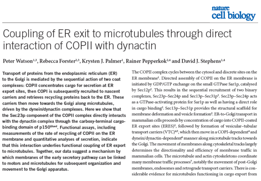
What is COPII coat directly linked to?
Dynactin = intermediate = link b/w dynein + cargo
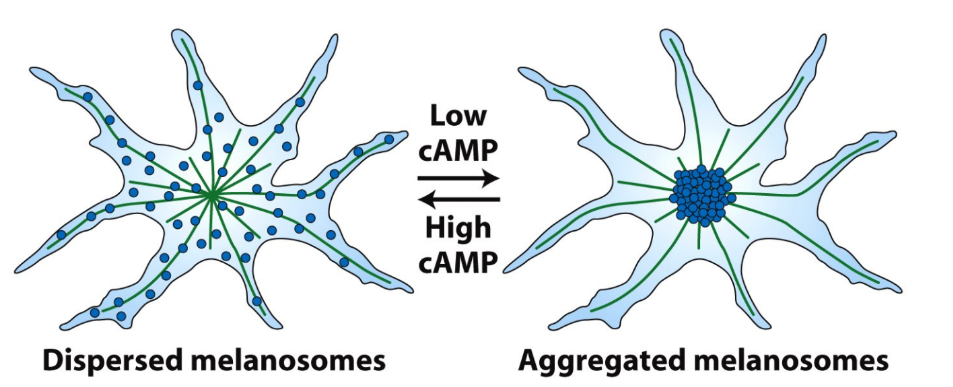
Explain melanosomes for organization look like for melanosome organization
A. What will melanosome organization look like if you treat cells with taxol?
B. What will melanosome organization look like if you treat cells with nocodazole?
C. What happens when you wash away the nozodazole or taxol?
A. Taxol → stabilizes MT → CANNOT depolymerize
B. Nocodazole → MT depolymerization
C. Regrowth of MTs → melanosomes = free to interact
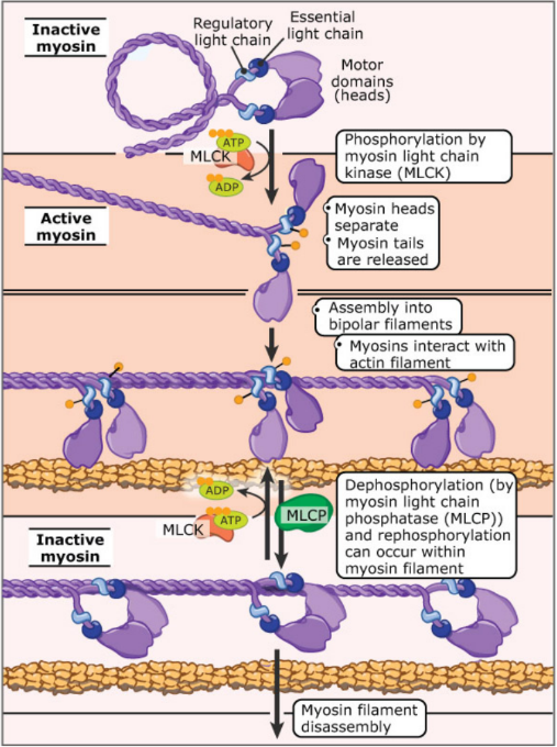
How phosphorylates non-muscle II light chains?
Myosin Light Chain Kinase (MLCK) = phosphorylates Myosin II light chains → functional myosin thick filament complex assembly
Inactive myosin = dephosphorylated
Active myosin = phosphorylated