AFFERENT PNS: HLSC exam 2
1/44
There's no tags or description
Looks like no tags are added yet.
Name | Mastery | Learn | Test | Matching | Spaced | Call with Kai |
|---|
No analytics yet
Send a link to your students to track their progress
45 Terms
what do photoreceptors detect and what is the modality of the detection
photoreceptors detect sensation information about vision and this is done through photons of light
what do chemoreceptors detect and by what modality? (five different things)
taste: via chemicals dissolved in saliva
smell: via chemicals dissolved in mucus
pain: via chemicals in extracellular fluid
blood oxygen: via oxygen dissolved in plasma
blood pH: via free hydrogen ions in plasma
what do thermoreceptors detect and by what modality
warm receptors: detect warmth, specifically when there is an increase in temperature between 30 and 43 degrees
cold receptors: detect cold, specifically when there is a decrease in temperature between 35 and 20 degrees
what do mechanoreceptors detect and how? (three)
baroreceptors: detect blood pressure by the stretch of specific blood vessel walls
osmoreceptors: detect osmolarity of extracellular fluid
hair cells: detect sound via sound waves, and balance and equilibrium via acceleration
what is transduction and how do receptors work?
transduction is the conversion of stimulus energy (modality) into electrical energy by the receptor. receptors are specific to a modality, but also respond in lesser degrees to other modalities.
when a stimulus is detected by a receptor, a graded potential called a receptor potential generates.
if axon hillock reaches threshold, an action potential in the afferent neuron will be generated. (afferent neuron may be attached to or apart of sensory receptor or not)
what happens with continuous stimulus on sensory receptors?
not all receptors send the same number of action potentials to the CNS.
adaptation: refers to a decrease in the amplitude of the receptor potential and corresponding decrease in the frequency of action potentials over time. adaptation decreases the perception of the stimulus by the receptor
phasic receptors such as for smell and touch, adapt quickly
tonic receptors such as for proprioceptors and muscle stretch receptors, do not adapt or adapt very slowly.
what is the pathway for sensory information to the brain
travels along a discrete somatosensory pathway (labelled lines) each pathway is specific for one modality
from periphery, action potentials travel along a first-order sensory neuron.
this then synapses with a second-order sensory neuron that may cross over or desecrate the midline either in spinal cord or medulla. this signal goes to the thalamus for processing.
a third-order neuron projects to a particular part of the somatosensory cortex.
what is a sensory unit and its receptive field?
the sensory unit refers to a single afferent neuron and all of its associated receptors. within one unit, all receptors are specific for the same modality.
the receptive field of a unit refers to the physical region over which stimuli can be detected by that particular sensory unit.
what effects sensory coding
sensory coding occurs according to stimulus type, intensity, and location.
each receptor specific for one modality, but combination of info from various receptors and receptor pathways that are stimulated at any given time that is integrated by CNS and perceived
stimulus intensity is coded in terms of frequency of action potentials (frequency coding) and the number of receptors activated or recruited (either on same afferent neuron or on multiple afferents aka population coding)
stimulus location is coded in terms of size and degree of overlap of the receptive field. the smaller the receptive field, the greater the acuity (sharpness). lateral inhibition can increase acuity if there are overlapping receptive fields
what are four kinds of somatosensory receptors
mechanoreceptors: some are rapidly adapting such as the Pacinian corpuscle, Meissner’s corpuscle, and hair follicle receptors. some are slowly adapting such as free nerve endings, Merkel’s disks, and Ruffini’s endings.
thermoreceptors
nociceptors (of skin): all free nerve endings and respond to mechanical, thermal, or chemical stimulu or combos of all three (polymodal)
proprioceptors (of muscles and joints): detect positions of the body in space. they are continuously active and DO NOT ADAPT. examples are muscle spindles, Golgi tendon organs, and free nerve endings
what is the dorsal column-medial lemniscal pathway
pathway that transmits information about both proprioception and the somesthetic sensation of touch and pressure to the CNS. the pathway decussates (crosses over) to the contralateral side at the medulla oblongata
what is the spinothalamic tract
transmits somethetic info about pain and temp from thermoreceptors and nociceptors and decussates at level of spinal cord
which receptors detect pain and what is the pathway
pain is complex due to autonomic and emotional components, and detection is situationally dependent and influenced by previous experiences
nociceptors detect stimuli that are potentially damaging to tissue
pain stimuli follows the spinothalamic tract in to the cerebral cortex
non specific pathways also synapse on the reticular formation, hypothalamus, and limbic system
there are also behavioural and emotional responses to pain when stimuli reaches cerebral cortex
what is referred pain
referred pain is he sensation of pain that is felt at the surface of the body that originates from internal organs (visceral pain)
it is perceived to arise from a different location than the actual origin this is because of afferent neurons sharing the same second-order neuron
body parts that experience this are the heart, the liver and gall bladder, appendix, colon, uterers, kidneys, stomach, and esophagus
what is the gate control theory of pain
describes the inhibitory pathways involved in the transmission of pain
modulated pain transmission ( presynaptic inhibition ) involves a modification of the signal by inhibitory interneurons that are excited by collaterals of sensory afferents responding to mechanoreceptors in the skin resulting in a decrease in pain signal
described by a “gate” at the dorsal horn of spinal cord that acts as a gatekeeper for pain signals
examples that support this theory include 1. the effectiveness of massage therapy and 2. the soothing nature of rubbing a bumped knee or a stubbed toe
how does presynaptic inhibition suppress pain
the brain has a built in pain suppressing analgesic system:
when nuclei in the periaquaductal grey matter or reticular foramen are stimulated, endogenous opiate neurotransmitters are released and attach to opiate receptors and prevent the release of the neurotransmitter substance.
since less pain inducing neurotransmitter is released, less pain is perceived
what are the three tissue layers of the eye
sclera: outer layer of connective tissue that forms the visible white part of the eye and the anterior transparent surface (cornea)
choroid: contains the blood vessels. anterior portion forms ciliary body and iris
retina: inner pigmented layer that contains photoreceptors
what are the two fluid filled cavities of the eye and what fluid do they contain
posterior cavity: contains vitreous humour
anterior cavity: contains aqueous humour
stimulation of which iris muscles controls the amount of light that can enter the eye through the pupil? and which division of the nervous system controls the muscles?
stimulation of the circular or constrictor muscles and the radial muscles control the amount of light that enters the eye.
the autonomic nervous system controls controls these muscles
in bright light or for near vision, the parasympathetic nervous system stimulates the circular or constrictor muscles of the iris to reduce the size of the pupil so that less light can enter.
in dim light or far vision, the sympathetic nervous system stimulates the radial muscles of the iris to increase the size of the pupil so that more light can enter.
what is accommodation in terms of the eye and it’s association with near vision
accommodation refers to the eyes ability to increase the curvature of the lens to focus light on the retina. this is important in near vision, which also includes contraction of the ciliary muscle by the parasympathetic nervous system
far vision does not normally required accommodation, thus the ciliary muscle remains relaxed
what does the term emmetropia mean
emmetropia refers to the ability to focus light on the retina for both near and far vision
length of eyeball or strength of lens can prevent the focusing of light on the retina for near or far vision resulting in common vision disorders known as hyperopia (farsightedness) which requires convex lenses, or
myopia (nearsightedness) which requires concave lenses
what cell layers does light travel through when it is focused on the retina
ganglion cells make up inner layer
bipolar neurons make up middle layer
photoreceptors (rods and cones) make up outer layer
in lateral layers (between the three different cell types listed), amacrine cells are between the inner and middle layers (ganglion and bipolar), and horizontal cells are between the outer and middle layers (bipolar and photoreceptors).
the axons of ganglion cells form the optic nerve (CNII)
explain phototransduction
photoreceptors in retina do phototransduction
starts by light stimulating photopigments (conformational change) which inhibits the release of neurotransmitters from the synaptic terminal of receptor
the decreased release of inhibitory neurotransmitters results in a change in the graded potential of the bipolar cells
if this change is of significant magnitude, an action potential in the corresponding ganglions cell is generated
differentiate between the “macula lutea” and the “fovea”
macula lutea: the larger, central area of retina responsible for sharp , detailed, central vision
fovea: smaller, pit/depression in the centre of the macula lutea where visual acuity is highest has an even greater concentration of (only) cones
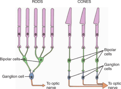
differentiate between cones and rods photoreceptors
cones: are excited b y bright daylight and are responsible for colour vision. cones have high acuity and synapse with ganglion cells in a ratio close to 1:1 (low degree of convergence)
rods: are highly sensitive and excited by small amounts of light (night vision). rods have lower acuity than cones due to a higher degree of convergence
what is the photopigment of rods and how does it change according to light vs dark adaptation
photopigment of rods is called rhodopsin
moving into a dark room from light, eyes switch from cone vision to rod vision. rhodopsin goes from maximally activated to being enzymatically returned to an inactive conformation before it is able to absorb light again
in light adaptation, the rhodopsin that was in its light sensitive inactive conformation in the dark will initially maximally absorb the light, resulting in a bleaching of rods (perception of being blinded by light). when all rods become saturated with light, rhodopsin is in active conformation can’t absorb any more light, and visual system begins to rely on cone vision for light info, and pupils constrict
where do nerve impulses travel along visual pathway
nerve impulses that originate from the medial (nasal) half of each eye cross over at the optic chiasm
nerve impulses that originate from the lateral (temporal) half of each eye do not cross over, but proceed ipsilaterally to the cerebral cortex
what are the three fundamental characteristics of sound waves
pitch or tone: determined by the frequency of the sound waves and is measured in cycles/second (hertz).
intensity or loudness: determined by the amplitude of the sound waves and is measured in decibels.
timbre (quality of sound that distinguishes it from another sound): depends on overtones that add richness and help us distinguish one voice from another and locate sound.
list the order of the small bones of the middle ear in order from most outer to most inner
MOST OUTER: malleus (next to ear drum)
MIDDLE: incus
MOST INNER: stapes
what route do sound waves travel
through the external auditory meatus of the external ear to the tympanic membrane
tympanic membrane then vibrates at the same pitch and amplitude as the incoming sound waves
this causes the ossicles (three ear bones: malleus, incus, stapes) to move. this amplifies sound so that the oval window of the cochlea vibrates the fluid that is within the cochlea
what are the three fluid filled chambers of the cochlea
scala vestibuli: filled with perilymph (resembles extracellular fluid: rich in Na+) to conduct sound and pressure waves ***in bony labyrinth***
scala tympani: filled with perilymph
cochlear duct: filled with endolymph (resembles intracellular fluid: rich in K+) to generate electrical signals ***in membrane labyrinth***
where and what are the actual auditory receptors
they are hair cells of the organ of Corti in the cochlear duct. these hair cells sit on the basilar membrane and can bend in relation to the overlapping tectorial membrane
what happens when the hair cells of the organ of Corti are bent
mechanically gated cation channels open resulting in depolarization of the cell and formation of a graded potential
this can translate into an action potential in the cochlear division of the vestibulocochlear nerve (CN VII)
the info travels to the medial geniculate nucleus of the thalamus (main relay centre for auditory information) and then to the auditory cortex of the temporal lobe
what does intensity coding for loudness involve
occurs in terms of the amplitude of the wave
coded for in terms of the degree of deflection and opening of the ion channels in the sterocilia (hair like non motile projections)
what does frequency coding for pitch involve
coded for in terms of the location of the deflection on the basilar membrane
where are the receptors for equilibrium and what do they detect
located in the inner ear in the vestibular apparatus
detect changes in motion and the position of the head
three kinds:
1. semicircular canals: detect rotational or angular acceleration or deceleration of the head
2. utricle: and 3. saccule: are otolith organs that provide info about the position of the head relative to gravity and the rate of linear motion
how does sound transduction by stereocilia in hair cells work in resting stage vs bending towards vs bending away from tallest sterocillium ( kinocilium ) (located in a gelatinous cupola above ampulla)
resting: partially depolarized state due to some mechanically gated K+ channels are open, allowing K+ to enter hair cells. also allows small amounts of Ca+ to enter cell through voltage gated channels which results in the release of of small amounts of neurotransmitter for communication with afferent neuron. this causes a low frequency of action potentials evident in afferent neuron under resting conditions
stereocilia bend in direction of tallest sterocillium, and more K+ channels are opened and more K+ enters cell thus greater depolarization. this means more Ca+ also enters cell thus more neurotransmitter and a higher frequency of action potentials
when stereocilia bend away from tallest sterocillium, more K+ channels close compared to resting state, less K+ enters, causing hyperpolarization. very small amounts of Ca+ enter, very little neurotransmitter is released at very low frequency of action potentials in afferent neuron.
what kind of head movement do the anterior, posterior, and lateral canals detect
anteior canal detects movement of the head up or down (ex. nodding yes)
posterior canal detects movement of the head from a vertical to a horizontal position (ex. touching ear to shoulder)
lateral canal detects movement of the head from side to side (ex. shaking head no)
axons from these hair cells join to form the vestibular branch of the vestibulocochlear nerve (CNVIII)
where are the olfactory receptors located and what other cells are present in the olfactory epithelium
located in a small patch of tissue in the roof of the nasal cavity
the axons project brought the cribiform plate of the ethmoid bone to form the olfactory nerve (CN1)
also found in the olfactory epithelium are supporting cells: to secrete mucus and basal cells: are the precursor cells for new receptor cells
how can an odourant be detected
odourant must be volatile and soluble in water
approx. 1000 diff types of olfactory receptors, therefore various parts of an odour are dissected and stimulate particular receptors
the more intense an odour is, therefore various parts more action potentials are produced
these action potentials are generated through a G-protein signalling cascade
what is the pathway to the brain for processing odours
initial processing of odours at the glomeruli within the olfactory bulb (humans can distinguish ~10,000 diff odours)
information then sent to either the limbic system ( amygdala and hippocampus ) or to the primary olfactory cortex (temporal lobe and partly frontal lobe) for interpretation
second order neurons in olfactory pathway are called mitral cells which form the olfactory tract
where are taste buds found
primarily found on the tongue, but some are found in pharynx
taste receptor cells are spindle shaped and surround a central taste pore and detect dissolved chemicals
what are the different tastant categories
sweet
sour
bitter
salty
umami
which cranial nerves are responsible for transmission of taste information to the brain stem, then thalamus, then cortical gustatory area of the cortex (or limbic system and hypothalamus)
facial (VII):
glossopharyngeal (IX):
vagus (X):