BIOL 1103 Week 5 - Nervous System
1/103
There's no tags or description
Looks like no tags are added yet.
Name | Mastery | Learn | Test | Matching | Spaced |
|---|
No study sessions yet.
104 Terms
The Peripheral Nervous System divides into the ____________ and __________________
Afferent system and Efferent system
The Efferent System divides into the ____________ and __________________
Somatic system and Autonomic system
The Autonomic system divides into the _____________________ and ____________________
Sympathetic system and parasympathetic system
Which types of nerves does the Peripheral Nervous System (PNS) contain?
Afferent nerves and efferent nerves
Afferent nerves are also known as _______________ nerves
Sensory
Which systems are efferent nerves a part of?
Efferent nerves are a part of the somatic and autonomic systems
Afferent nerves
Carry electrical impulses from the central nervous system to muscles and glands
Efferent nerves
Carry electrical impulses from the central nervous system to muscles and glands
Somatic nervous system
Deals with the parts of the body that you can move voluntarily
Autonomic nervous system
Regulates the functions of internal organs such as the heart, stomach, intestines, and some muscles
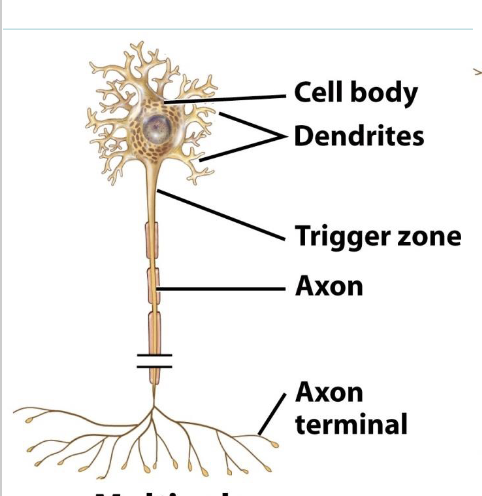
What are the parts of a neuron (nerve cell)?
Cell body, dendrites, axon
Nerve cell body
Main part of a neuron that contains most of the cell’s organelles and processes information received from dendrites
Dendrites
Branch-like extensions of a neuron that receive signals from other neurons and carry them toward the cell body
Axon
Neuron projection that carries electrical impulses away from the cell body to other neurons, muscles, or glands
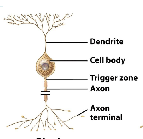
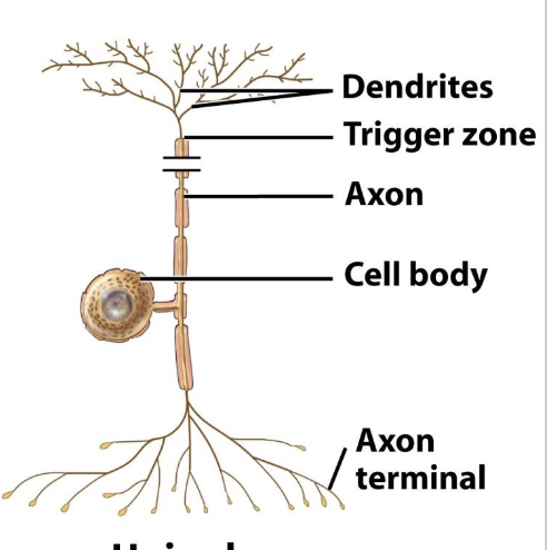
What are the three types of neurons?
Multipolar neuron, bipolar neuron, unipolar neuron
Multipolar neuron
Three or more processes -one axon and many dendrites. Found in most brain and spinal cord neurons.
Bipolar neuron
Two extensions – an axon and a dendrite. Found in retina, inner ear, olfactory area
Unipolar neuron
Single short process from cell body that divides T-like into proximal and distal branches. Found in sensory neurons for touch or stretch
Neuroglia
Supportive cells in the nervous system that protect, nourish, and insulate neurons, and help maintain the overall environment for nerve function
Myelin sheath
Axon covering formed by certain neuroglia that increase the speed of nerve impulse conduction
Demyelination
Loss or destruction of the myelin sheath
Unmyelinated neurons
Neurons without a myelin sheath
What is the name of the neuroglia in the PNS that forms a myelin sheath?
Schwann cells
Ganglion
Clusters of cell bodies
Nerves
Bundle of neuron fibers outside the CNS
Grey matter
Groups of cell bodies
Tract
Bundle of neuron fibers inside the cns
White matter
A group of tracts (it’s white due to the myelin sheath covering the axons)
How is white and grey matter distributed in the brain?
Grey matter outside, white matter inside
How is white and grey matter distributed in the spinal cord?
White matter outside, grey matter inside
Meninges
A set of connective tissue membranes that surrounds the central nervous system (brain and spinal cord)H
The three membranes in the meninges are (PAD!)
Pia mater
Arachnoid mater
Dura mater
How do the meninges protect the brain?
They form three layers that cushion the brain, anchor it in place, and help circulate cerebrospinal fluid for added protection and nourishment.
Brain ventricles
Four brain ventricles are continuous with one another and
with the cavity in the spinal cordFilled with cerebrospinal fluid
What are the functions of cerebrospinal fluid?
Forms a liquid cushion that supports and protects the brain, prevents it from crushing under its own weight, absorbs shocks from trauma, and nourishes the brain while carrying chemical signals throughout it.
What abilities does the cerebrum control?
Motor function, interpretation fo sensory information, reading, writing, speaking, thinking, remembering, feeling, and moving
What are the four external lobes in the cerebrum?
In the cerebellum, there are the rontal, parietal, temporal, and occipital lobes
What abilities does the thalamus control?
Sensory processing, attention, alertness, and awareness by relaying information from the senses (except smell) to the proper areas of the brain for interpretation.
Which part of the brain is associated with the interpretation of pain and pleasure?
The thalamus interprets pain and pleasure
What structure does the thalamus and hypothalamus form?
They form the diencephalon
What does the hypothalamus control?
Controls body temperature, hunger, thirst, sleep, and emotions; regulates the endocrine system by controlling the pituitary gland and maintaining the body’s internal balance (homeostasis).
Which structure in the brain is the principle intermediary between the nervous and endocrine systems?
The hypothalamus is the principle intermediary
What does the brain stem control?
Controls vital life functions such as breathing, heart rate, and blood pressure; it also regulates reflexes and connects the brain to the spinal cord.
What are the three parts of the brain stem?
The midbrain, pons, and medulla oblongata (in descending order)
Midbrain
Reflex center for head and eye movements in response to sight and sounds
What does the pons control?
Controls breathing rhythm, helps relay messages between different parts of the brain, and plays a role in sleep and arousal
What does the medulla oblongata control?
Controls involuntary vital functions like heart rate, breathing, blood pressure, and swallowing.
What does the cerebellum do?
Provides precise timing and coordination of skeletal muscle movements, maintains posture, and ensures balance by using sensory input from the inner ear.
How many pairs of nerves emerge from the brain?
12 pairs
Where do the cranial nerves come from?
10 pairs from the brain stem
1 pair from the cerebrum
1 pair from the thalamus
What are the classifications of cranial nerves?
Sensory, motor, or mixed (both sensory and motor)
How many pairs of each type of cranial nerves are there?
3 pairs are sensory nerves (olfactory, optic,
vestibulocochlear)5 pairs are motor nerves (signals sent to skeletal muscles,
e.g. oculomotor)4 pairs are mixed (both sensory and motor, e.g. facial)
How many pairs of spinal nerves are there?
31 pairs
Functions of the spinal cord
Carries messages between the brain and body and controls quick, automatic reflexes
Reflex arc
Fast, automatic, unplanned sequence of actions in response to a stimulus handled by the spinal cord
What makes a reflex arc happen so quickly?
The stimulus bypasses the brain and is processed by the spinal cord
How does the spinal cord process signals in a. reflex arc?
Sensory neurons carry signal from receptors → connects to interneuron which decides what to do → interneuron directly sends impulse to motor neuron → motor neuron carries signal to the effector causing an immediate reaction!!!!
What is the reflex arc named in skeletal muscles?
It is named the somatic reflex arc
What is the reflex arc named in smooth muscles?
It is named the autonomic (visceral) reflex
What does it mean for body divisions to be “antagonistic?”
It means that two parts (or systems) work in opposite ways to balance each other
What is the sympathetic nervous system responsible for?
It is responsible for the body’s “fight or flight” response. It increases heart rate, blood pressure, dilates pupils, releases adrenaline, etc
What is the parasympathetic nervous system responsible for?
It is responsible for the body’s “rest and digest” functions. It slows down heart rate and lowers blood pressure, constricts airways, stimulates digestion and nutrient absorption, and promotes relaxation and recovery
Resting membrane potential
Stable, negative (relative to outside the membrane) electrical charge across a cell’s plasma membrane in its non-excited state
How many molecules of Na+ and K+ are pumped in the sodium-potassium pump?
2K+ pumped in, 3 Na+ pumped out
How do intracellular proteins maintain a cell’s membrane potential
The intracellular protein anions can’t leave the cell, making the inside more negative
What is the numerical value for the resting potential?
-70 mV
Why is the plasma membrane more permeable to potassium (K⁺) than to sodium (Na⁺)?
Because there are more K⁺ leak channels than Na⁺ channels, allowing potassium to move out of the cell more easily.
What is the significance of potassium being more permeable than sodium?
It causes more K⁺ to leave the cell, making the inside of the cell negative and establishing the resting membrane potential,
Is there more potassium inside or outside the cell?
There is more potassium inside the cell
Leaky channel
A membrane protein channel that is always open, allowing ions like K+ to passively move across the channel
Excitable cells
Excitable cells are cells that can generate and transmit electrical signals by changing their membrane potential in response to a stimulus
Ligand-gated channels
Membrane channels that open or close when a specific molecule (ligand) binds to them, allowing ions to pass
Mechanically-gated channels
Membrane channels that open or close in response to mechanical stimulation
Voltage-gated channels
Membrane channels that open and close in response to membrane potential
When gated channels open…
– Ions move quickly across the membrane
– Movement is down their electrochemical gradients
– Voltage changes across the membrane
Action potential
A rapid change in membrane potential that travels along excitable cells, allowing signal transmission
Does the action potential require energy?
No, since it relies on ions moving passively through voltage-gated channels to change the charge
During an action potential, the inside of the membrane becomes _____________________ charged
positively
Resting state (of an action potential)
Cell’s stable, negative membrane potential when not firing (~–70 mV).
During depolarization, what happens?
Voltage-gated Na+ channels open, Na+ rushes into the cell, and the membrane potential increases to +30mV~
During depolarization, what happens?
Voltage-gated Na+ channels close and voltage-gated K+ channels open, making K+ rush out of the cell
What causes a dip in mV during hyperpolarization?
Na+ channels are reset, but voltage-gated K+ channels remain open for slightly longer, causing extra K+ to leave the cell
First phase of action potential
Resting state (resting membrane potential)
Second phase of action potential
Depolarization (Na+ gates open)
Third phase of action potential
Depolarization (Na+ gates close, K+ gates open)
Fourth phase of action potential
Hyperpolarization (K+ gates close, Na+/K+ pump takes over)
How does the action potential propagate across neurons?
The cell body (soma) end of the axon depolarizes, spreads down the axon, and travels to the axon terminal
Why does the action potential only travel in one direction?
Due to the refractory period which desensitizes sodium voltage-gated channels
Why does the refractory period exist?
After sodium voltage gated channels close, they are desensitized for a period of time while the gate “resets” itself
What causes saltatory conduction?
Myelin sheaths surrounding axons
How does saltatory conduction allow for faster transmission of the nerve impulse?
The action potential jumps between myelin sheaths in myelinated axons, which transmits the signal faster and uses less energy
What causes the action potential to “skip” along myelinated axons?
The depolarization spreads passively under the myelin to the next node, “jumping” over the insulated sections.
Nodes of ranvier
A gap in the myelin sheath of a nerve
Synaptic transmission
The process where an action potential triggers neurotransmitter release from a neuron, which crosses the synaptic cleft and activates the next neuron.
What happens when a nerve impulse reaches the axon terminal?
Voltage-gated Ca2+ channels open, and Ca2+ rushes into the axon bulb, triggering neurotransmitter release
How are neurotransmitters released into the synaptic cleft?
Ca2+ causes vesicles to move to the presynaptic membrane and undergo exocytosis, releasing neurotransmitters into the synaptic cleft
How does the neurotransmitter trigger an action potential in the next neuron?
Neurotransmitters bind to ligand-gated Na+ channels on the postsynaptic neuron → Na+ enters → depolarization triggers a new action potential
Neuromuscular junction (NMJ)
Specialized synapse where a motor neuron connects with a muscle fiber to trigger muscle contraction
Where is acetylcholine released in the body?
It’s released at many synapses in the brain and PNS, released at the neuromuscular junction
Is acetylcholine excitatory or inhibitory?
It can be excitatory or inhibitory, depending upon which part of the nervous system is using