ECG Interpretation; Arrhythmias - Pathophysiology and Pharmacology (Class 4/5)
 Call Kai
 Learn
Learn Practice Test
 Spaced Repetition
 Match
Match Flashcards
 Knowt Play
1/47

There's no tags or description
Looks like no tags are added yet.
Name | Mastery | Learn | Test | Matching | Spaced | Call with Kai | Chat |
|---|
No analytics yet
Send a link to your students to track their progress
48 Terms
What are the three major arteries that supply blood to the heart?
1. Right coronary artery (RCA)
- Primarily supplies the right side of the heart
2. Left anterior descending artery (LAD)
- Supplies blood to the left side of the heart
- Specifically the front and bottom of the left ventricle and the front of the septum
3. Circumflex artery
- Supplies blood to the left side of the heart
- Specifically the left atrium, side, and back of the left ventricle
Describe how the electrical current is generated and conducted in the heart.
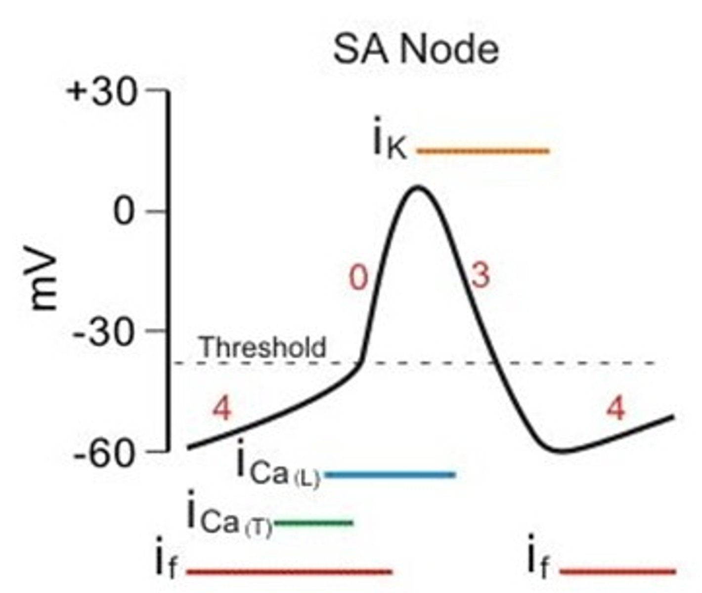
1. Electrical activity is initiated at the sinoatrial (SA) node
- Spontaneously generates action potentials
2. Wave of depolarization spreads through the atria
- Atrial contraction occurs
3. Current reaches atrioventricular (AV) node
- AV node slows conduction
- Ensures atrial contraction is completed before ventricles are activated
4. Continues down through Bundle of His (AV bundle)
- Splits into right and left bundle branches
5. Bundle branches further divide into Purkinje fibers
- Spread through ventricular walls, resulting in ventricular contraction
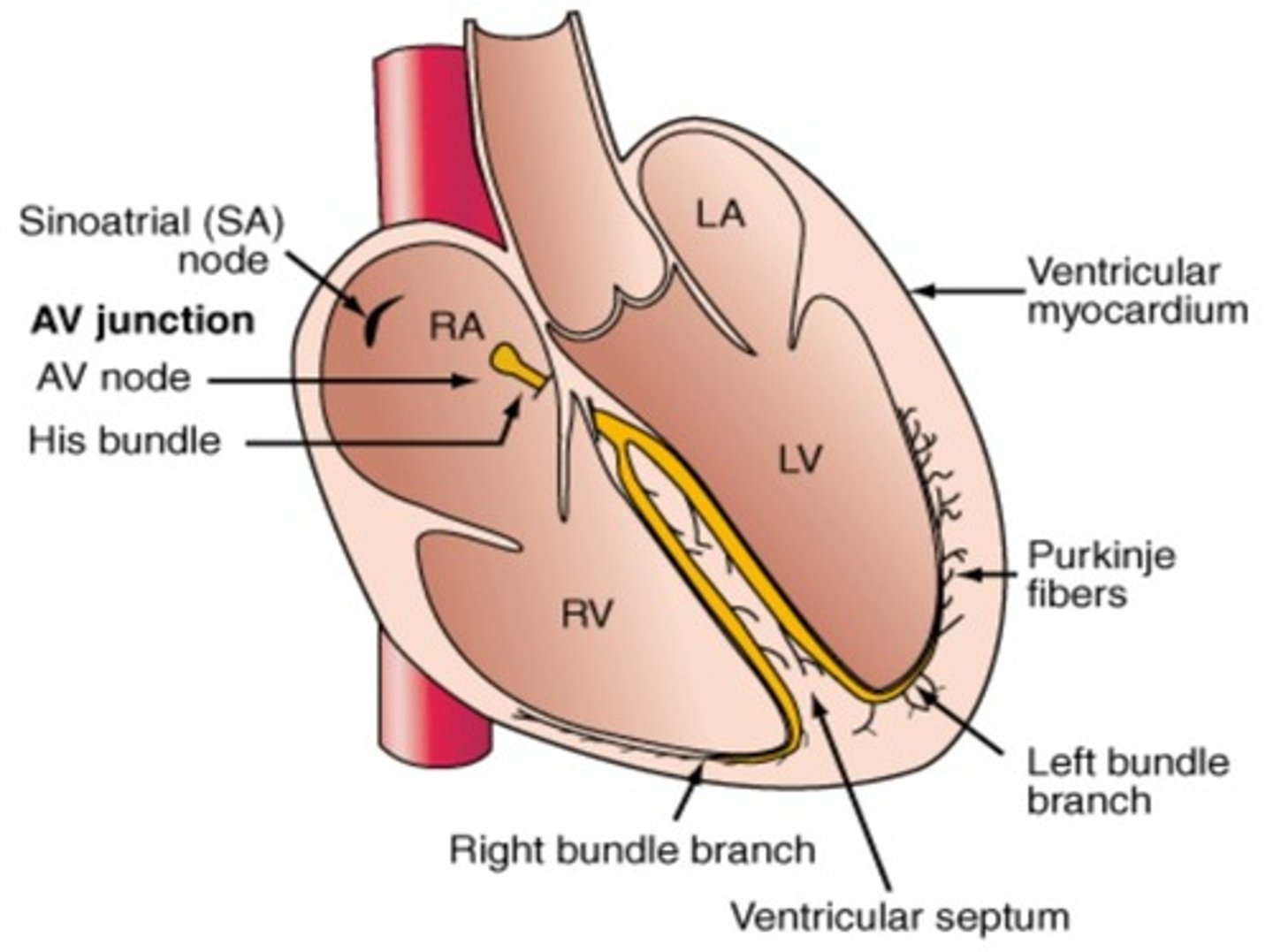
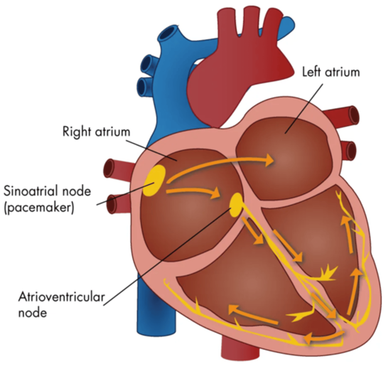
What is the general direction of normal electrical conduction in the heart?
Right to left and superior to inferior
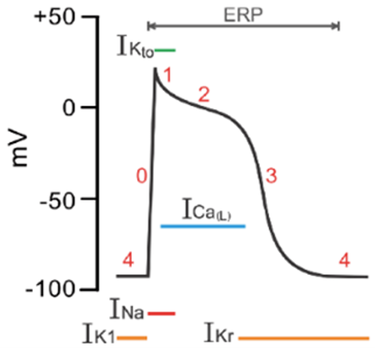
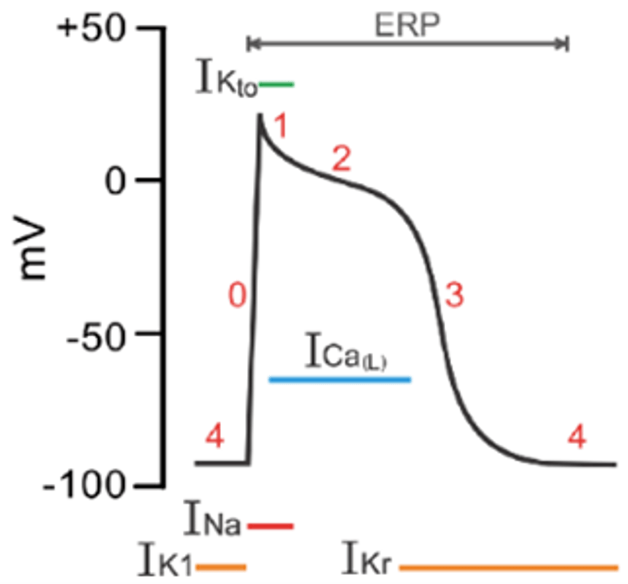
What triggers depolarization in atrial/ventricular tissue (i.e., non-nodal tissue)?
Rapid depolarization current through Na+ channels
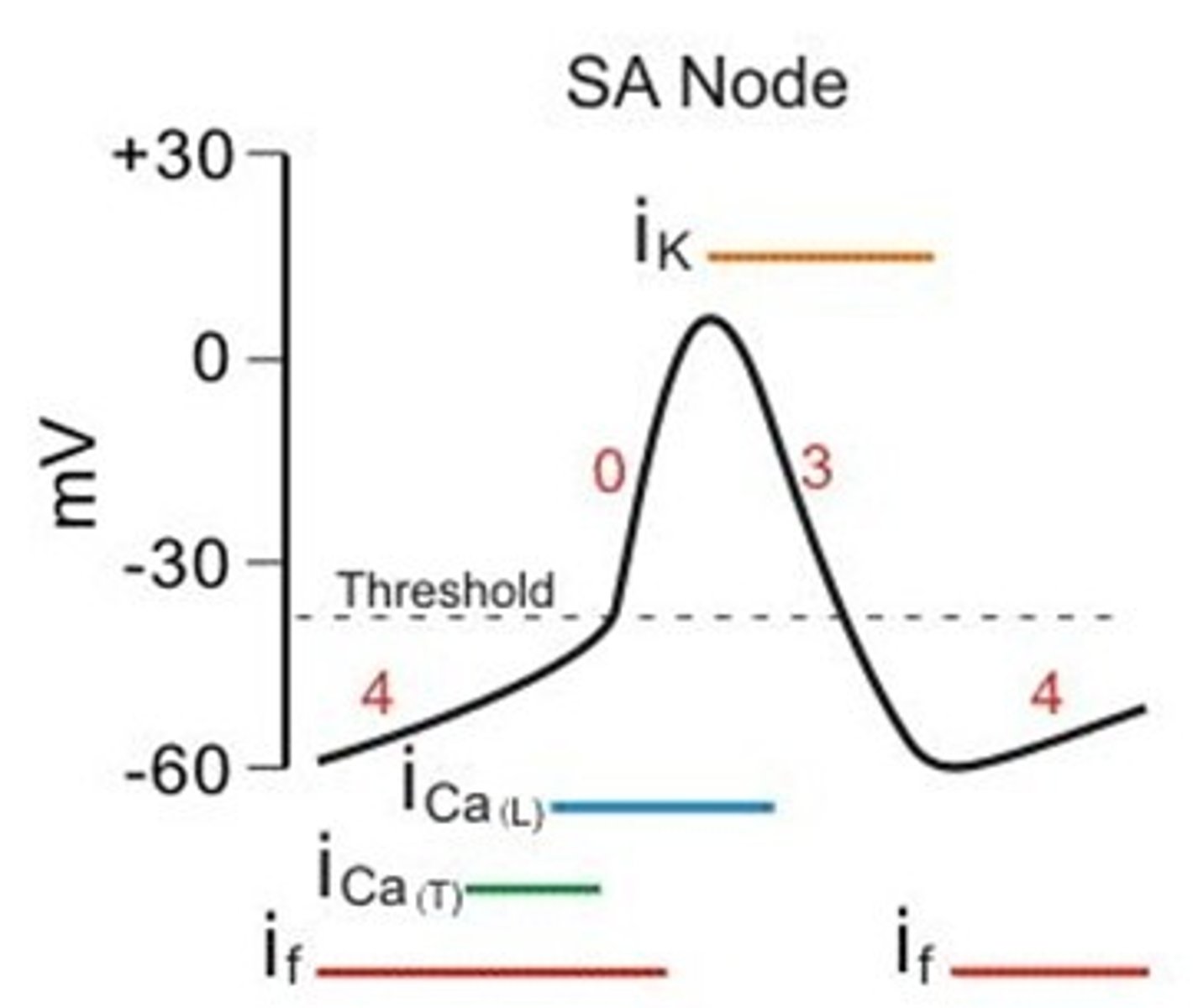
What triggers depolarization in SA/AV nodal tissue?
Slow depolarizing current through Ca2+ channels
What does the P wave on an ECG represent?
- Atrial depolarization
- Initiated by SA node
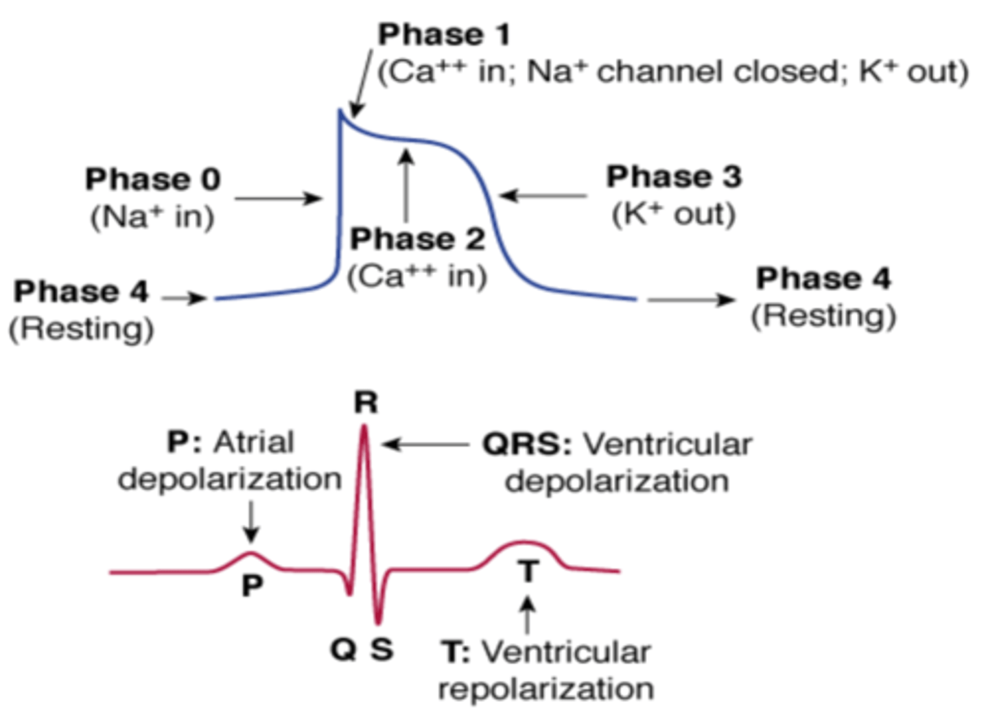
What does the PR interval on an ECG represent?
Impulse processing at AV node
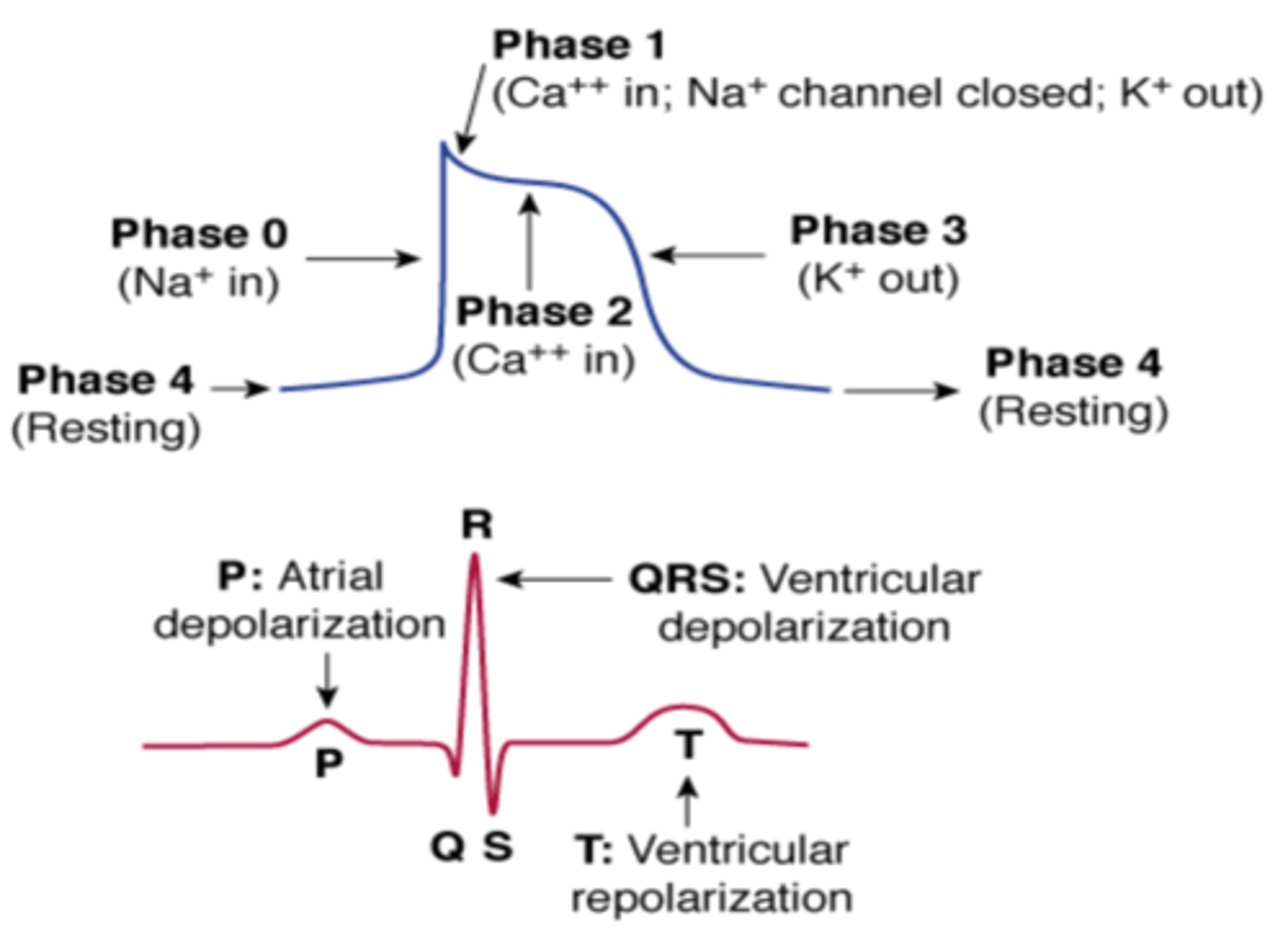
What does the QRS wave on an ECG represent?
- Ventricular depolarization
- Also atrial repolarization
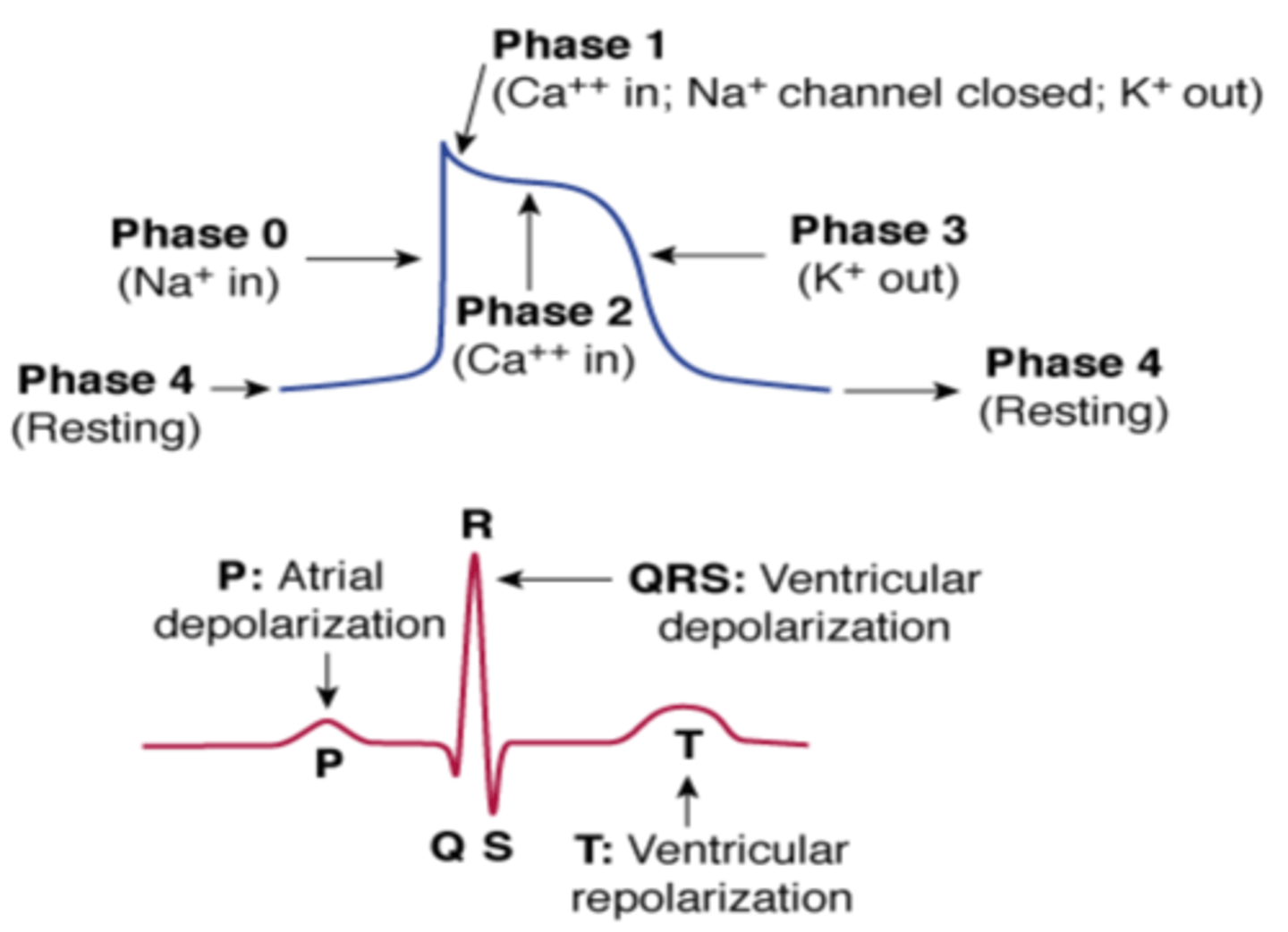
What does the ST segment on an ECG represent?
- Plateau phase
- Early ventricular repolarization
- Changes in blood supply to the heart by coronary arteries affect this segment
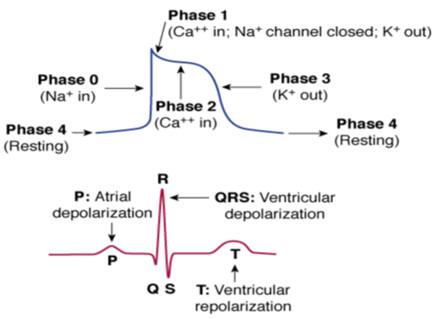
What does the T wave on an ECG represent?
Ventricular repolarization
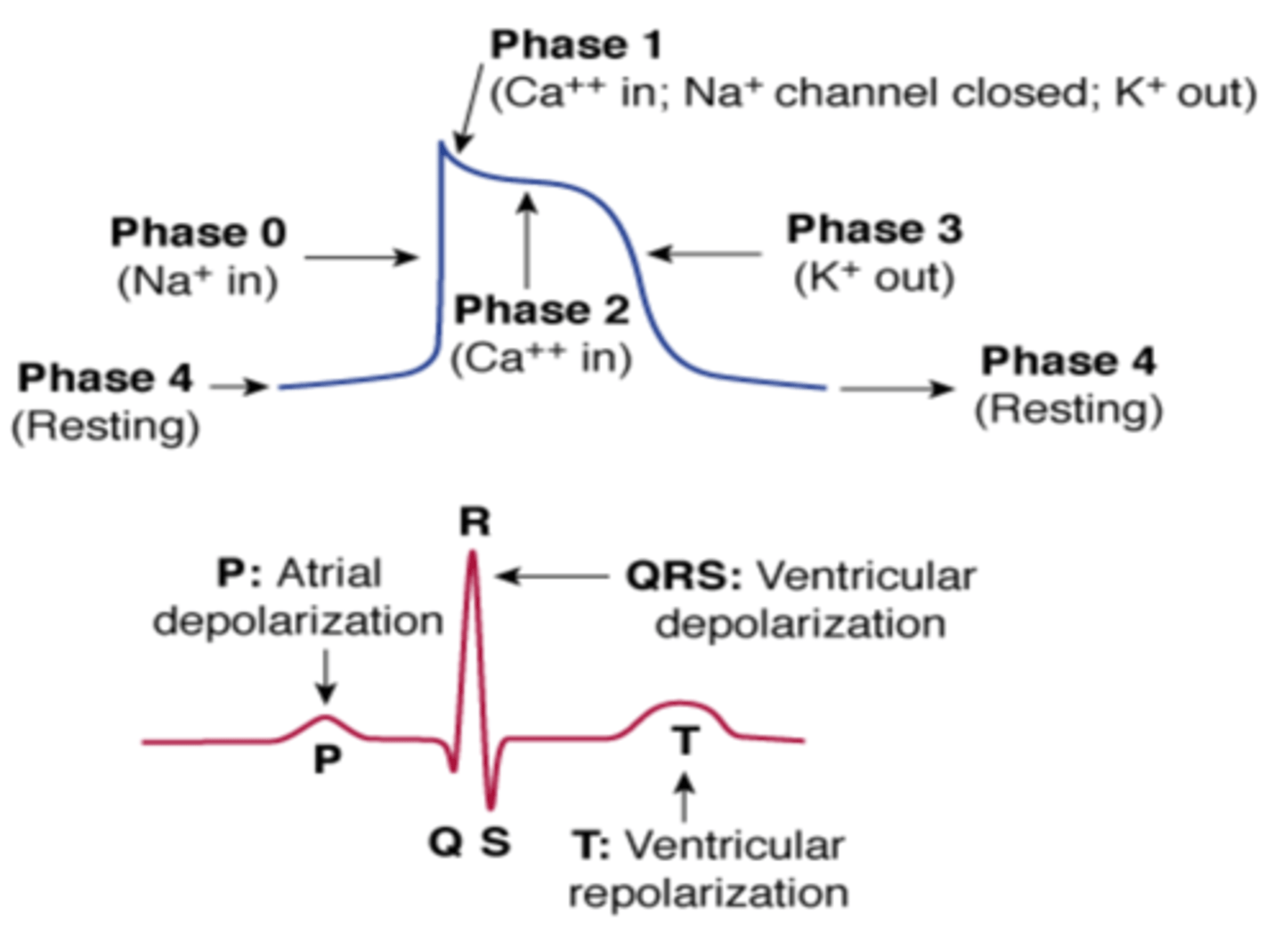
What is an electrocardiogram (ECG, EKC)? What do the squares represent?
Test that records a composite of all the electrical activity of the heart
ECG printed on a grid:
- One small square = 0.04 seconds
- One large square = 0.2 seconds
How can an ECG be used to estimate heart rate?
10-second method
- Used for irregular and regular rhythms
- Count # of R waves on standard 10-second ECG rhythm strip, then multiply by 6
300 method
- Only used for regular rhythms
- Count # of large boxes between 2 successive R waves, then divide by 300
1500 method
- Only used for regular rhythms
- Count # of small squares between 2 consecutive R waves, then divide by 1500
How many heart beats per minute indicate bradycardia and tachycardia in an adult?
Bradycardia: < 60
Normal: 60-100
Tachycardia: > 100
What segment on an ECG will be affected if there is ischemia of the heart?
ST segment depression
- Seen in subendocardial ischemia (i.e., ischemia affecting the inner layer of the heart wall)
- Sign of NSTEMI (non-ST elevation myocardial infarction)
ST segment elevation
- Seen in transmural ischemia (i.e., ischemia affected the entire thickness of the heart muscle)
- Sign of STEMI (ST elevation myocardial infarction)
Profound ST elevation/depression in multiple leads usually indicates very severe ischemia
What ECG leads make up the anterior viewing area? What major artery is primarily shown with this view?
Leads: V1, V2, V3, V4
View: Anterior wall of the left ventricle and interventricular septum
Supplied by: Left anterior descending artery (LAD)
What ECG leads make up the lateral viewing area? What major artery is primarily shown with this view?
Leads: I, AVL, V5, V6
View: Lateral wall of the left ventricle
Supplied by: Left circumflex artery (LCx)
What ECG leads make up the inferior viewing area? What major artery is primarily shown with this view?
Leads: II, III, and AVF
View: Inferior wall of the left ventricle
Supplied by: Right coronary artery (RCA)
Which lead is most commonly used for rhythm strips?
Lead II
Rhythm strip of any of the leads may be included at the bottom of a 12-lead ECG result to help identify patterns more easily
Explain three primary pathophysiologic mechanisms for cardiac arrhythmias.
Automatic tachycardias (i.e., increased automaticity)
- Abnormal generation of APs at tissues other than SA node
- Tissues compete with SA node for cardiac rhythm dominance/pacemaker control
- Steeper slope of phase 4 increases the rate of spontaneous depolarization
Triggered automaticity
- Spontaneous depolarizations that occur during or after phase 3 repolarization, but prior to phase 4
- Early after-depolarizations (EADs) – occur during phase 3
- Late-after depolarizations (LADs)/delayed after-depolarizations (DAD) – occur after phase 3
Reentry tachycardias
- Self-sustaining electrical loop where an impulse continues to depolarize tissue by re-entering it after the refractory period
Describe the general MOA for Class I antiarrhythmics.
Block fast Na+ channels (effects on non-nodal tissue)
Reduce conduction velocity in non-nodal tissue
- Via reducing slope of phase 0 and peak of AP
- Slowing conduction eliminates reentry
Some agents also alter the AP duration (APD) and effective refractory period (ERP)
- Via actions on K+ channels (phase 3)
Suppress automatic properties in abnormal (non-nodal) Na+-dependent pacemaker cells
- Decrease slope of phase 4 depolarization (or elevate threshold potential)
How does pH influence the ionization and binding properties of Class I?
Class I functions as weak bases, blocking Na+ channels in ionized form
In acidosis (i.e., ischemic cells):
- More Na+ blockade
- Example: Lidocaine (fast on-off) acts similar to IA agents (intermediate on-off) in ischemia/acidosis
In alkalosis:
- Less Na+ blockade
What medications fall into Class IA of the Vaughan-Williams classification system?
- Disopyramide (Norpace)
- Quinidine (Quinidex)
- Procainamide (Pronesytl)
What effect do Class IA antiarrhythmics have on ion channels? Explain basic electrophysiologic outcomes.
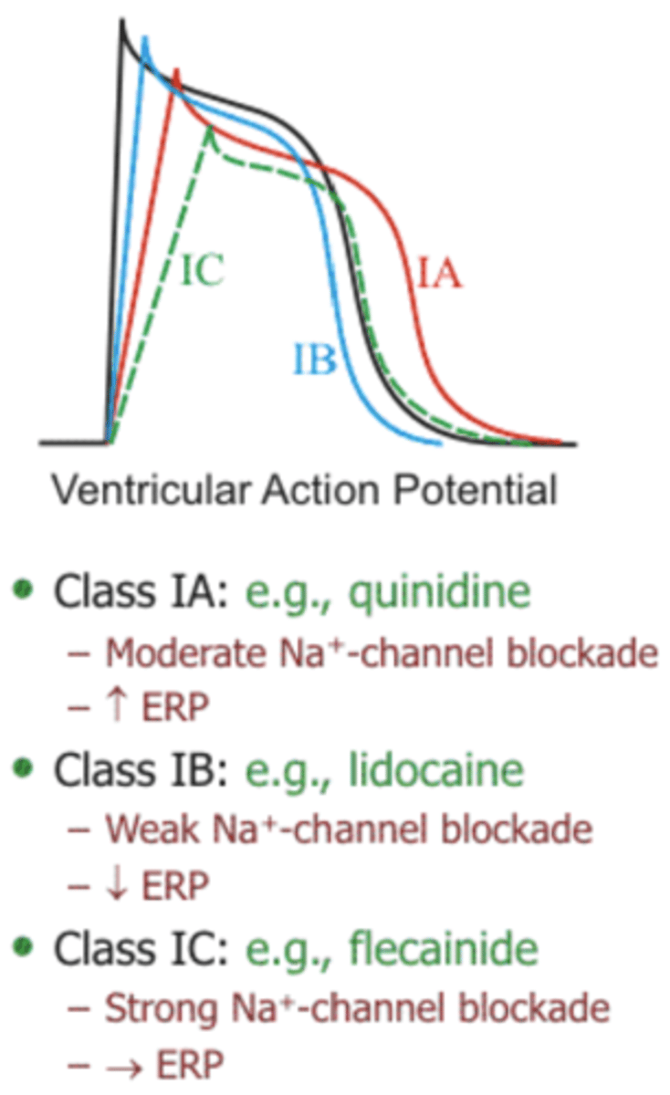
Moderately slow conduction velocity
- Moderate Na+ channel blockade
- Also block K+ channels (and thereby increase ERP)
What adverse effects may occur with Class IA antiarrhythmics?
- GI effects
- Drug-induced lupus-like syndrome (with procainamide)
- Anticholinergic/antimuscarinic effects (disopyramide > quinidine > procainamide)
- Proarrhythmic (QTc prolongation can lead to TdP)
- Can exacerbate or worsen HF
What medications fall into Class IB of the Vaughan-Williams classification system?
- Lidocaine (Xylocaine)
- Mexiletine (Mexitil)
What effect do Class IB antiarrhythmics have on ion channels? Explain basic electrophysiologic outcomes.
Minimally slow conduction velocity (least potent)
- Loosely bind to Na+ channels (fast on-off)
- Weak Na+ channel blockade
- Decrease ERP
What medications fall into Class IC of the Vaughan-Williams classification system?
- Flecainide (Tambocor)
- Propafenone (Rythmol)
What effect do Class IC antiarrhythmics have on ion channels? Explain basic electrophysiologic outcomes.
Substantially slow conduction velocity (most potent)
- Tightly bind to Na+ channels (slow on-off)
- Strong Na+ channel blockade
- Also minimally block K+ channels (and thereby have minimal to no effect on ERP)
- Note: Pro-arrhythmia is related to strong Na+ channel blockade
Compare and contrast the subclasses of Class I with regard to strength of Na+ channel blockade and effect on ERP.
Na+ blockade: Ic (strongest) > Ia > Ib (weakest)
Effect on ERP:
- Increased by Ia
- Minimal to effect from Ic
- Decreased by Ib
Describe the MOA for Class II antiarrhythmics.
Block β-adrenergic receptors (effects on nodal tissue)
- Indirectly interfere with Ca2+ entry
- Reduced sympathetic stimulation decreases Ca2+ channel activation
Decreased Ca2+ influx reduces depolarization of SA node, resulting in:
- Decreased heart rate, contractility, and conduction velocity
- Increased ERP
- Suppressed automaticity of nodal tissue
What medications fall into Class II of the Vaughan-Williams classification system?
Cardioselective β blockers:
- Esmolol (Brevibloc)
- Metoprolol (Lopressor, Toprol)
- Propranolol (Inderal)
- Acebutolol (Sectral)
Describe the general MOA for Class III antiarrhythmics.
Block K+ channels and slow repolarization (effects on non-nodal tissue)
- Results in increased APD and ERP (prolonged refractoriness in atrial and ventricular tissue can help to abolish reentry tachycardias)
- May cause QT prolongation and arrhythmias (e.g., TdP)
What medications fall into Class III of the Vaughan-Williams classification system?
Potassium channel blockers:
- Amiodarone (Cordarone, Pacerone)
- Dofetilide (Tikosyn)
- Dronedarone (Multaq)
- Ibutilide (Corvert)
- Sotalol (Betapace, Sorine)
How does amiodarone exert its antiarrhythmic effects?
Properties from every class (I, II, III, and IV)
- Blocks Na+ channels with fast on-off kinetics
- Nonselective β blocker effects
- Blocks K+ channels (predominant effect)
- Small degree of Ca2+ antagonism
- Also acts as a vasodilator
What adverse effects may occur with amiodarone?
- Pulmonary fibrosis
- Thyroid dysfunction
- Corneal microdeposits
- Hepatotoxicity
- Bradycardia, AV block
- Tremors, ataxia, peripheral neuropathy
- Photosensitivity
- Skin discoloration (blue-gray)
How does dronedarone compare to amiodarone?
Structurally similar to amiodarone except:
- No iodine component (allows for less thyroid toxicity)
- Addition of methane-sulfonyl group (allows for shorter half-life and decreased lipophilicity)
Less effective for maintaining normal sinus rhythm in AFib
Less adverse effects/discontinuation
Has additional contraindications:
- Symptomatic HF (with recent decompensation requiring hospitalization or class IV symptoms)
- Permanent AFib (i.e., normal sinus rhythm cannot be cardioverted)
Which Class III antiarrhythmics act as pure K+ channel blockers?
- Dofetilide (Tikosyn)
- Ibutilide (Corvert)
How does sotalol exerts its antiarrhythmic effects?
Racemic mixture of d- and l-sotalol
- Both isomers have K+ channel blockade effects (i.e., prolong repolarization and increase ERP)
- l-isomer has non-selective β-blocker effects (i.e., slows heart rate and decreases AV nodal conduction)
Describe the MOA for Class IV antiarrhythmics.
Block L-type calcium channels to directly interfere with Ca2+ entry (effects on nodal tissue)
Decreased Ca2+ influx reduces depolarization of SA node, resulting in:
- Decreased AV node conduction velocity
- Increased ERP
- Suppressed automaticity of nodal tissue
What medications fall into Class IV of the Vaughan-Williams classification system?
- Diltiazem (Cardizem, Cartia, Dilacor, Diltia)
- Verapamil (Calan, Covera, Isoptin, Verelan)
What other antiarrhythmic drugs exist outside of the Vaughan-Williams classification system?
- Adenosine (Adenocard, Adenoscan)
- Magnesium
- Digoxin (Digitek, Digox, Lanoxin)
What is the MOA for adenosine?
Binds to A1 adenosine receptors in the AV node, resulting in…
- Increased K+ efflux (leads to hyperpolarization)
- Decreased Ca2+ influx (inhibits depolarization)
- Transient inhibition of AV nodal conduction and increased refractory period
What is the MOA for digoxin?
Enhances parasympathetic vagal tone
- Slows AV nodal conduction
- Useful in atrial arrythmias
Inhibits Na+/K+-ATPase
- Leads to an increase in intracellular Ca2+
- Positive inotropic effects (i.e., increased contractility)
What factors guide antiarrhythmic drug selection?
1. Underlying structural heart disease
2. Renal or hepatic dysfunction
3. Risk of QT prolongation
4. Side effect profile and drug interactions (antiarrhythmics have many)
Which antiarrhythmic medications are contraindicated in heart failure?
- Class IA: Disopyramide, quinidine, procainamide
- Class IC: Flecainide, propafenone
- Dronedarone
What considerations should be made when selecting an antiarrhythmic drug in renal dysfunction?
- Sotalol: Do not use if CrCl < 40 mL/min
- Dofetilide: Do not use if CrCl < 20 mL/min
What drug-drug interaction occurs between amiodarone/dronedarone and digoxin?
- Increase digoxin levels (via decreasing its metabolism)
- Requires digoxin dose to be reduced by 50%
What medications are associated with increased risk of QTc prolongation/Torsades de pointes?
Antiarrhythmics
- Class IA: Disopyramide, quinidine, procainamide
- Class IC: Flecainide)
- Class III: Amiodarone, dronedarone, dofetilide, ibutilide, sotalol
Antibiotics
- Macrolides: Azithromycin, clarithromycin, erythromycin
- Fluroquinolones: Ciprofloxacin, levofloxacin, moxifloxacin
Antipsychotics
- Haloperidol
- Chlorpromazine
- Thioridazine
Antidepressants
- SSRIs: Citalopram, escitalopram
Others
- Donepezil
- Methadone