Anatomy Ch.2 & 3
 Call Kai
 Learn
Learn Practice Test
 Spaced Repetition
 Match
Match Flashcards
 Knowt Play
1/60

There's no tags or description
Looks like no tags are added yet.
Name | Mastery | Learn | Test | Matching | Spaced | Call with Kai | Chat |
|---|
No analytics yet
Send a link to your students to track their progress
61 Terms
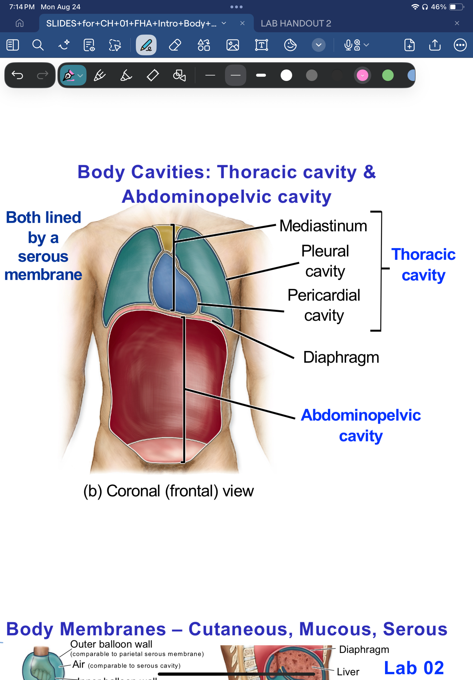
Serous Membrane
a thin membrane that lines body cavities and covers organs. it secretes serous fluid to reduce friction
Peritoneum
Serous membrane of abdominopelvic cavity
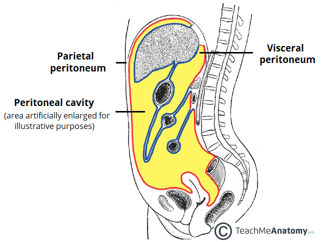
Parietal peritoneum
lines the inside of the abdominal cavity wall
Visceral Peritoneum
wraps and covers abdominal organs

what is serous fluid
fluid within the serous membrane reducing friction when organs move during breathing and digestion
Greater Omentum
A mesentry (specialized peritoneal fold) under the ribcage midline on top of belly button
Mesentery
A double fold of the peritoneum that holds the intestines, and carries the blood vessels and nerves needed to hold structure
Pleura
serous membrane covering the lungs
pericardium
serous membrane covering the heart
peritoneum
serouns membrane covering abdominal organs
parietal layer
outer layer of serous membrane
visceral layer
serous membrane’s layer holding an organ

Organ system’s purpose:
To maintain homeostasis so cells have a stable internal environment and can survive.
interstital fluid and plasma: ____
body internal environments
How do cell shape and size affect function?
Cell structure is specialized for its job (structure determines function)
What is the plasma membrane composed of?
A phospholipid bilayer with proteins, cholesterol, and carbohydrates
What are the functions of the plasma mebrane?
physical barrier, selective permeability, electrochemical gradients, communication
what forms the plasma membrane barrier?
lipids
what is the role of proteins in the plasma membrane?
Transport, signaling, receptors, pumps, enzymes, and cell recognition
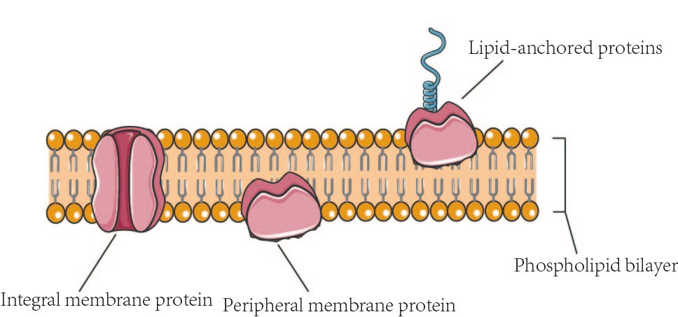
Integral proteins
proteins embedded in the phospholipid layer
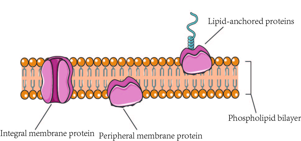
peripheral proteins
Attached to EITHER the inside or the outside of the phospolipid layer (not through)
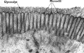
glycocalyx
A carbohydrate coat on the outside of the plasma membrane that helps with protection, cell adhesion and cell recognition (allowing right cells to be in right places)
Passive transport
no energy needeed; follows a concentration gradient
active transport
requires atp, can move against the concentration gradient; opposite direction from diffusion
What are the types of passive transportation?
diffusion, facilitated diffusion, and osmosis
Ion Pump
A membrane protein that uses ATP to move ions against their concentration gradient (active transport).
endocytosis
Bringing materials into the cell; removes membrane from the plasma membrane.
exocytosis
Bulk transport of waste OUT of cell; fusion of membranes is active transport requiring energy
What types of transportation does exocytosis and endocytosis use?
active transportation
Apoptosis
Controls the contents of a dying cell with no inflammation
Necrosis
Swelling and explosion of waste contents due to lack of apoptosis
Necrosis
Swelling and explosion of waste contents due to lack of apoptosis
Passive transport
Diffusion mechanism to transport across the plasma membrane
Osmosis
Passive transport of water
Aquaporins
Rapid water movement during Osmosis
H2O
Small molecule; partial charge → slow movement
Transport Proteins
Regulate gradients which are used to power co-transport (controls selective permeability)
Covalent Bonds
Strongest and longest bonds(long lasting)
Coupled transport
Ion pump (1st protein) builds the Na+ gradient, 2ndary transport protein uses it to actively transport the amino acid
Primary active transport
The building of a Na+ gradient with an ion pump
Secondary Transport Protein
Using gradient (already built) to actively transport the amino acid, and passively diffuse the Na+ i concentration direction (primary active transport builds against the gradient
Microvilli
Maximize surface area to maximize rate of exchange
Gastrulation
Movement of cells from epiblast into the interior
Embryo builds extra amniotic membranes
Epiblast
An opening on the amnion; The ultimate embryo
Amnion
Extra embryonic membrane
Primitive streak
The opening from the invagination of epiblast cells
Invagination
cells move to middle and drop creating primitive streak and forming: 1st; endoderm, mesoderm, and what doesn’t go through the primitive streak becomes ectoderm.
What replaces the hypoblast?
The endoderm
Gastrulation - Trilaminar Embryo Test diagram
Do all three germinal layers move through the primitive streak?
No. The part that doesn’t go through it becomes the ectoderm
ectoderm makes up:
Nervous tissue and sensing organs, epidermis of skin and epidermal derivatives, pituitary gland, adrenal medulla, enamel of teeth, lens of eye, and some bones of skull
Endoderm makes up:
Epithelial lining of respiratory tract (GI, urinary tract, reproductive tract tympanic cavity, auditory tube, liver, gallbladder, pancreas, saltine tonsils, parathyroid glands, thymus
Mesoderm makes up:
Epithelial lining (vessels and serous membranes), muscle tissue, connective tissue, adrenal cortex, heart, kidneys, ureters, internal reproductive organs, spleen
Epithelium
A lot of cells right next to each other (identifiable by nucleus), with little extracellular matrix

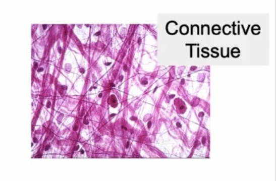
Connective tissues
Few scattered cells (identifiable by nuclei) making fibers (tissue) with a lot of background extracellular matrix (space)
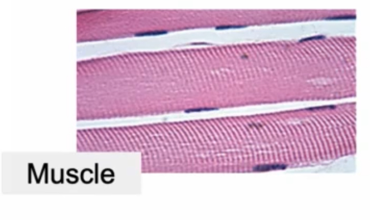
Muscle tissue
Composed of bunch of stripes (mostly) with scattered cells (identifiable by the scattered nuclei dark dots)
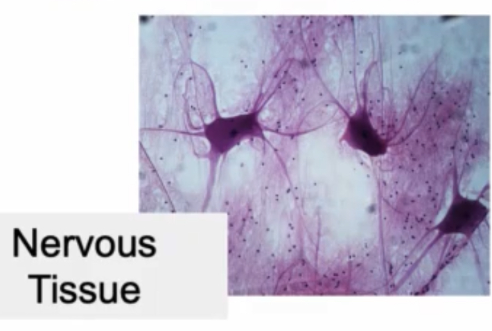
Nervous Tissue
Composed of neurons with a bunch of glia; bigger glial nuclei than other types; not a lot of nervous tissue compared to the big glial nuclei
What serous membrane covers most of the liver/stomach/intestines
Visceral peritoneum
Pleura
serous membrane covering lungs
Pericardium
serous membrane covering heart