Elective 1: Dysphagia Lecture Flashcards
 Call Kai
 Learn
Learn Practice Test
 Spaced Repetition
 Match
Match Flashcards
 Knowt Play
1/214

Earn XP
Description and Tags
Comprehensive vocabulary flashcards covering anatomy, physiology, neural control, models of swallowing, and etiology of dysphagia based on lecture notes.
Name | Mastery | Learn | Test | Matching | Spaced | Call with Kai | Chat |
|---|
No analytics yet
Send a link to your students to track their progress
215 Terms

Anatomy of the Oral Cavity
The structural region comprising the teeth, hard palate, soft palate, retromolar trigone, tongue (front two-thirds), lips, gingiva (gum), uvula, tonsil, buccal mucosa, and floor of mouth.

Cheek and Lip Musculature
Facial muscles including the buccinator and orbicularis oris that control oral contents during mastication and swallowing.
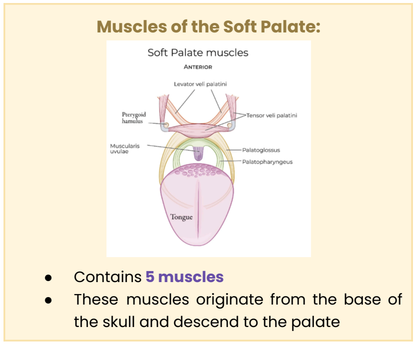
Soft Palate Muscles (Anterior View)
Anterior structural arrangement showing the levator veli palatini, tensor veli palatini, palatoglossus, palatopharyngeus, and muscularis uvulae relative to the tongue and pterygoid hamulus.

Soft Palate Muscles (Posterior View)
Posterior anatomical view displaying the tensor veli palatini, levator veli palatini, palatopharyngeus, palatoglossus, muscularis uvulae, palatine aponeurosis, pharyngotympanic tube, and pterygoid plates.

Extrinsic Tongue Muscles
Muscles that originate outside the tongue and insert into it (palatoglossus, styloglossus, genioglossus, hyoglossus), primarily controlling tongue position and movement.

Intrinsic Tongue Muscles
Muscles located entirely within the tongue (superior longitudinal, transverse, vertical, inferior longitudinal) that alter the shape of the tongue.

Pharyngeal Constrictors (Lateral View)
Lateral anatomical view of the superior, middle, and inferior pharyngeal constrictors along with the stylopharyngeus and salpingopharyngeus muscles.

Divisions of the Pharynx
Anatomical division of the pharyngeal space into three parts: nasopharynx, oropharynx, and laryngopharynx.
Pharyngeal Constrictors (Posterior View)
Posterior view showing the thyropharyngeus, cricopharyngeus, and the intervening Killian's dehiscence region.

Innervation of the Pharynx Diagram
Anatomical chart outlining the sensory and motor nerve distributions supplying the pharyngeal constrictor and longitudinal muscle groups.
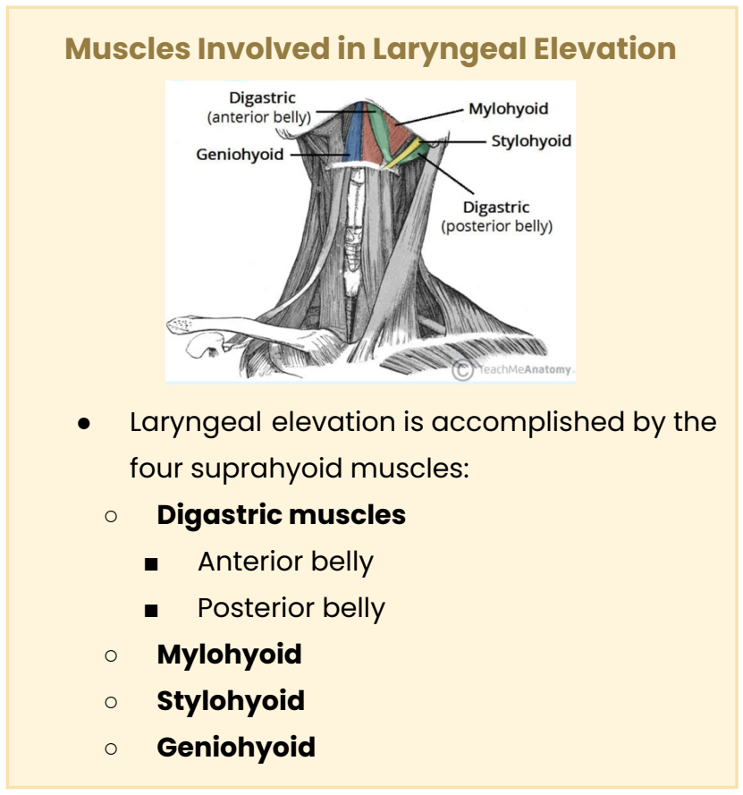
Muscles Involved in Laryngeal Elevation Diagram
Anterior neck muscular diagram depicting the suprahyoid group (digastric anterior/posterior bellies, mylohyoid, stylohyoid, geniohyoid) responsible for elevating the hyolaryngeal complex.

Glottis Diagram
Anatomical illustration showing the vocal folds, rima glottidis space, and surrounding intrinsic laryngeal sphincter musculature.

Epiglottis Anatomy Diagram
Posterior view showing the leaf-like/heart-shaped epiglottic cartilage positioned above the laryngeal inlet.

Oral Phase of Swallowing Diagram
Five-step visual sequence illustrating food entry, preparation, bolus formation, anterior-to-posterior transport, and bolus arrival at the oropharynx.

Esophageal Phase of Swallowing Diagram
Five-step visual sequence detailing bolus entry, primary peristalsis wave, bolus propulsion, lower esophageal sphincter (LES) relaxation, and peristalsis termination.

Process Model Stage 1 Diagram
Illustration of Stage 1 Transport showing initial food ingestion into the mouth followed by lateral rotation onto post-canine teeth.

Process Model Stage 2 Diagram
Illustration showing Stage 2 Transport, bolus propulsion through the fauces, and bolus accumulation in the valleculae prior to pharyngeal swallow trigger.

Bolus Transport in the Esophagus Diagram
Detailed diagram demonstrating primary peristaltic wave progression, sphincter dynamics at UES/LES, and gravity assistance during the esophageal stage.

The Patient's Medical Chart Categories
Overview of key medical record components including Medical History (H&P), Laboratory Tests (BMP, CBC), and Diagnostic Tests (ECG, Echo, Ultrasound, CT, MRI, X-ray).
Swallowing
A physiologic function involving coordinated muscle contractions that propel food and liquids from the oral cavity to the stomach.
Number of Muscles in Swallowing
Over 30 paired muscles.
Traditional Three-Phase Model
The standard swallowing framework consisting of the Oral Phase, Pharyngeal Phase, and Esophageal Phase.
Oral Preparatory Phase
The sub-phase of swallowing that begins when solid or liquid food enters the oral cavity to be manipulated and formed into a bolus.
Oral Propulsive Phase
The sub-phase of swallowing that begins when the bolus is propelled along the palate by the tongue towards the oropharynx.
Oral Cavity
The space between the upper and lower dental arches where bolus formation occurs.
Orbicularis Oris Origin
Maxilla and mandible.
Orbicularis Oris Insertion
Inserts around the lips/mouth.
Orbicularis Oris Function
Controls entrance into and exit from the oral cavity, allowing the lips to close, purse, and compress.
Lips
Structures surrounding the mouth that form a sphincter through the orbicularis oris muscle.
Lips External Covering
Specialized skin.
Lips Internal Lining
Mucous membrane.
Lips Innervation
Buccal branch of the facial nerve (CN VII).
Lips Contents
Superior and inferior labial muscles, blood vessels, and nerves.
Cheeks
Lateral walls of the oral cavity continuous with the lips, covered externally by skin and internally by mucous membrane.
Cheeks Contents
Buccinator muscle and buccal fat pads superficial to the buccinator.
Buccinator Muscle Origin
Posterior maxilla and mandible.
Buccinator Muscle Insertion
Blends with the orbicularis oris muscle.
Buccinator Muscle Function
Compresses the cheek and holds food between the teeth during mastication and swallowing.
Buccinator Muscle Innervation
Buccal branch of the facial nerve (CN VII).
Hard Palate
Forms the anterior 2/3 of the palate; a bony, concave roof of the oral cavity covered by mucous membrane.
Soft Palate
Forms the posterior 1/3 of the palate; a mobile fibromuscular fold separating the nasal cavity from the nasopharynx.
Uvula
A structure hanging from the free, curved margin of the soft palate.
Fauces
The space between the oral cavity and the pharynx.
Number of Soft Palate Muscles
5 muscles.
Origin of Soft Palate Muscles
Originate from the base of the skull and descend to the palate.
Levator Veli Palatini Origin
Petrous part of the temporal bone and cartilaginous portion of the pharyngotympanic (auditory/Eustachian) tube.
Levator Veli Palatini Insertion
Middle portion of the palatine aponeurosis.
Levator Veli Palatini Action
Elevates the soft palate, moving it upward and backward during swallowing.
Tensor Veli Palatini Origin
Scaphoid fossa of the medial pterygoid plate, spine of the sphenoid bone, and cartilage of the pharyngotympanic tube.
Tensor Veli Palatini Insertion
Pterygoid hamulus and palatine aponeurosis.
Tensor Veli Palatini Action
Tenses the soft palate and opens the pharyngotympanic tube during swallowing.
Palatopharyngeus Origin (Soft Palate)
Hard palate.
Palatopharyngeus Action (Soft Palate)
Elevates the pharynx and larynx during the pharyngeal phase, and depresses the soft palate at the end of the oral phase.
Palatopharyngeal Arches
Paired ridges of mucous membrane on lateral pharyngeal walls (posterior faucial pillars) marking the boundary between oral cavity and oropharynx.
Key Soft Palate Swallowing Sequence
Levator veli palatini pulls soft palate up and backward; palatopharyngeus brings/depresses soft palate downward at the end of the oral phase.
Palatoglossus Insertion (Soft Palate)
Posterolateral tongue.
Palatoglossus Action (Soft Palate)
Elevates posterior tongue, draws soft palate toward tongue, and narrows the diameter of the oropharyngeal isthmus.
Palatoglossal Arches
Paired ridges of mucous membrane also known as the anterior faucial pillars.
Musculus Uvulae Origin
Posterior nasal spine of the hard palate and palatine aponeurosis.
Musculus Uvulae Insertion
Connective tissue of the uvula.
Musculus Uvulae Action
Shortens the uvula and pulls the uvula superiorly during swallowing.
General Soft Palate Innervation
Pharyngeal branch of the vagus nerve (CN X).
Tensor Veli Palatini Innervation Exception
Mandibular division of the trigeminal nerve (CN V3).
Tongue
Mobile muscular structure located partly in the oral cavity and oropharynx, covered by mucous membrane.
Extrinsic Tongue Muscles Definition
Muscles that primarily control the position and movement of the tongue.
Intrinsic Tongue Muscles Definition
Muscles located entirely within the tongue with no external attachments that alter tongue shape.
Palatoglossus Extrinsic Origin
Inferior surface of the palatine aponeurosis.
Palatoglossus Extrinsic Function
Elevates the posterior tongue and draws the soft palate onto the tongue.
Styloglossus Origin
Apex of the styloid process, adjacent to origin of stylomandibular ligament.
Styloglossus Insertion
Merges with the hyoglossus and inferior longitudinal muscle of the tongue.
Styloglossus Function
Elevates and retracts the tongue.
Hyoglossus Description
Thin, quadrilateral muscle in the upper neck and floor of the mouth.
Hyoglossus Origin
Hyoid bone, entire length of the greater cornu, and front of the body of the hyoid.
Hyoglossus Insertion
Side of the tongue, between styloglossus laterally and inferior longitudinal muscle medially.
Hyoglossus Function
Depresses and retracts the tongue.
Genioglossus Description
Fan-shaped extrinsic muscle that makes up much of the bulk of the tongue.
Genioglossus Origin
Superior mental spine of the symphysis menti (posterior surface of midline mandible).
Genioglossus Insertion
Entire tongue mass and body of the hyoid bone.
Genioglossus Function
Depresses and retracts the tongue.
Superior Longitudinal Origin
Submucosal connective tissue of the posterior tongue.
Superior Longitudinal Insertion
Submucosal connective tissue and mucosa along the tongue margins.
Superior Longitudinal Function
Curls the apex and lateral sides upward; works with inferior longitudinal to shorten and retract protruding tongue.
Inferior Longitudinal Origin
Root of the tongue and hyoid bone.
Inferior Longitudinal Insertion
Apex of the tongue.
Inferior Longitudinal Function
Curls the apex and lateral sides downward; works with superior longitudinal to shorten/retract protruding tongue.
Transverse Intrinsic Muscle Origin
Median septum of the tongue.
Transverse Intrinsic Muscle Insertion
Submucosal fibrous layer of the lateral tongue margins.
Transverse Intrinsic Muscle Function
Elongates and protrudes the tongue.
Verticalis Intrinsic Muscle Origin
Submucosal connective tissue on the dorsum of the tongue.
Verticalis Intrinsic Muscle Insertion
Connective tissue in the ventral regions of the tongue.
Verticalis Intrinsic Muscle Function
Flattens and broadens the tongue.
Hypoglossal Nerve (CN XII) Tongue Rule
Innervates all intrinsic and extrinsic tongue muscles except Palatoglossus.
Palatoglossus Tongue Innervation Exception
Pharyngeal branch of the vagus nerve (CN X).
Pharynx
A muscular tube-shaped passageway located behind the nose and mouth serving as a shared pathway for air and food.
Pharyngeal Wall Layers
Internal longitudinal muscles and external longitudinal/constrictor muscles.
Pharynx Boundaries
Extends from cranial base to inferior border of cricoid cartilage anteriorly and C6 vertebra posteriorly.
Nasopharynx Span
From posterior of nose to superior of soft palate.
Oropharynx Span
From soft palate to hyoid bone.
Laryngopharynx Position
Posterior to the larynx.
Superior Pharyngeal Constrictor Origin
Pterygoid hamulus, pterygomandibular raphe, posterior end of mylohyoid line, transverse muscle of tongue.