week ten
 Call Kai
 Learn
Learn Practice Test
 Spaced Repetition
 Match
Match Flashcards
 Knowt Play
1/23

There's no tags or description
Looks like no tags are added yet.
Name | Mastery | Learn | Test | Matching | Spaced | Call with Kai | Chat |
|---|
No analytics yet
Send a link to your students to track their progress
24 Terms
breast screening
a cancer detection program set in place to allow early detection of breast tumours
screening mammography
offered to asymptomatic, average risk women biennialy
ages 40-74
available every 2 years for patients over the age of 40
screening program contraindications
patient is pregnant
patient is lactating (wait 3 months after breastfeeding)
have had breast cancer
(may need a diagnostic mammo or breast ultrasound of if symptomatic patient is below 40)patient has breast implants
patient has a new lump on the breast
high risk of breast cancer
breast cancer gene (BRCA1 or BRCA2)
parent, sibling or child has had breast cancer or carries the gene
2 or more relatives on same side of family has had breast cancer before the age of 50
3+ relatives on same side of family had breast cancer at at least one was under the age of 50
they have had radiation to the chest
Mammos are recommended every year for people in this categories
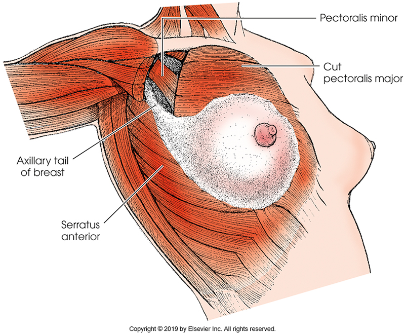
breast location
attach anteriorly at ribs 2-7
extend from the lateral margin of the sternum to the axillary plane and form a tear drop shape
the axillary tail- extends from the upper lateral aspect of the breast to the armpit region
(composed of various structures that all them to change in shape and composition as the body changes)
lobes
each breast contains 15-20 of these
contain milk secreting components (lobyles) controlled by hormones
composed of glandular tissue
become very dense during pregnancy or hormonal therapy
adipose tissue
fat tissue that fills spaces betwene lobes
gives shape to breasts
replace glandular tissue as the aging process progresses and contribues to a homogenous appearance in later life
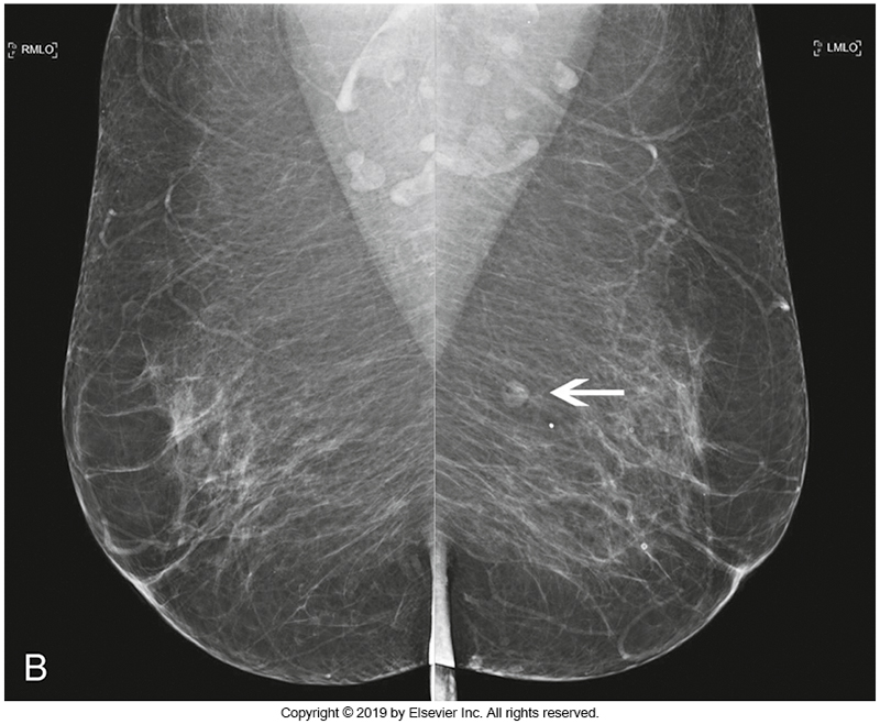
pectoralis major muscle
triangular in shape and originates from the clavicle, sternum and ribs
inserts into the lateral border of the bicipital groove of the humerus and lies between the breast tissue and ribs
shows as white trainagular densitis
lactiferous ducts
transport milk from the lobes / lobules to the nipples
these are a common site of cancer
retro-mammary space
composed of fibrous connective tissue and is located between the posterior breast fat and the pectroalis muscle
lymph nodes
there are 12-30 of these located in each axillary space
drain intercellular fluid from the chest and arms
these are biopsied for positive malignant breast cells and for sentiel node assessment
we include the axilla and pectroalis to include this region and rule out any potential patholoyg present
compression
a technique used for all mammograms that creates a uniform thickness of the breast tissue
if the overall thickness is reduced, there is a decrease in radiation dose to patient
decreases motion and distortion
spreads out SI structures into a single layer
should be able to see the pectoralis muscle and the nipple directed forward in profile
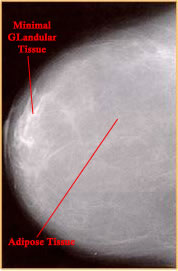
BI-RADS A
fatty breast consists of mainly adipose tissue with minimal glandular tissue
(remember density can only be determined on a mammogram as the look and feel of a breast does not indicate the ratio of fatty to fibroglandular tissue
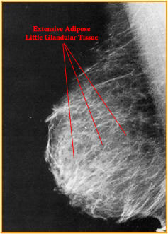
BI-RADS B
these breasts have a small amount of glandular tissue and extensive adipose tissue
BI-RADS C
mixed density breasts are composed of glandular tissue infiltrated with adipose tissue
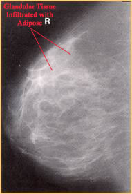
BI-RADS D
the dense breast consists of primarily connective tissue with very little adipose tissue
minimal tissue differentiation between the two types of tissue
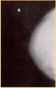
lactating breast
this breasts consists of extensive glandular tissue with minimal adipose tissue since the glands are actively producing milk
the breast is extrenely dense and radioopaque
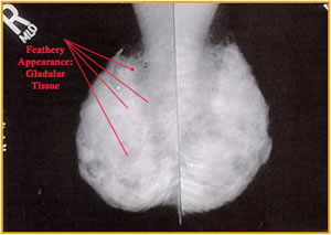
25-32
the kVp used for mammo:
(AEC is typically used for mAs selection)
a lower one of kVp permits high contrast imaging - will result in a higher absorbed radiation dose than conventional radiogrpahy- byt the rad must be able to visualize the contrast of the breast
50-80
what is the SID used
(this number is needed as the kVp is low)
might affect image sharpness and distortion, a small focal spot and compressed (decreased OID) will aidn in this
Molybdenum
the most common alloy used for the anode | has a low atomic number and therefore lower energy photons are produce
(may also be Rhodium and Tungsetn is a better fit to penetrate thick and extremely dense breast)
anode rotates and is anhled +10 degrees to -9
cathode is nearest to the chest wall for anode heel effect
filtration
typically constructued of beryllium (Be)
the units must be kept low
units are often equipped with added __ made of Mo or Rh
different ones are used for different anode targets
(also the focal spot size are 0.3-0.4 for screening and 0.1 to 0.15mm for diagnostic)
a small focal spot will compensate when magnification is needed as mag will reduce the resolution
patient prep
no deodorant or powder (mimic micro-calcification)
reduce or refrain from caffeine intake (causes th ebody to retain fluid and the breats tissue is a common place for accmulation)
avoid any artifacts (includding clipping hair back)
gown that opens from the front
complete history questionnaire is filled out
try to relax (when patient is tense, compression is difficult)
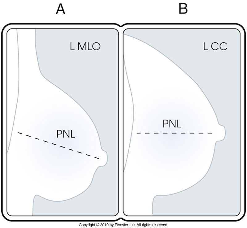
posterior nipple line
an imaginary line drawn from the nipple to the edge of the image, chest wall or pectoralis muscle (whichever comes first)
used to measure the depth of the breast in both CC and MLO images
determines whether sufficient breast tissue was imagied
(compared between both CC and ML images and should be within 1cm of each projection
should demonstrate the inferior aspect of the pectoralis muscle extending to or below the line for the MLO
images will inclyde the pectroalis muscle 30% of the time for CC
digital breast tomosynthesis
a 3D imaging technology being used by several sites
machine acquires images of the breasts at multiple angles instead of 90-degrees
tube moves in an arc, taking a series of low dose images to allow view of breast tissue slices in as thin as 1mm
allows for better visualization of breast abnormalities, as it separates breast tissue with each slide