Repair and Regeneration of Nervous System; Adult Neurogenesis
1/32
There's no tags or description
Looks like no tags are added yet.
Name | Mastery | Learn | Test | Matching | Spaced |
|---|
No study sessions yet.
33 Terms
Stroke or injury patients do have the ability to regain function over time:
Movement in paralyzed limbs can improve (especially with physical therapy)
Speech impairments will diminish with speech therapy
Stroke recovery is not thought…
to reflect regrowth of replacement of damaged neurons
Undamaged brain regions become…
reorganized to support functions that were disrupted due to the injury
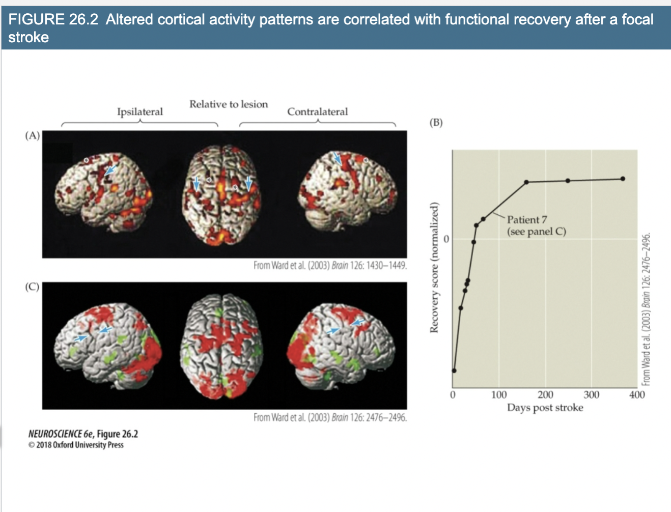
Explain Figure 26.2- Altered cortical activity patterns are correlated with functional recovery after a focal stroke
(A) Compilation of about 20 people that show where declines are occurring in the brain. Red= loss of activity and green= areas that increase activity to help compensate for this loss
(B) and (C) correspond to a specific patient, motor deficits in hand, shows that patient recovers that function over time. Increase in activity right after stroke.
3 types of neuronal repair that could occur
Regrowth of axons (peripheral)
Restoration of damaged central nerve cells (injured but survive)
Generation of New Neurons (replace those that have been lost)
3 Components of Regrowth of axons (peripheral ganglia or peripherally projecting axons)
Requires reactivation of the developmental processes for axon growth and guidance and initial synapse formation
Seen primarily when sensory or motor nerves are damaged in the periphery; nerve cell bodies are intact
Peripheral nerve regeneration is the most easily accomplished type of repair in the nervous system and the most clinically successful
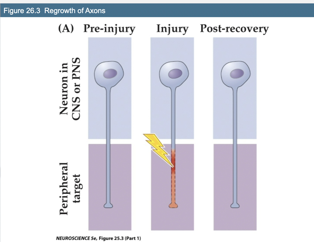
Explain Figure 26.3- Regrowth of Axons
Shows neuron injury, gets damaged, then goes back to being repaired
Recovery seen in periphery but not in CNS
Restoration of damaged central nerve cells (injured but survive)
Requires that nerve cells are capable of restoring their damaged processes and connections to some level of functional integrity
Several developmental mechanisms must be re-engaged
Appropriate regulation of polarity in order to distinguish axon and dendrites
Trophic signaling
Regrowth of neuronal and glial elements in a more complex
environmentLocal overgrowth of glial cells and production of signals that inhibit neuron growth
Inflammatory molecules may suppress reactivation of cellular mechanisms for axonal and dendritic growth
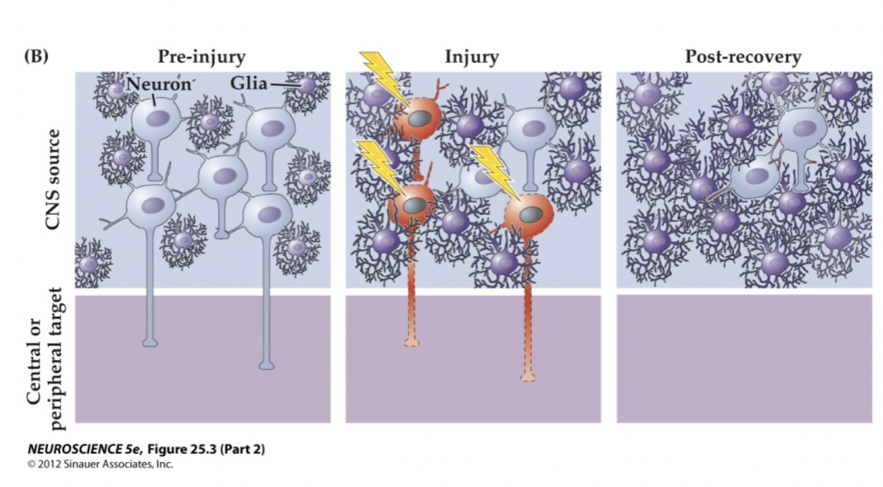
Figure 25.3- Restoration of damaged CNS Cells
CNS cells get damaged but unlike PNS cells, they do not get repaired and are instead lost, because not able to get trophic support they need, unable to remake connections.
Could occur but does not
Generation of New Neurons (replace those that have
been lost)
Adult neuronal genesis occurs rarely
For such repair to occur the following must be met:
Retention of a population of neural stem cells able to give
rise to all cell types in the brain region that has been
damagedNeural stem cells must be present in a “niche” that provides
the appropriate environment for genesis and differentiation of
new neurons and gliaRegenerating tissue must retain the capacity to recapitulate
migration, outgrowth of processes, and synapse formation to
form local and distant functional networks
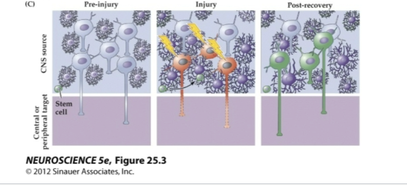
Explain this image
If stem cells are there, they can proliferate and differentiate to join network and replace damaged cells
Not done in CNS
This in theory could occur but in actuality does not
Head’s experiment 1905 - Peripheral nerve regeneration
Early 1900’s – it was clear that damage to a peripheral nerve resulted in a gradual but incomplete restoration of sensory and motor function; the speed and precision of
recovery could be facilitated by surgically connecting the two endsBritish neurologist Henry Head decided to perform a nerve transection on himself; this was documented in a paper published in 1905.
First indication of recovery was the return of general sensitivity to pressure and touch
Sensitivity to light touch, temperature discrimination, pinprick, two-point discrimination, and fine motor control
were slower to recover and did not fully recover after 2
years
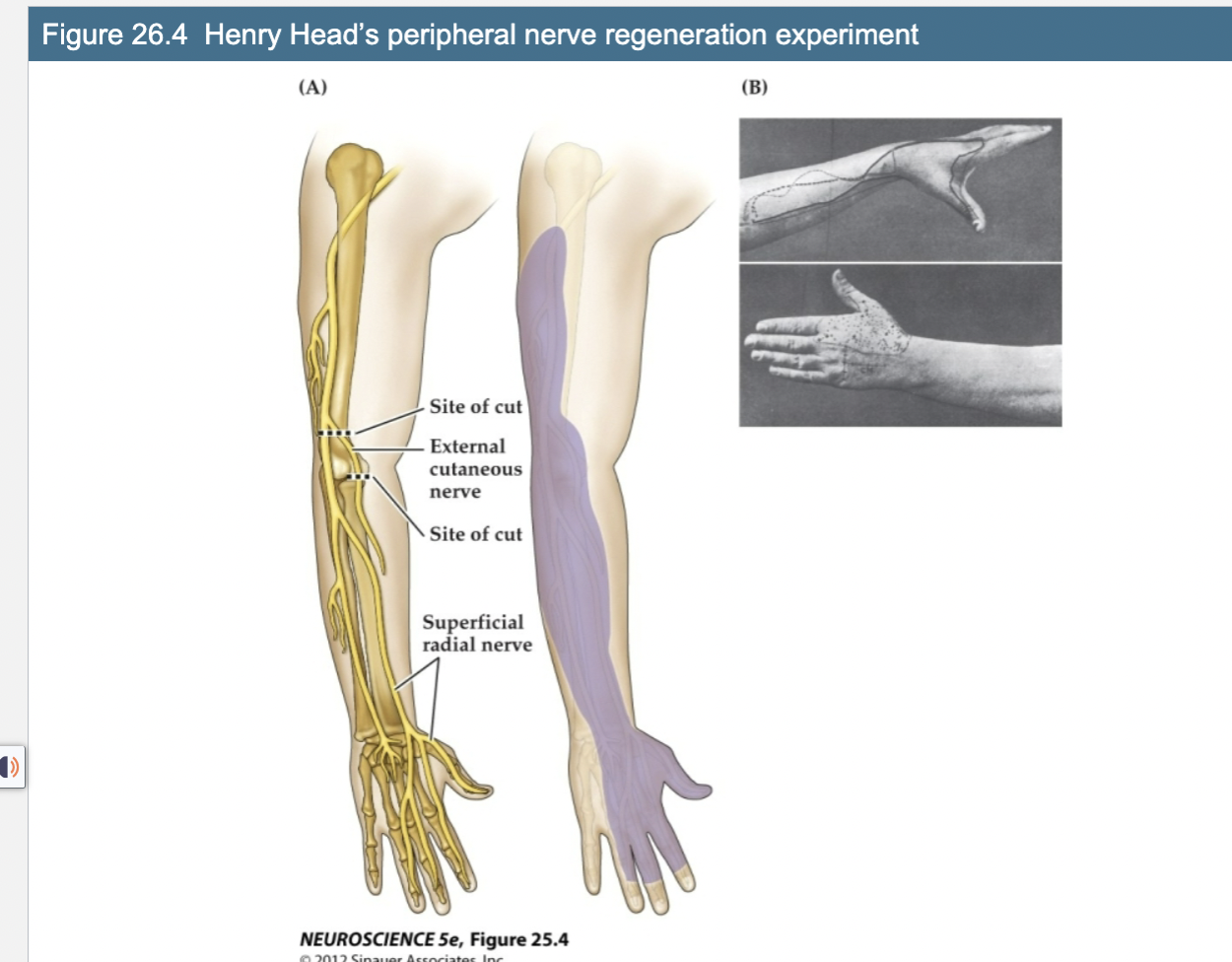
Figure 26.4 Henry Head’s peripheral nerve regeneration experiment
Cut nerve and then joined them back together
Areas of deficit, hand and thumb regained sensation, less or more simulation response
2 Major elements that contribute to peripheral axon regrowth and reinnervation of target
Schwann Cells
Macrophages
These two only present in peripherary and not CNS
Schwann cells
glial cells that myelinate peripheral
axons
Macrophages
immune cells that clear the degenerating remains of of severed axons
In addition, these schwann cells and macrophages secrete molecules that are…
essential for successful regeneration; mimic the
environment that supports axon guidance and
growth during early development
Regeneration is more efficient after…
crushing vs. cutting a nerve
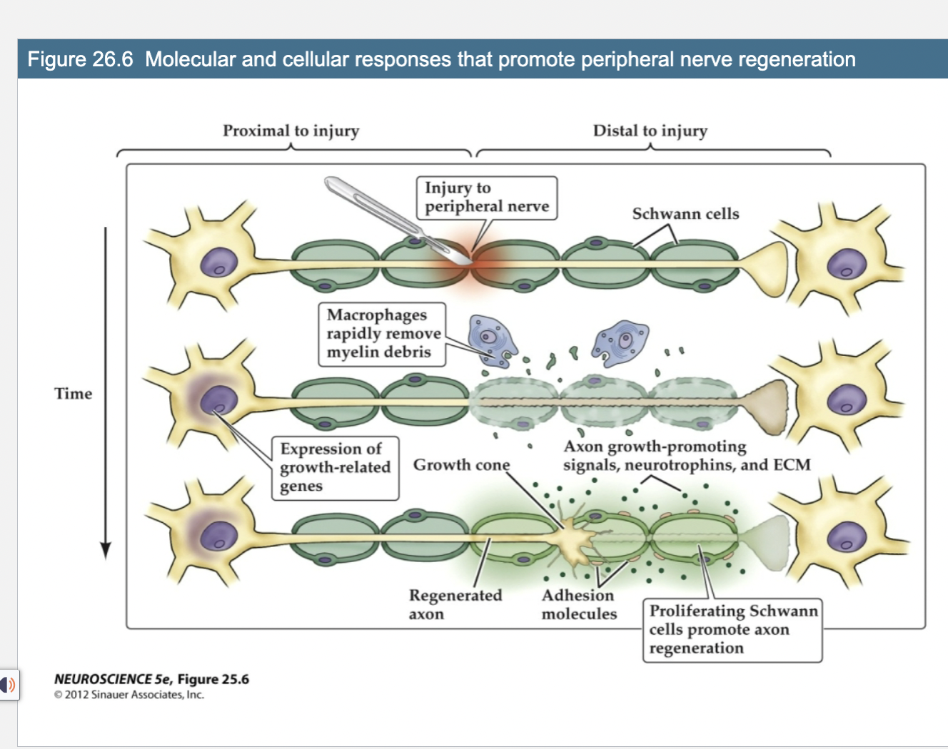
Figure 26.6- Molecular and Cellular Response that promote peripheral Nerve Regeneration
Shows injury to peripheral nerve
After damage occurs, damaged parts of axon need to be removed and macrophages remove debris
Increase transcription for genes that are related to growth of the axons
Peripheral nerve graft of schwann cell, basal lamina (extracellular matrix), and connective tissue will promote growth of…
Why doesn’t this happen normally in the CNS?
CNS axons
Reestablishment of synaptic connections is necessary
for successful recovery of function
Most thoroughly characterized at the neuromuscular junction.
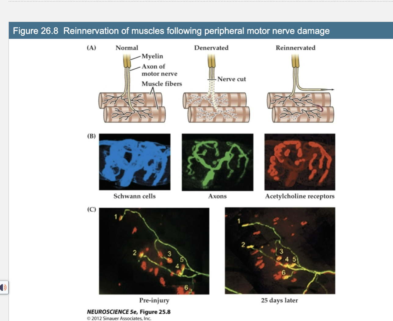
Figure 26.8 Reinnervation of muscles following peripheral motor nerve damage
From experiment that shows motor neurons that innervate muscle fibers, if nerve is cut, it gets degenerated, but overtime the axons can regrown and find target muscle fibers, and synapse needs to be reestablished to regain function
Axon= green
Red= Ach receptors (on muscle fibers itself for junction)
25 days later, reestablished back to normal- in periphery
not seen in CNS
Very little axon growth and reestablishment of functional
connections within the…
central nervous system following injury
3 ways damage occurs in the CNS
external physical trauma
hypoxia – lack of oxygen often created by lack of blood flow (ischemia) due to stroke
neurodegenerative diseases
All result in some amount of neuronal death either immediately or over time
CTE
Deposits of Tau protein that accumulate that indicate brain damage
overwhelm the ability of the
skull and the fluid-filled cushion created
by the subarachnoid space to protect the
brain from shearing forces. These forces
can cause acute bleeding around the
meninges or within brain tissue.
One of the most striking differences in the consequences of central versus peripheral nerve cell damage is the extent of cell death that occurs after direct damage to the brain.
Neuronal cell death in the CNS is seen regardless of the type of damage (traumatic, hypoxic, or degenerative).
Factors that lead to limited regeneration in the CNS
Damage to brain tissue tends to engage mechanisms that lead to necrotic and apoptotic cell death of nearby neurons
Cellular changes at the site of injury do not recapitulate developmental signaling that supports growth
Glial growth and proliferation and microglial activity actively inhibit growth
Upregulation of growth inhibiting molecules
Neuronal cell death in the CNS is seen regardless of the type of damage; studied most extensively in brains where hypoxia has occurred due to local vascular occlusion (stroke)
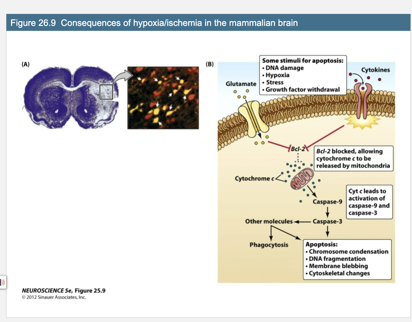
Figure 26.9 Consequences of hypoxia/ischemia in the mammalian brain
White= cells that have died
Yellow= stain for apoptotic protein
Glutamate is released alot when ther is cns injury, stimulates apoptosis pathways
Bd2 promotes capsapse to promote apoptosis
Some stimuli for apoptosis
DNA damage
Hypoxia
Stress
Growth Factor Withdrawal
Apoptosis
Chromosome condensation
DNA fragmentation
Membrane blebbing
cytoskeletal changes
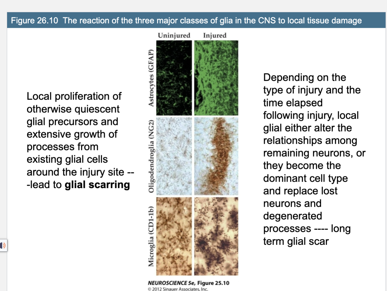
Figure 26.10 The reaction of the three major classes of glia in the CNS to local tissue damage
Injured vs uninjured
Glial cells, astrocytes, microglia overproduced at injury site to get int he way, which prevents axons to find target because of glial scarring, can also affect axons that are near
Growth inhibitng
Local proliferation of otherwise quiescent
glial precursors and extensive growth of
processes from existing glial cells around the injury site -- -lead to glial scarringDepending on the type of injury and the time elapsed following injury, local glial either alter the relationships among remaining neurons, or they become the dominant cell type and replace lost neurons and degenerated processes ---- long term glial scar
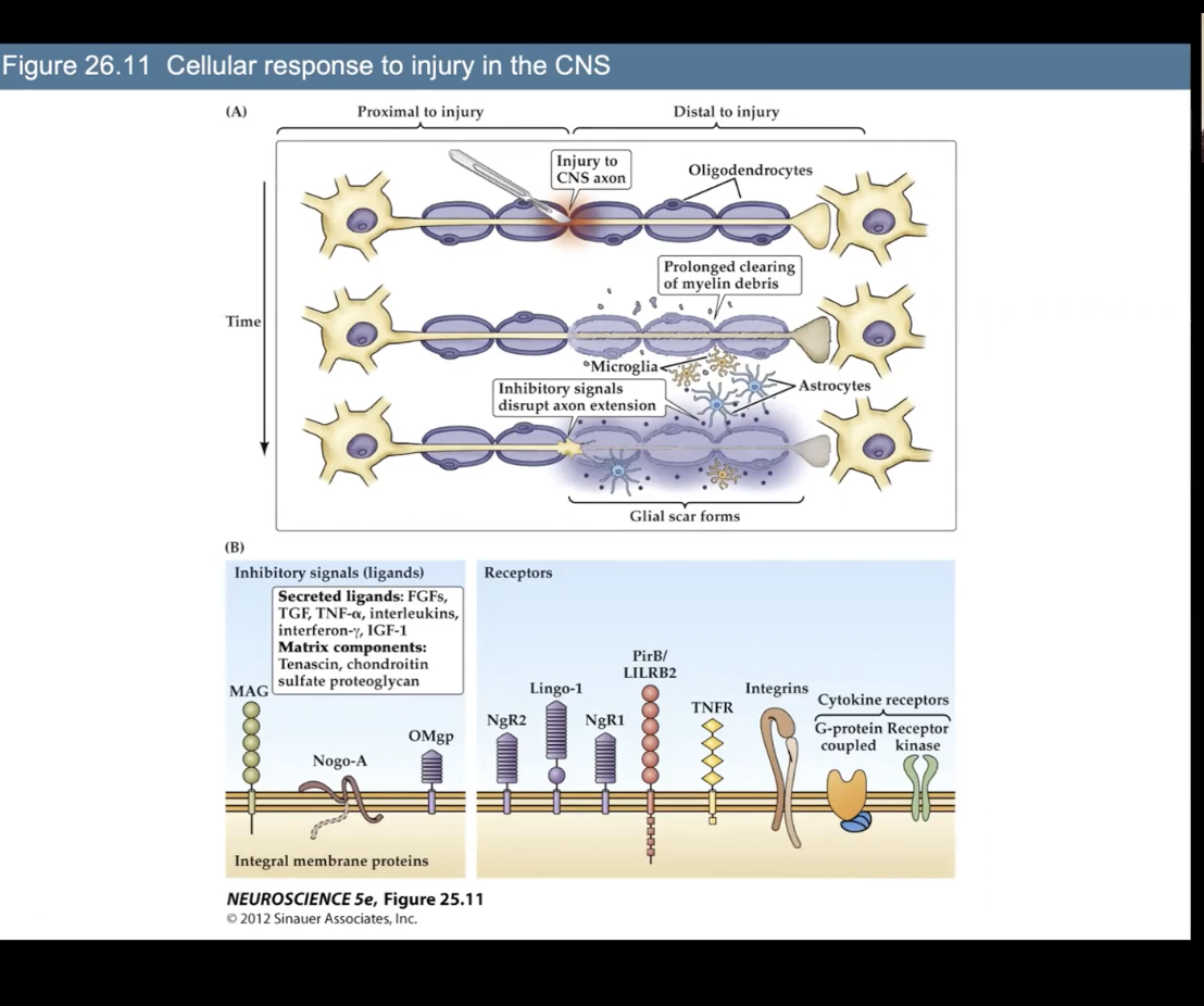
Figure 26.11 Cellular response to injury in the CNS
In CNS, has oligodendrocytes
Microglia can be there to sort of act as macrophages, but inactuality do not really clear away. debris
Microglia and Asrtocytes release signlas that inhibit growth of axon, glial scar occurs, nothing left to promote growth, and neuron ends up dying
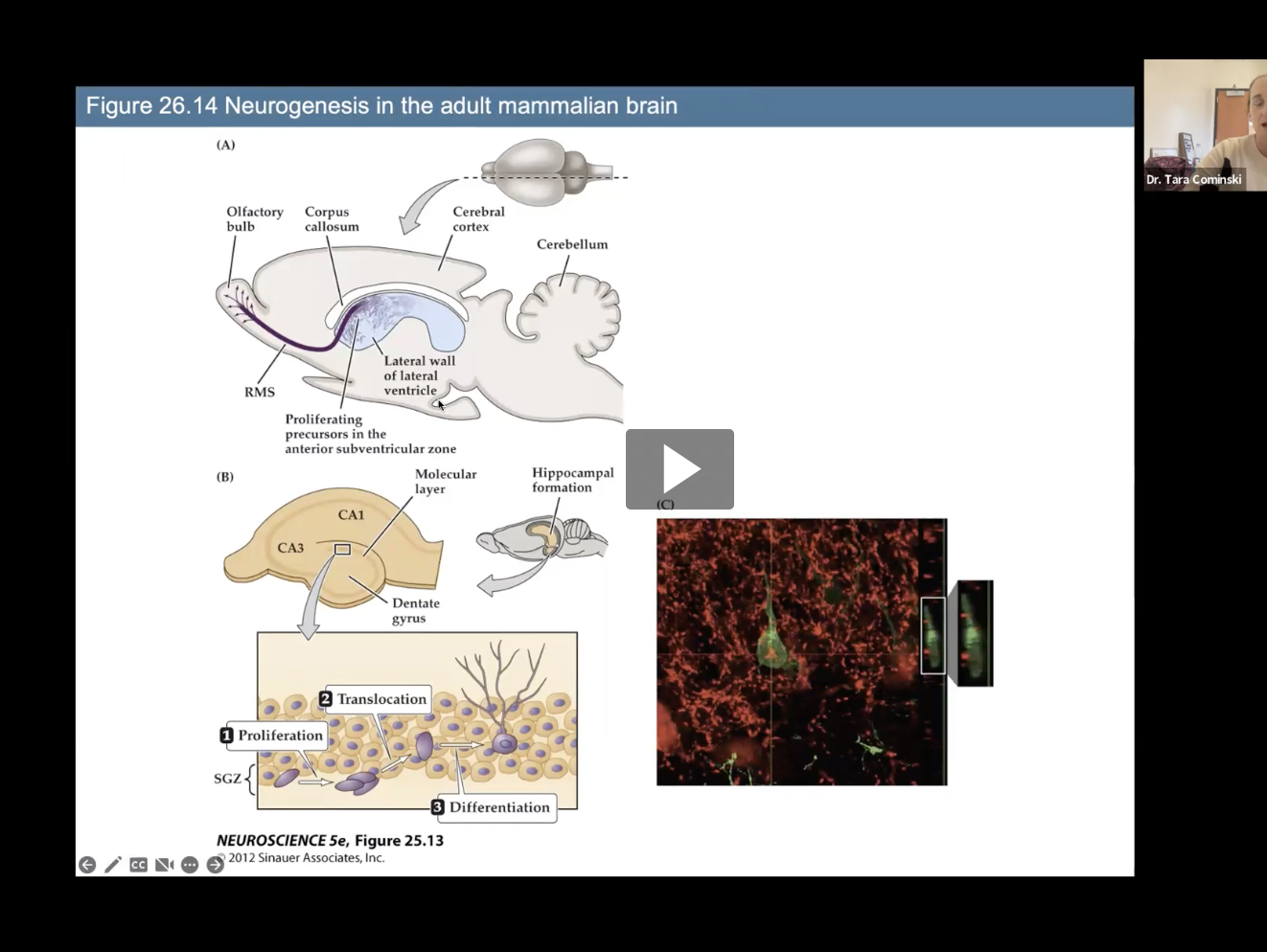
Fig 26.14- Neurogenesis regions in mammalian brain
Subventicular Zone
Dentate Gyrus of hippocampus
3 Factors that impact neurogenesis in the Dentate Gyrus
Exercise and anti-depressants increase cell proliferation in the DG
Stress decreases cell proliferation in the DG
Drugs of abuse, including opiates, decrease cell proliferation in the DG