Development Lecture 3
1/46
There's no tags or description
Looks like no tags are added yet.
Name | Mastery | Learn | Test | Matching | Spaced | Call with Kai |
|---|
No analytics yet
Send a link to your students to track their progress
47 Terms
How do we find out what is present in egg cells
Must all be from what mother put in:
Systematically create mutations in the maternal genome
screen for mutations that lead to the production of defective eggs and embryos
→ identify all the genes concerned
NEXT: show how their products operate in egg cells
Experiment hypothesis and test
HYpothesis: the egg contains fine-grained map of future cell fates in the form of cytoplasmic determinants
Experiment: test the genome of the mother→ she is responsible for making the egg cell→ use mutations
What are these mutants called
Maternal Effect Mutations
mutations that have their effect only when they are present in the mother
NO effect if they are carrier by the father
Female mutation x male wild type=
Mutation/wild type defected embryo
Female wild type x male mutation=
mutation/wild type normal embryo
Results
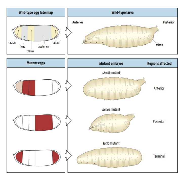
Not a fine-grained map
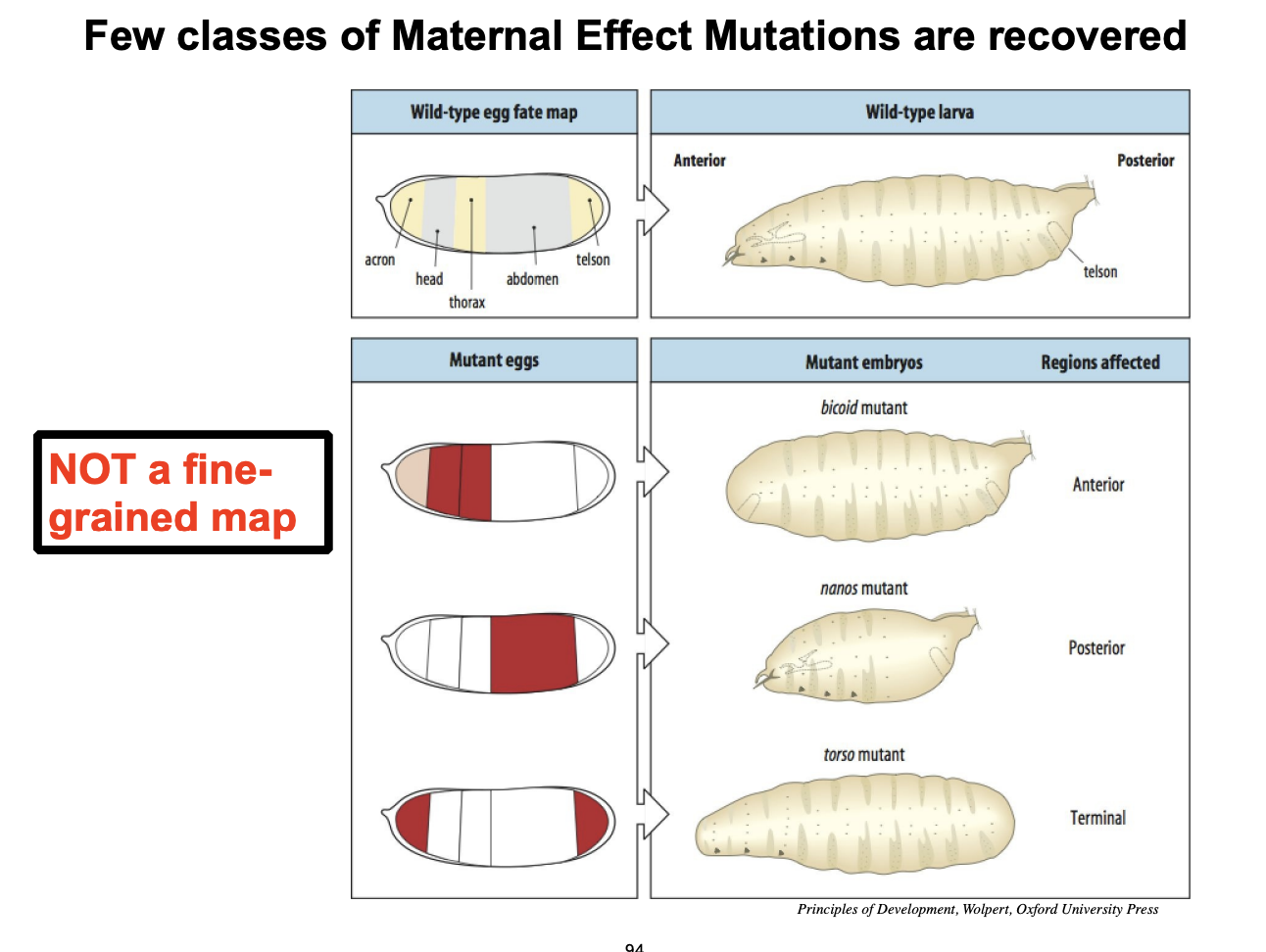
Examples of mutant phenotypes recovered in a screen for maternal mutations in Drosophila
Bicoid→ anterior strucutures
nanos→ posterior strucutures
torso→ terminal
In each case→ embryos/larvae produced LACK these particular strucutures
What does the bicoid encode for
maternal determinant for anterior strucutures
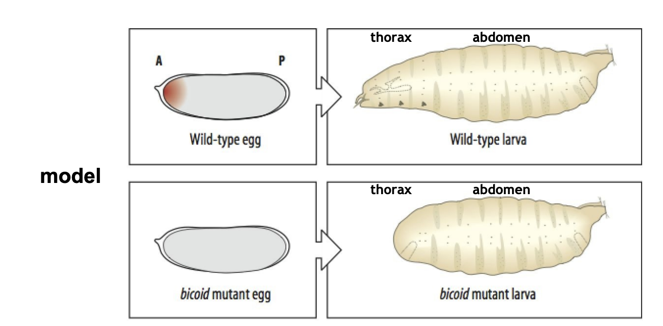
How can we test how genes like bicoid act
Cytoplasm transfer
How this works
transplnatation of anterior cytoplasm
→ rescues and induces anterior strucutures in bicoid mutants

How we know that bioid encodes something for the anterior?
if anterior transferred to the middle of the bicoid mutant egg
the head forms in the middle!
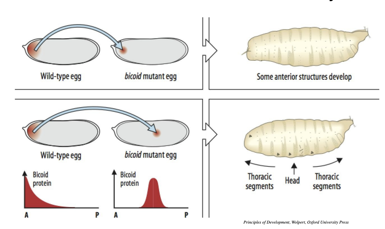
What do the gradients in the diagram suggest?
SHow the active substance→ bicoid protein
localised in the anterior part of the egg cell
this shows that not only is bicoid protein necessary but also sufficient in forming anterior/head in the cell

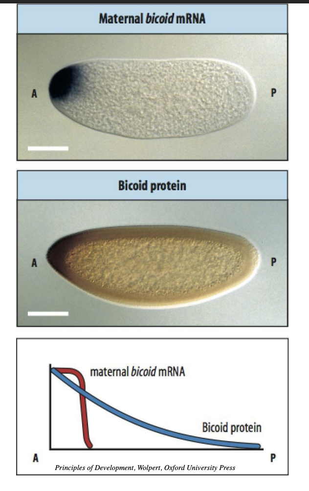
How was it confirmed that it is localised to the anterior part of the egg cell?
Visualisation (in situ ybrisiation)
of bicoid RNA and protein in the egg cell
→ RESULT: localised to the anterior pole and diffusing away from it in a graded fashion
Shows that bicoid works as a switch. Once there is enough of it to get to a threshold→ switches on the hunchback gene to half of the cell

Overall how does bicoid work?
Bicoid codes for transciption factor
maternalling generated bicoid mRNA deposited at anterior tip
translation leads to gradient of Bicoid protein
Bicoid protein leads to activation anteriorly of zygotic gene→ hunchback (i.e working as a trasnciption factor)
Bicoid is ina gradient
over a line→ causes a straight line of hunchback
If bicoid is sufficient to trigger anterior formation, how many other such factors does the maternal genome encode and put into the egg?
Does the egg contain a fine map of determinants for all the future strucutures of the larva?
Do a maternal screen for mutations:
RESULT: not such map, instead:→ 4 classes of mutants
What are the 4 classes of determinants
Formation of germ cells→ oskar
anterior-posterior axis→ biocoid
Dorso-ventral axis→ dorsal
termini of the embryo
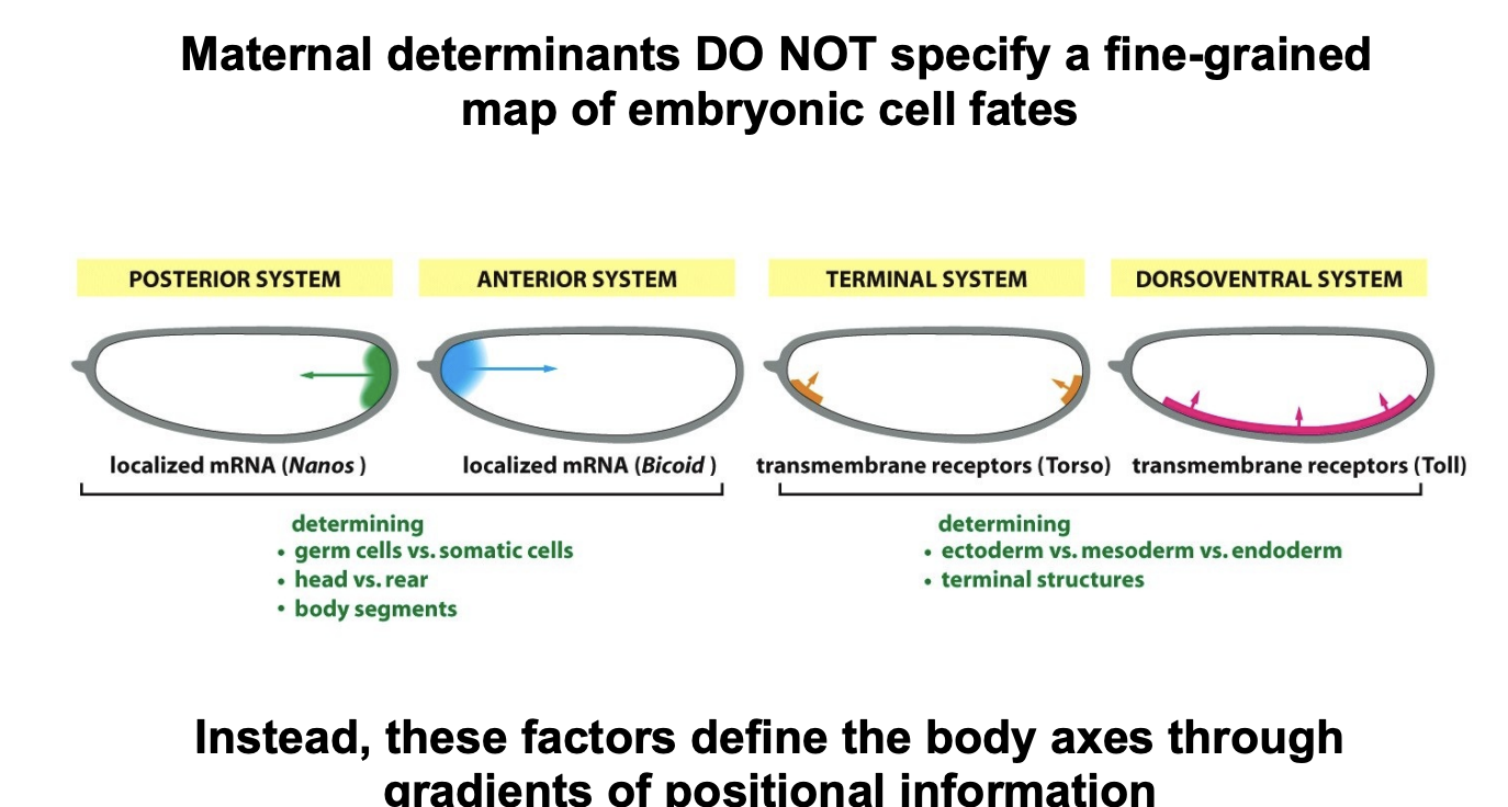
Overall, what does this say about the role of the maternal genome?
sets coordinate for future development
laying out the a-p and d-v axese
When are the finer details (assignment of cells to form particular strucutures), made?
After the Mid-Blastula Transition
as the zygotic genome becomes active!
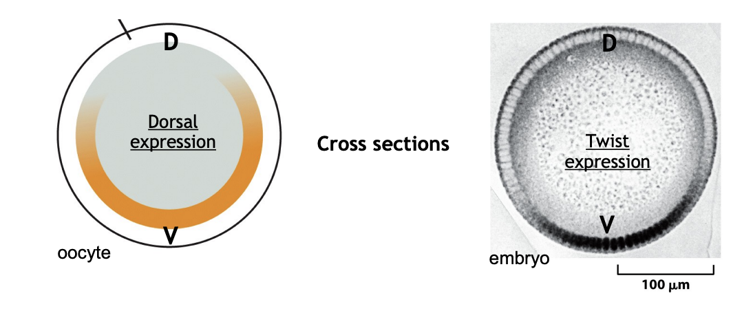
How does maternal productions form dorsal-ventral axis in Drosophila
Trasnciption facotr Dorsal
leads to activation of the zygotic gene twist
in the nuclei of the most ventral cells
Expression of twist→ ventral cells to form medoerm
But where do these transcription factors know where to go?
need to look back in the maternal material
and how the egg is made
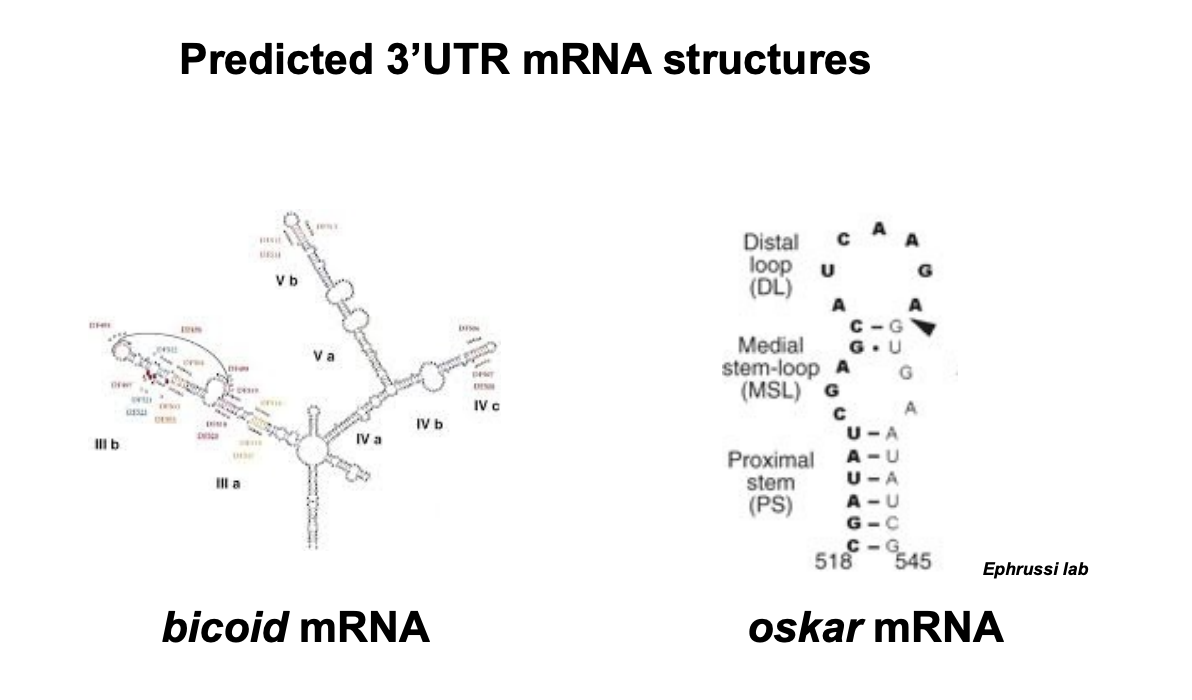
How are bicoid and askar used at different ends? (how are they localised)
the mRNA of both segregate to oppsotide ends
via microtubules of the oocyte
bicoid mRNA→ anterior
oskar→ mRNA to the posterior
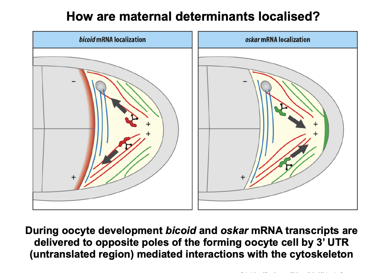
What does the specificity of this interaction depend on?
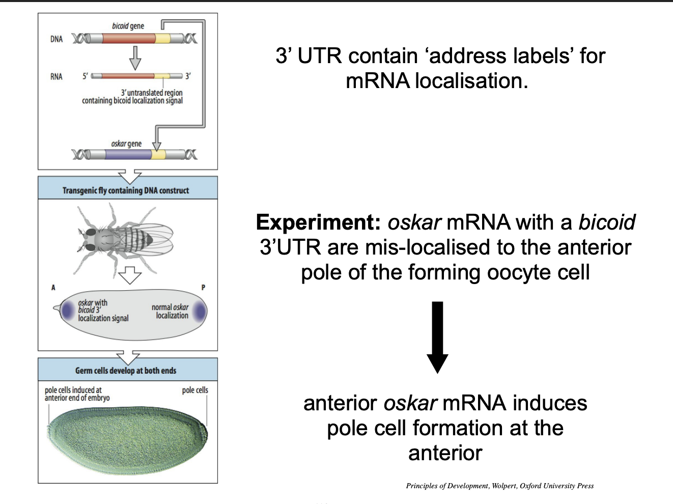
3’ untranslated regions (3’ UTR) of the transpits
How do we know this?
Experiment:
oskar transgene fitted with bicoid 3’ UTR sequences
Result:
oaskar mRNA localised with bicoid mRNA
pole cells then form at the anterior end
How do you polarise an egg?
how do you start to get the singularities in an otherwise uniform cell
from which subsequence differeneces then unfold
Example 1→ Fucus
Example 2→ Xenopus
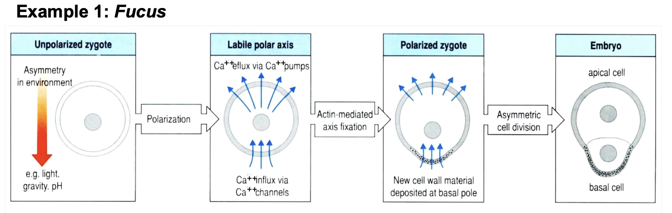
E.g 1: Fucus→ facts
Common sea weed Fucus
Sheds eggs into sea water→ which are fertilised
E.g 1 Fucus→ what happens to the egg?
uniform sphere for the first few hours
polarises:
Rresult after first cell division:
one Apical cell→ produce the frond on the sea weed
one Basal cell→ make the hold fast

E.g 1→ how is this apico-basal polarity set?
cell responds to environmental cues
Light, pH. gavity
redistributing pumps and leaks of Ca2+
OVERALL→ uniform flux of Ca2+ across the membrane→ beomes focussed as the current flowing in the apino-basal axis
Small initial differences in membrane charge can be amplified by positive feedback loops
E.g 1: Fucus polarity
light (or other external signal)
causes electrical depolarisation→ causes the apical to become negative and the basal to become positive
Accumulation of charge Ca2+ pumps and leaks
Ca2+ enters the basal and leaks out of the apical
Overall reinforces itself→ amplification
Waves cause recruitment of material at the basal pole
Causes asymmetric cell division
OVERALL→ differentiated directly,
12 hours→ fast!
Example 2: Xenopus→ the two axes
Animal-vegetal axis
Ventral-dorsal
→ Seen on the egg as it has camoflage a bit like the frog itself→ dark on top and light on the bottom
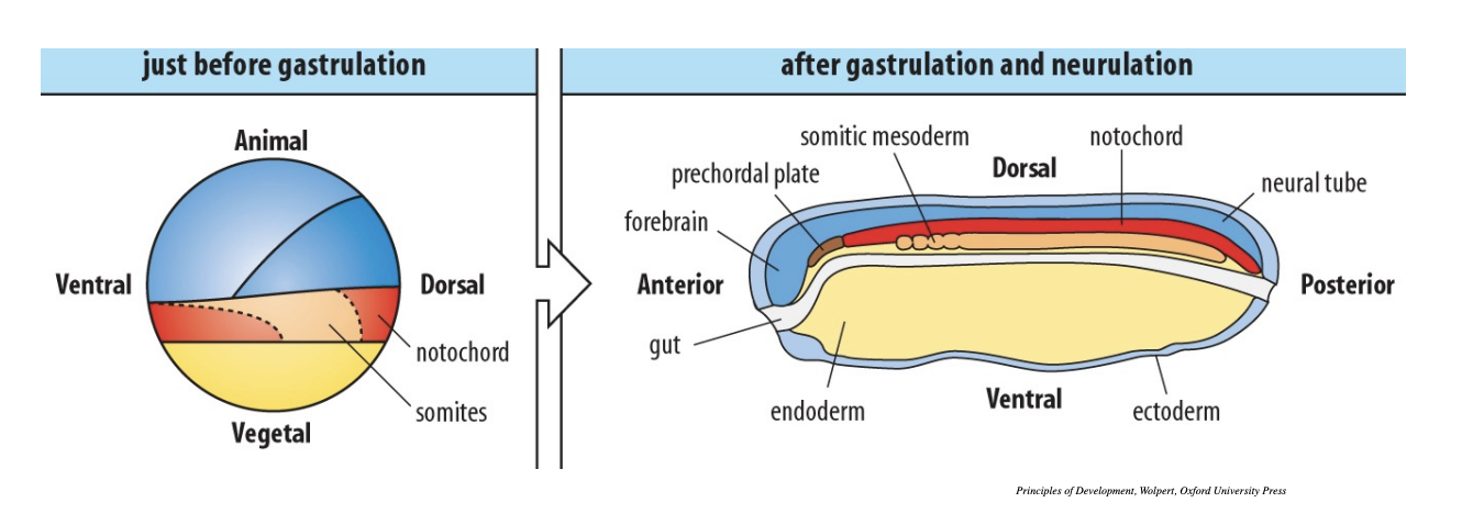
Axes before and after gastrulation
Before gastrulation:
dorsla-and ventral look like the anterior and posterior but they are what will become the dorsal and ventral!
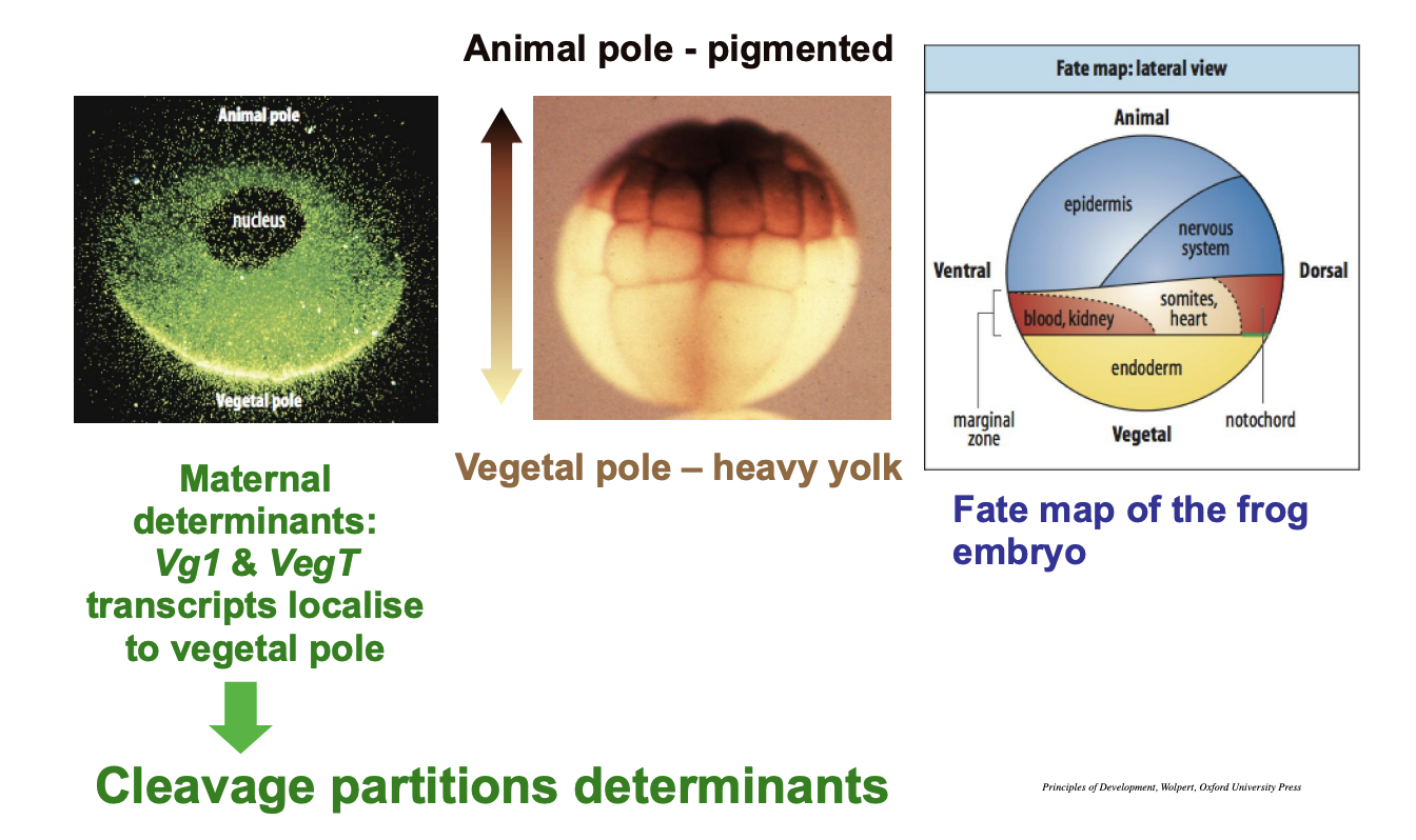
Animal-vegetal A-V axis
Animal hemisphere
hald of an egg/ embryo that contains less yolk
divides more rapidly compared to vegetal
In eggs with considerable yolk
animal hemisphere will be the upper half
Why?→ what is the environmental cue?
Gravity
Egg floats in the water
heavy yolk falls to the lower pole
les yolky cytoplasm towards the animal pole
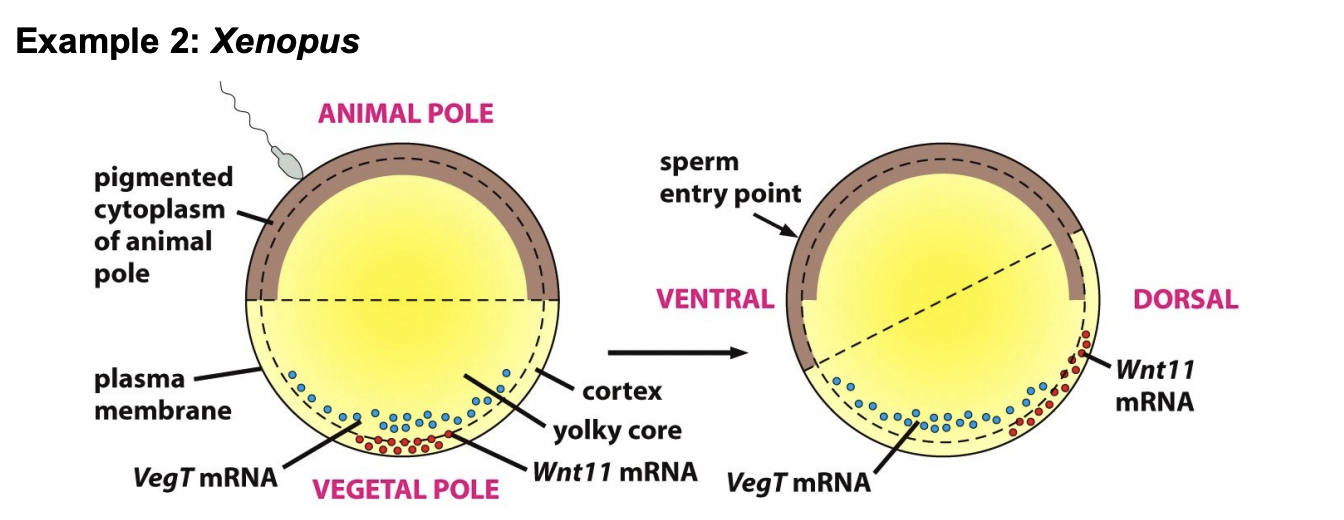
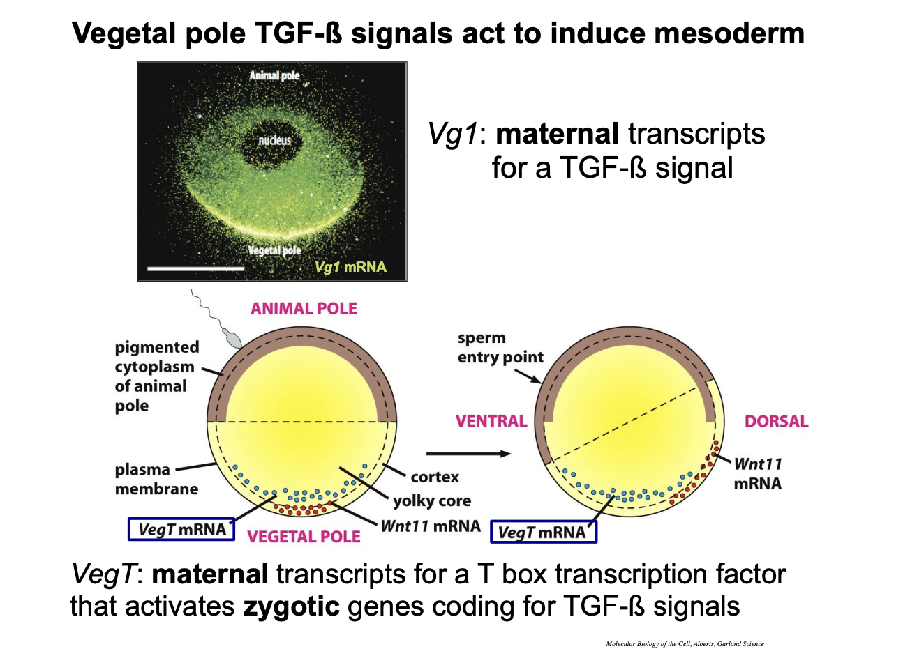
What actually sets the animal-vegetal axis
mRNAs localising due to gravity
How is this shown in experiments?
Centrifuging or shaking eggs
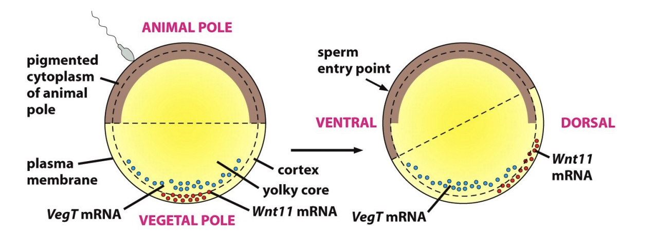
transcipts like VegT RNA→ associated in the region with the yolk at the vegetal pole

What did this experiemnt also show?
Wnt11 RNA→
closer to the plasma membrnae in the cortical cytoplasm
more freely able to move
→ This hints to its use in polarising role for the doral central axis!
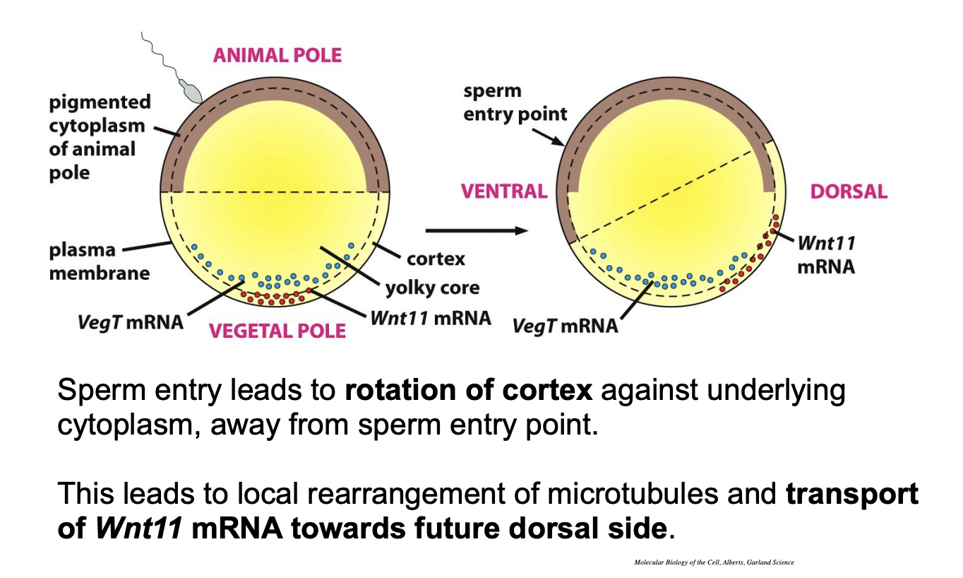
How is the dorsal ventral axis polarity made?
Environmental stimulus→ Entry of the sperm
triggers mechanism→ rotates the cortical cytoplasm 30 degrees away from sperm entry
vegetally located transcipts like Wnt11 are shifted away from entry point
Asymmetric redistribution→ sets future dorso-ventral axis
How?→ through the overlaying of the different signals
dorsalising signals→ cortex is just inside the cytoplasm
but the vegt does not move
roates to be opposite side from where sperm enter
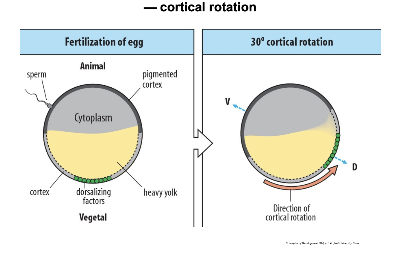
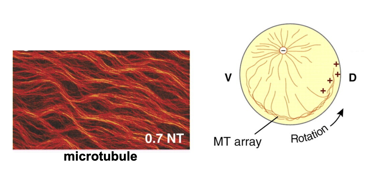
Which parts rotate→ how is this shown?
The cortex against the underlying cytoplasm
BUT
The stuff above does not (the yolky core)
Shown by→ microtubules
Many mitotbules at the bottom for rotation→ not so mant at the apex→ which stays the same
The diagram shows how the bottom Wnt11 rotates
BUT
the VegT do not roate ontop of this inthe yolky core
Microtubules
What are the finer details of thegene expression dictated by?
by the zygote
once the embryp has cellularised
What dictates the patterns of cell differentiation at this stage?
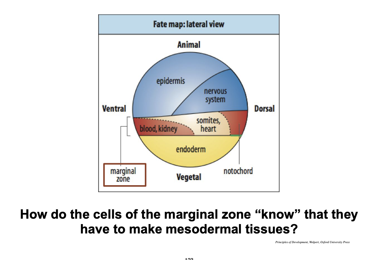
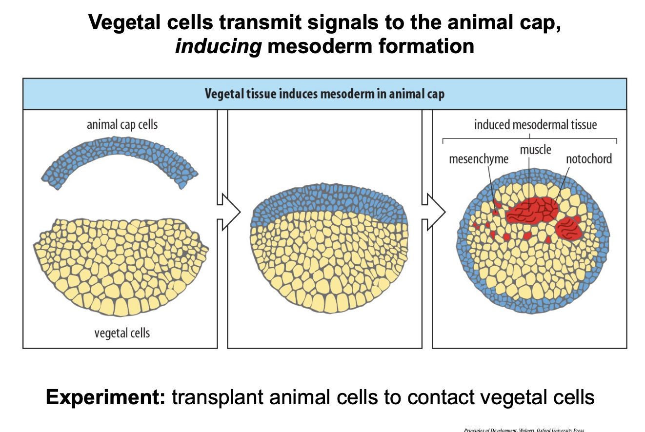
See image→ how do the cells in the marginal zone (which makes blood, somites heart etc)→ know that they have to make mesodermal tissues??
cell-cell interactions→ shown in the fate map of amphibians
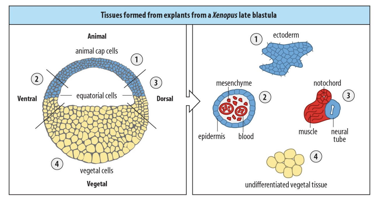
Experiment→ took apart each part of the egg cell
Conclusions
Cells that are found are in contact with animal and vegetal cells
Form into the mesoderm cells
→ The ones on their own→ just ectoderm or endoderm
Suggested that there are no mesodermal determinants but that they are just incontact?
Mesoderm only when two halves of the embryo come into contact→ INDUCTION→ EXTRINSIC!
Next experiment to test this
Experiment
Put animal cap cells directly onto vegetal cells
transplant
Result
cells from the most animal pole can be
Induced mesodermal tissue!
Conclusion:
vegetal cells are the source of signals that ‘tells’ animal cells to make mesoderm
What are the signals involved?
e.g Vg1 (used earlier to transcitp the vegetal half)
encodes a signal of the TGF beta family of growth factors
What is the blastocoel
fluid filled cavity in the embryo
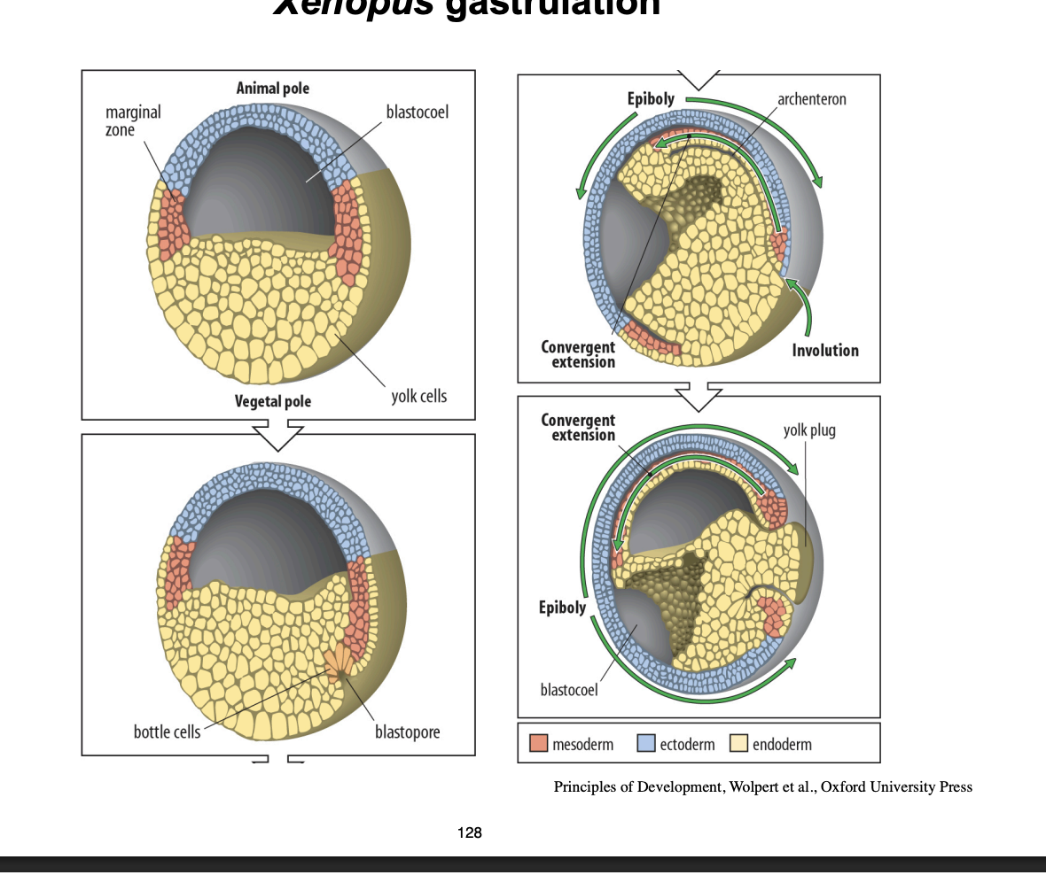
Gastrulation
Tissue movements which turn 2D→ 3D
Bottle cells move inward to form the doral lip of the blastopore
archenteron and cell migrate into the embryo
Moves inside out!
Displaces the blastocoel
Well established cellular movements in frog gastrulation:
involution
conergent extension
converge back to back which enxtends them
epiboly
AT THE END:
blastocoel gone
embryo surrounded by ectoderm
endoderm is internalised
mesoderm is between these
Overall result of gastrulation
shifts the germ layers into 3D shape:
ectoderm on outside
then mesoderm
then endoderm interntalised
No more blastocoel
Archenteron made→ forms the gut