Vital Signs : Pulse
 Call Kai
 Learn
Learn Practice Test
 Spaced Repetition
 Match
Match Flashcards
 Knowt Play
1/57

There's no tags or description
Looks like no tags are added yet.
Name | Mastery | Learn | Test | Matching | Spaced | Call with Kai | Chat |
|---|
No analytics yet
Send a link to your students to track their progress
58 Terms
The rhythmic dilation of the arteries and pulsation of blood flow that occurs with each contraction of the left ventricle.
Pulse
The intrinsic pacemaker of the heart, consisting of a small group of specialized cells in the right atrium that produces electrical impulses.
Sinoatrial (SA) node
A node located near the ventricles of the heart that receives electrical impulses sent from the SA node.
Atrioventricular (AV) node
Mechanism of heart rate generation
Electrical impulses generated by the SA node travel to the AV node and cause the heart muscle to contract, thereby establishing the pulse.
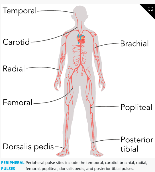
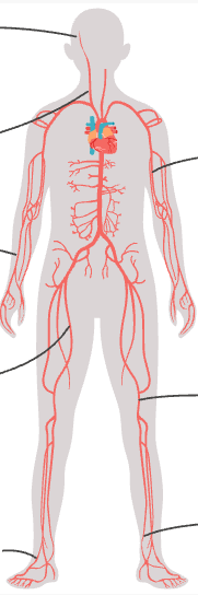
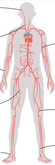
Peripheral pulse sites
temporal, carotid, brachial, radial, femoral, popliteal, dorsalis pedis, and posterior tibial
temporal
along the hairline of the temple
carotid
either side of the neck


brachial
antecubital area

radial
on the thumb side of wrist


femoral
groin area

popliteal
behind the knee


dorsalis pedis
top of foot
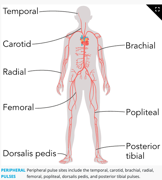

posterior tibial
behind the inner ankle

Bradycardia
A heart rate that is less than the expected reference range. (Low pulse)
tachycardia
Heart rate above the expected reference range. (high pulse)

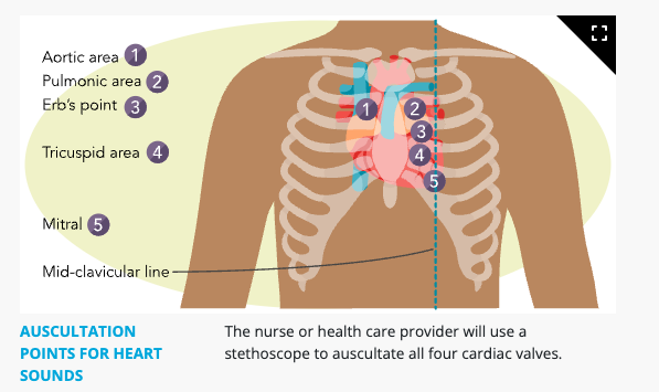
name the auscultation Points for Heart Sounds
The intrinsic pacemaker of the heart, consisting of a small group of specialized cells in the right atrium that produces electrical impulses.
Sinoatrial (SA) node
A node located near the ventricles of the heart that receives electrical impulses sent from the SA node.
Atrioventricular (AV) node
Mechanism of heart rate generation
Electrical impulses generated by the SA node travel to the AV node and cause the heart muscle to contract, thereby establishing the pulse.
Expected pulse reference range: Newborn (birth to 4 weeks)
110 to 160/min
Expected pulse reference range: Infant (1 month to 12 months)
90 to 160/min
Expected pulse reference range: Toddler (1 to 2 years)
80 to 140/min
Expected pulse reference range: Preschooler (3 to 5 years)
70 to 120/min
Expected pulse reference range: School-age (6 to 12 years)
60 to 110/min
Expected pulse reference range: Adolescent (13 to 18 years)
50 to 100/min
Expected pulse reference range: Adult (18 years and older)
60 to 100/min
Peripheral pulse sites
Locations where peripheral pulses can be palpated, including temporal, carotid, brachial, radial, femoral, popliteal, dorsalis pedis, and posterior tibial pulses.
An irregular heart rhythm or a pulse rate that falls outside of the expected reference range.
Arrhythmia
Nursing action for an irregular pulse
Auscultate the apical pulse for 1 full minute to confirm the irregularity and then notify the healthcare provider for further evaluation.
The heart rate heard or felt over the apex of the heart, also known as the point of maximal impulse.
Apical pulse
Apical pulse location in adults and adolescents (7 years of age and older )
Located at the fifth intercostal space at or just medial to the left midclavicular line.
Apical pulse location in infants and young children (<7 years age)
Located at the fourth intercostal space, lateral to the left midclavicular line.
A dull, low-pitched 'lub' sound created by the closure of the mitral and tricuspid valves; best auscultated using the bell of the stethoscope.
S1 heart sound
A shorter, higher-pitched 'dub' sound created by the closure of the aortic and pulmonic valves; best auscultated using the diaphragm of the stethoscope.
S2 heart sound
Aortic valve auscultation site
Second intercostal space (ICS) on the right side of the chest at the right sternal border.

Pulmonic valve auscultation site
Second intercostal space (ICS) on the left side of the chest at the left sternal border.

Tricuspid valve auscultation site
Fourth intercostal space (ICS) on the left side of the chest at the left sternal border.

Mitral valve auscultation site
Fifth intercostal space (ICS) at the left midclavicular line.
same as Apical pulse/APEX of the heart
Auscultation points for heart sounds
The specific anatomical landmarks used to listen to cardiac valves: Aortic (2nd ICS, right sternal border), Pulmonic (2nd ICS, left sternal border), Tricuspid (4th ICS, left sternal border), and Mitral/Apical (5th ICS, left midclavicular line).
Pulse deficit
The difference between the apical pulse rate and a peripheral pulse rate (usually radial) measured over 1 minute.
Causes of a pulse deficit
Aortic rupture, coronary artery disease, or atrial fibrillation.
Procedure for assessing a pulse deficit
Two nurses count simultaneously for 1 minute—one auscultates the apical pulse while the other palpates the radial pulse—and then compare the results.
Finding that confirms a pulse deficit
A radial pulse rate that is lower than the apical pulse rate.
Nursing action when a pulse deficit is detected
Document the findings in the client's medical record and notify the healthcare provider.
Physiological indication of a pulse deficit
A decrease in ventricular contraction or peripheral perfusion.
Formula for calculating a pulse deficit
Apical Pulse Rate−Radial Pulse Rate=Pulse Deficit
Client factors to assess prior to measuring pulse rate
Ensure the client is relaxed and has not exercised or used nicotine in the past few minutes.
Technique for palpating a peripheral pulse
Use the pads of two or three fingers to apply gentle pressure over the pulse site.
Effect of applying excessive pressure during pulse palpation
Obliterates the pulse, making it nonpalpable.
Pulse strength rating: 0
Absent / nonpalpable pulse
Pulse strength rating: +1
Weak / diminished pulse
Pulse strength rating: +2
Normal pulse
Pulse strength rating: +3
Increased / strong pulse (expected finding)
Pulse strength rating: +4
Bounding pulse
Assessment technique for bilateral peripheral pulses
Compare findings from both sides of the client's body simultaneously to determine if pulses are equal bilaterally.
Physical manifestations of impaired peripheral circulation
Cool skin temperature or alterations in skin color.
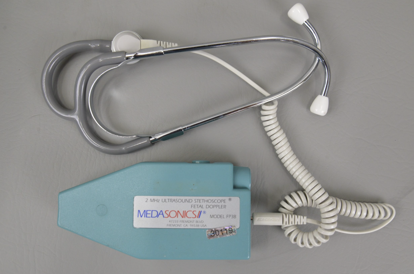
A stethoscope fitted with an audio unit and a transducer that amplifies vascular or other sounds of the body. Used when there is difficulty finding a pulse
Doppler ultrasound stethoscope (DUS)