IMED1001 - Muscle Anatomy I
1/23
There's no tags or description
Looks like no tags are added yet.
Name | Mastery | Learn | Test | Matching | Spaced |
|---|
No study sessions yet.
24 Terms
Skeletal Muscle
- muscles attached to the bones of the skeleton
- msucle cells are called muscle fibres
- striated muscle
- mostly under voluntary control but involuntary movements can occur with skeletal muscle e.g twitching
- multi-nucleated (multiple nuclei in the periphery)
DIAGRAM
Cardiac Muscle
- only found in the walls of the heart
- striated muscle with intercolated disks
- under involuntary control
ADD DIAGRAM
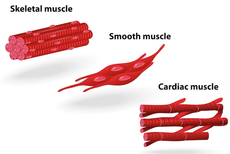
Smooth Muscle
- found mainly in the walls of the hollow organs e.g stomach
- no striations and single nucleus per muscle cell
- under voluntary control
ADD DIAGRAM
Somatic Nervous System
- innervates the skeletal muscles (voluntary control)
Autonomic Nervous System
- innervates viscera (smooth and cardiac muscle) (unconscious control)
Muscle functions (no need to name all)
- facilitate movement
- stabilise joints
- maintain posture
- propel substances around (and in/out of) the body
- generate heat
- protect some visceral organs
- glycaemic control
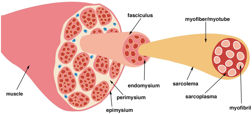
Skeletal Muscle Composition
- each muscle is comprised of muscle fascicles, which are formed of muscle fibres (myocytes = muscle cells)
- In addition to muscle fibres, skeletal muscle is formed of: blood vessels (an artery and vein/s, i.e it is vascular. It is also formed from nerves i.e its innervated by the nervous system. Also made of connective tissue
Skeletal Muscle Coverings
1. Epimysium: firbous CT sheath surrounds the entire muscle. Outer surface grades into the fascia. Its inner surface projects between fascicles to form the perimysium
2. Perimysium: This is a thicker CT sheath that wraps muscle fibres together in bundles. Passage for nerves and blood vessels as well as stretch receptors (muscle spindles)
3. Endomysium: Thin loose CT surrounding individual muscle fibres (myocytes). Creates room for capillaries and nerve fibers to stimulate and nourish each cell. The endomysium is external to the sarcolemma (plasma membrane).
Ways that skeletal muscles can be anchored
- Directly: epimysium is fused to the periosteum of the bone or perichondrium of cartilage
- Indirectly: CT coverings (epimysium) extends beyond the muscle. Creates rope like tendon or sheetlike aponeurosis, which attach the muscle to bone or ligament.
Tendons and aponeuroses may also attach the muscle to a fascia
- Retinaculum: also form to help hold tendons (and therefore the muscle) closer to the bone
- Tendon: a flexible (but inelastic) cord of fibrous collagen tissue attaching a muscle to a bone
- Aponeurosis: a flattened tendon of collagen fibres arranged in a regular weave
- Fascia: collagenous fibres arranged in an irregular weave. Withstands stress in multiple directions
- Retinaculum: restraining band or ligament
ADD IMG
Myocyte Composition
- Organelles (lots of mitochondria)
- Nuclei (multiple, peripheral)
- Bundles of contractile myofilaments (formed of actin and myosin)
- Myofilament arrangement gives striated appearance
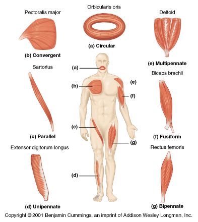
Fascicle Organisation
- Fascicles = bundle of muscle fibres. Due to fascicle arrangement, muscles are classified as:
- Circular (orbicularis oris)
- Convergent (pectoralis major)
- Parallel (Straight, Rectus)
- Pennate (Multipennate = deltoid, Unipennate)
- Fusiform (rectus femoris)
PUT DIAGRAM

Circular (Fascicles)
arranged in concentric rings; close by contracting; called sphincters
e.g oculi, orbicularis, orbicularis oris
Convergent (Fascicles)
Broad origin. Fascicles converge toward a single tendon for insertion. Triangular or fan-shaped
e.g pectoralis major

Parallel (Fascicles)
- fascicles arranged parallel to long axis of the muscle
e.g sartorius and rectus abdominis
ADD DIAGRAM
Pennate (Fascicles)
short fascicles attach obliquely and attach to a central tendon
- Unipennate: fascicles insert only one side of tendon. e.g digitorum longus
- Bipennate: fascicles insert on opposite sides of tendon e.g rectus femoris
- Multipennate: many fascicles together. e.g deltoid muscle
ADD DIAGRAM FROM SLIDES 20, 21, 22

Fusiform (Fascicles)
- Muscles are thick in the middle and tapered at the end
How Skeletal Muscle is named
based on:
- location (may indicate bone or body region where muscle is located, e.g temporalis)
- shape (distinctive shape may be captured by name. e.g orbicularis, rhomboideus major)
- size (maximus/major (largest), minimus/minor (smallest), longus (longest), brevis (shortest), e.g pectoralis major)
- direction of muscle orientation (Rectus (striaght), oblique, transversus (transverse), e.g rectus femoris)
- number of origins (Bi = two, tri = three, quad = four)
- location of attachment ("origin" = first part of name, "insertion" = last part of name, e.g sternocleidomastoid is attached from sternum up to clavicle and back of the skull)
- muscle action (flexor, extensor, opponens (opposable thumb), abductor, adductor)
Agonist
muscle that provides the major force for specific movement
e.g quadriceps femoris extends the leg at the knee joint
Antagonist
muscle that oppose or reverse a particular movement. e.g biceps femoris flex the leg at the knee joint
Agonist and Antagonist
- muscles that work together. Located on opposite sides of a joint. As one contracts the other must relax
Synergists
muscle that produce power to aid the agonist or modify the direction of the movement
Fixators
muscle that stabilise a movement by holding bones in place
Great resource
Through UWA Library (OneSearch) use the Visible Body App Human Anatomy is the one to use. there are some quizzes for IMED1001
Another Great Resource
Register for Lippincott Health Library (Premium Basic Science)