spinal code
 Call Kai
 Learn
Learn Practice Test
 Spaced Repetition
 Match
Match Flashcards
 Knowt Play
1/38

There's no tags or description
Looks like no tags are added yet.
Name | Mastery | Learn | Test | Matching | Spaced | Call with Kai | Chat |
|---|
No analytics yet
Send a link to your students to track their progress
39 Terms
cervical region
(C1-C8)This region is responsible for innervating the neck, shoulders, arms, and hands. It plays a crucial role in motor control and sensory perception for the upper body.
thoracic region
(T1-T12)This region innervates the chest and abdominal muscles. It is involved in the control of the trunk and the autonomic functions of the body.
lumber region
(L1-L5)This region is responsible for innervating the lower back, hips, and legs. It plays a significant role in motor control and sensory perception for the lower body.
Sacral Region
This region innervates the pelvic organs and lower limbs. It is crucial for functions related to bowel, bladder, and sexual function.
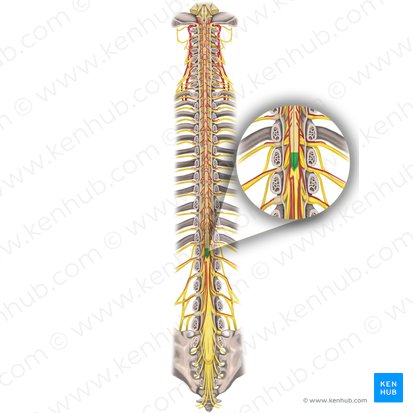
medullary cone
is the tapered, cone-shaped terminal end of the spinal cord. In adults, it is typically located between the L1 and L2 vertebrae,
Motor/sensory for lower limbs, pelvis, perineum; parasympathetic for pelvic organs
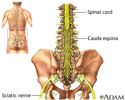
Cauda equina
Below L1, in lumbar cistern
Motor, sensory, and parasympathetic innervation to lower limbs, pelvis, and perineum
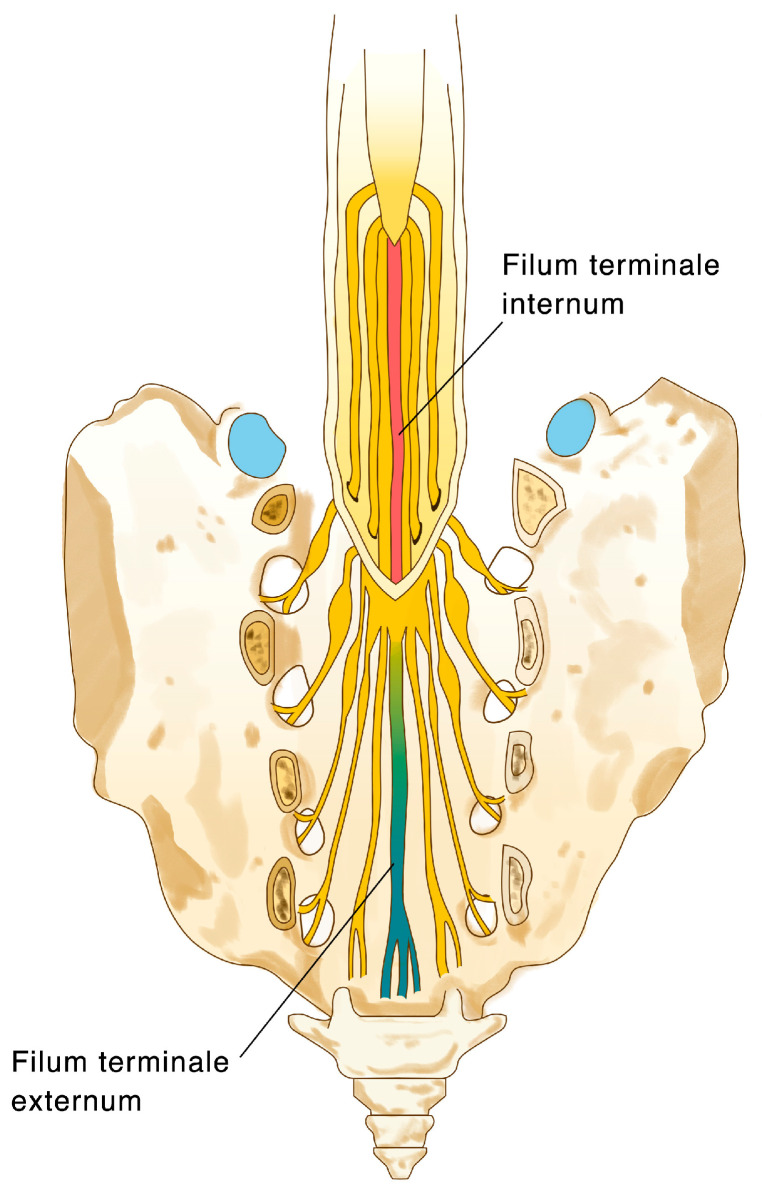
Filum terminale
From conus tip to coccyx
Anchors and stabilizes the distal spinal cord
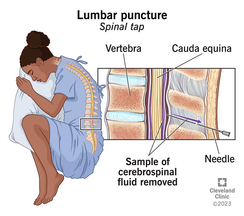
spinal tap
A spinal tap, also called a lumbar puncture (LP), is a medical procedure in which a needle is inserted into the lower back, into the space between two lumbar vertebrae, to collect a small sample of cerebrospinal fluid (CSF) for testing or to measure its pressure
low risk, cause needle cant reach spinal code, the needle is made to be less trauma into the back.
gray matter for spinal code
the core of the nerve. has axon, dendrites, and glial cells. it has horns.
white matter of the spinal code
outer side of the code. has columns
ganglion
PNS. Ganglia serve as relay stations for sensory or autonomic signals.
Nucleus
A nucleus is a group of neuron cell bodies. CNS. ex/ hypothalamic
Nerve
bundle of axon in the PNS. ex/ Optic nerve, Sciatic nerve
Tract
bundle of axon in CNS that transmit info. ex/ spinothalamic tract
Dorsal root
Contain afferent (sensory) fibers that carry information from the periphery (skin, muscles, joints) into the spinal cord.
Ventral root
Contain efferent (motor) fibers that carry motor commands from the CNS to skeletal muscles
Dorsal Root Ganglion
Neurons are first-order somatosensory neurons that detect touch, vibration, proprioception, pain, and temperature.
Spinal Nerve Formation
Spinal nerves exit the vertebral canal via intervertebral foramina.
They are mixed nerves (somatic motor, sensory, and autonomic fibers) that connect the CNS to the periphery
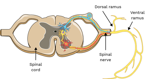
Dorsal ramus
Supplies motor innervation to deep (intrinsic) back muscles and sensory innervation to skin of the posterior head, neck, and back.
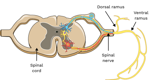
ventral ramus
Supplies motor innervation to most trunk and limb muscles and sensory innervation to skin of the anterior and lateral trunk and limbs.anterior of spinal nerve
Endoneurium
Provides insulation and support to the axon, and contains endoneurial fluid—a low-protein liquid similar to cerebrospinal fluid in the CNS—that bathes the axon and helps maintain a blood-nerve barrier. covers each indivisual axon
Perineurium
Protects the fascicle, regulates its internal environment, and contributes to the blood-nerve barrier.
Epineurium
: Provides structural support, cushions the nerve against mechanical injury, and supplies nutrients to the inner layers.
entire nerve
Ascending tract
Ascending tracts are responsible for transmitting sensory information from peripheral receptors to higher brain centers for processing, including touch, pain, temperature, vibration, and proprioception
so send twords brain.
First-order neurons
Located in the dorsal root ganglion, receive input from sensory receptors.
Second-order neurons
Found in the dorsal horn of the spinal cord or brainstem; their axons often decussate (cross to the opposite side).
Third-order neurons
Located in the thalamus, projecting to the primary sensory cortex for conscious perception
Dorsal column-medial lemniscus system
Carries fine touch, vibration, and proprioception.
Spinothalamic tracts
Transmit pain, temperature, and crude touch.
Spinocerebellar tracts
Convey unconscious proprioception to the cerebellum for coordination
Found in the white matter of the spinal cord, organized into dorsal, lateral, and anterior funiculi
Descending tracts
Descending tracts carry motor commands from the brain to the spinal cord, controlling voluntary and involuntary movements
twords muscle
Upper motor neurons
Originate in the motor cortex or brainstem and project to lower motor neurons.
Lower motor neurons
Located in the anterior horn of the spinal cord; directly innervate skeletal muscles
Corticospinal tracts
Control voluntary fine motor movements.
Rubrospinal, reticulospinal, vestibulospinal, and tectospinal tracts
Rubrospinal, reticulospinal, vestibulospinal, and tectospinal tracts: Regulate posture, balance, and reflexive movements
Found in the white matter of the spinal cord, primarily in lateral and anterior funiculi
somatic reflex
receptor, sensory neuron, interneuron, motor neuron, and effector, which work together to produce a rapid, involuntary skeletal muscle response.
monosynaptic
single synapse
polysynaptic
multiple synapses
Muscle spindles
Muscle spindles are encapsulated stretch receptors located within the belly of skeletal muscles, running parallel to the main muscle fibers