Ch 4 - Pt 2: Connective Tissues
 Note
Note Call Kai
 Learn
Learn Practice Test
 Spaced Repetition
 Match
Match Flashcards
 Knowt Play
1/52

Earn XP
Description and Tags
A comprehensive vocabulary review of connective tissue classes, cells, fibers, extracellular matrix components, and tissue characteristics.
Name | Mastery | Learn | Test | Matching | Spaced | Call with Kai | Chat |
|---|
No analytics yet
Send a link to your students to track their progress
53 Terms
Connective Tissue
The most abundant and widely distributed primary tissue in the body, serving functions in binding, support, protection, insulation, energy storage, and transport.
Extracellular Matrix
The nonliving portion of connective tissue composed of ground substance and fibers in which living cells are suspended, allowing tissues to bear weight and withstand tension.
Mesenchyme
An embryonic tissue from which all classes of connective tissue originate.
Ground Substance
Unstructured gel-like material that fills the space between cells and fibers, forming a structural component of the extracellular matrix.
'-blast' Cells
Immature connective tissue cell forms that actively secrete ground substance and matrix fibers.
'-cyte' Cells
Mature connective tissue cell forms that maintain matrix health and can revert to '-blast' cells to repair and regenerate matrix.
Fibroblasts
Immature cells in connective tissue proper that actively secrete matrix components before becoming fibrocytes.
Chondroblasts
Immature cartilage cells found in growing cartilage that secrete matrix before becoming chondrocytes.
Osteoblasts
Immature bone cells that secrete bone matrix components before maturing into osteocytes.
Hematopoietic Stem Cells
Immature stem cells that give rise to blood cells.
Adipocytes
Fat cells in connective tissue that store energy in the form of fat droplets.
Leukocytes
White blood cells, including neutrophils, eosinophils, and lymphocytes, that respond to tissue injury.
Mast Cells
Connective tissue cells that detect foreign microorganisms and initiate local inflammatory responses using secretory granules.
Macrophages
Phagocytic cells that devour foreign materials and dead cells, existing either fixed in tissues or migrating freely.

Proper Loose Areolar Connective Tissue

Proper Loose Adipose Connective Tissue
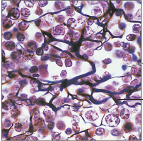
Proper Loose Reticular Connective Tissue
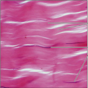
Proper Dense Regular Connective Tissue
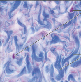
Proper Dense Irregular Connective Tissue

Proper Dense Elastic Connective Tissue
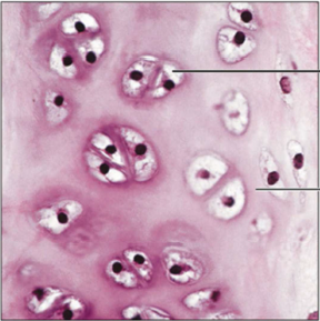
Hyaline Cartilage

Elastic Cartilage

Fibrocartilage
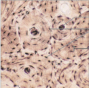
Bone Connective Tissue

Blood
Classes of Connective Tissue
Connective Tissue Proper
Loose - areolar/adipose/reticular
Dense - regular, irregular, elastic
Cartilage
Hyaline
Elastic
Fibrocartilage
Bone (compact and spongy)
Blood

Connective Tissue Proper: Cells, Matrix Components, and Features
Cells: Fibroblasts, fibrocytes, defense cells, adipocytes
Matrix: Gel-like ground substance with all three fiber types (collagen, reticular, elastic)
General Features: Consists of 6 subclasses (loose: areolar, adipose, reticular; dense: regular, irregular, elastic); functions as binding tissue, resists tension/mechanical stress, stores energy, and acts as a reservoir for water and salts
Cartilage: Cells, Matrix Components, and Features
Cells: Chondroblasts (found in growing cartilage), chondrocytes
Matrix: Gel-like ground substance with collagen fibers (and elastic fibers in some types)
General Features: Avascular tissue that resists compression due to high water content; cushions and supports body structures
Bone Tissue: Cells, Matrix Components, and Features
Cells: Osteoblasts, osteocytes
Matrix: Gel-like ground substance calcified with inorganic salts, containing collagen fibers
General Features: Hard tissue consisting of compact and spongy subclasses; resists both compression and tension to support and protect body structures
Blood: Cells, Matrix Components, and Features
Cells: Red blood cells (erythrocytes), white blood cells (leukocytes), platelets
Matrix: Plasma (fluid matrix, lacks fibers)
General Features: Fluid connective tissue contained within blood vessels; functions to transport O2, CO2, nutrients, wastes, and hormones
Three Primary Structural Elements of Connective Tissue
Ground substance, fibers, and cells (with ground substance and fibers together forming the extracellular matrix).
three type of connective tissue fibers and their functions
Collagen Fibers: The strongest and most abundant fibers, providing high tensile strength.
Elastic Fibers: Long, thin elastin fibers that form networks allowing stretch and recoil.
Reticular Fibers: Short, fine, highly branched collagenous fibers that form flexible supportive networks.
Proper Loose Areolar Tissue
Description: Gel-like matrix with all three fiber types (collagen, elastic, reticular) and cells including fibroblasts, macrophages, mast cells, and white blood cells.
Function: Wraps and cushions organs, phagocytizes bacteria, plays an important role in inflammation, and holds/conveys tissue fluid.
Location: Widely distributed under epithelia of body (e.g., forms lamina propria of mucous membranes), packages organs, and surrounds capillaries.
Proper Loose Adipose Tissue
Description: Extracellular matrix similar to areolar tissue but very sparse; contains closely packed adipocytes (fat cells) with nuclei pushed to the side by large fat droplets.
Function: Provides reserve fuel, insulates against heat loss, and supports and protects organs.
Location: Under skin in subcutaneous tissue, around kidneys and eyeballs, within abdomen, and in breasts.
Proper Loose Reticular Tissue
Description: Loose network of reticular fibers in a gel-like ground substance with reticular cells lying on the fibers.
Function: Supports, binds, and cushions organs while facilitating immune defense and nutrient diffusion
Location: Lymphoid organs (lymph nodes, bone marrow, and spleen).
Proper Dense Regular Tissue
Description: Primarily parallel collagen fibers with a few elastic fibers; major cell type is the fibroblast.
Function: Attaches muscles to bones or to muscles, attaches bones to bones, and withstands great tensile stress when pulling force is applied in one direction.
Location: Tendons, most ligaments, and aponeuroses.
Proper Dense Irregular Tissue
Description: Primarily irregularly arranged collagen fibers with some elastic fibers; major cell type is the fibroblast.
Function: Withstands tension exerted in many directions and provides structural strength.
Location: Fibrous capsules of organs and joints, dermis of the skin, and submucosa of the digestive tract.
Proper Dense Elastic
Description: Dense regular connective tissue containing a high proportion of elastic fibers.
Function: Allows tissue to recoil after stretching, maintains pulsatile flow of blood through arteries, and aids passive recoil of lungs following inspiration.
Location: Walls of large arteries, within certain ligaments associated with the vertebral column, and within the walls of the bronchial tubes.
Hyaline Cartilage
Description: The most abundant, glass-like cartilage type; Amorphous but firm matrix with an imperceptible network of collagen fibers; chondroblasts produce matrix and mature into chondrocytes in lacunae.
Function: Supports and reinforces, serves as a resilient cushion, and resists compressive stress.
Location: Forms most of the embryonic skeleton, covers ends of long bones in joint cavities, forms costal cartilages of ribs, and forms cartilages of nose, trachea, and larynx.
Elastic Cartilage
Description: Similar to hyaline cartilage, but contains a higher proportion of elastic fibers in matrix.
Function: Maintains the shape of a structure while allowing great flexibility.
Location: External ear (pinna) and epiglottis.
Fibrocartilage
Description: Matrix similar to but less firm than hyaline cartilage; thick collagen fibers predominate with chondrocytes in lacunae. Strongest type of cartilage.
Function: High tensile strength allows it to absorb compressive shock.
Location: Intervertebral discs, pubic symphysis, and discs of knee joint.
Bone Tissue
Description: Hard, calcified matrix containing many collagen fibers; osteocytes lie in lacunae; very well vascularized.
Function: Supports and protects organs, provides levers for muscles to act on, stores minerals/fat, and houses marrow for blood cell formation (hematopoiesis).
Location: Bones.
Blood
Description: A fluid connective tissue consisting of red blood cells (erythrocytes), white blood cells (leukocytes), and platelets suspended in a fluid matrix (plasma); lacks fibers.
Function: Transports respiratory gases (O2 and CO2), nutrients, wastes, and other substances (such as hormones).
Location: Contained within blood vessels.
What characteristics of Loose Areolar Tissue make it ideal for its function as the most widely distributed proper connective tissue?
Universal “packing material”
Because it contains all three fiber types (collagen, elastic, and reticular) and a mix of many basic cell types (like fibroblasts and immune cells) in roughly equal ratios, it functions as the standard baseline structure for connective tissue proper
Watery gel matrix: Acts like a sponge to store fluids and pass nutrients easily to other tissues.
Loosely woven fibers: Creates a stretchy framework that cushions organs and lets them move without tearing.
Tough collagen threads: Provides the strength needed to fasten skin and organs securely in place.
Open, airy spaces: Leaves plenty of room for immune cells to patrol and fight off infections.
What characteristics of Proper Loose Adipose Tissue make it ideal for its function?
Its unique structure of tightly packed fat cells allows it to act as a shock-absorbing cushion to protect vital organs and an insulating blanket to trap body heat.
What characteristics of Proper Loose Reticular Tissue make it ideal for its function?
Forms a delicate, 3D spiderweb-like framework made of branching fibers that holds soft organs together without blocking fluid flow. This open, mesh-like structure slows down passing fluids so that trapped immune cells can easily filter out pathogens and foreign particles.
What characteristics of Proper Dense Regular Tissue make it ideal for its function?
Thick collagen fibers packed tightly together in perfectly parallel bundles give it immense tensile strength to resist pulling forces along a single direction
What characteristics of Proper Dense Regular Tissue make it ideal for its function?
Thick collagen fiber bundles woven together in a random, interlocking pattern allows it to withstand tension from multiple directions without tearing
What characteristics of Hyaline Catrilage make it ideal for its function?
Glassy, smooth extracellular matrix rich in collagen fibers and bound water provides exceptional shock absorption and a frictionless surface for joint movement
What characteristics of Elastic Catrilage make it ideal for its function?
Packed with a dense network of dark-staining elastic fibers embedded within its matrix, giving it extraordinary flexibility and the ability to repeatedly bend and snap back to its original shape.
What characteristics of Fibrocatrilage make it ideal for its function?
Packed with alternating layers of thick, dense collagen fiber bundles and cartilage cells that allows it to absorb severe compression shocks and resist tearing under heavy pressure
What characteristics of bone tissue make it ideal for its function?
Hard, calcified extracellular matrix reinforced with tough collagen fibers provides the skeleton with exceptional compressive strength, rigidity, and weight-bearing support.
What characteristics of blood make it ideal for its function?
Specialized cells suspended in a liquid extracellular matrix called plasma make it perfectly adapted for rapid transit and body-wide communication. Its fluid nature allows it to continuously circulate, transporting oxygen, nutrients, hormones, and metabolic wastes efficiently to and from every cell.