CH3 PROTEINS MCBWhat is the term for the portion of each amino acid that gives the amino acid its unique properties?
 Call Kai
 Learn
Learn Practice Test
 Spaced Repetition
 Match
Match Flashcards
 Knowt Play
1/19

There's no tags or description
Looks like no tags are added yet.
Name | Mastery | Learn | Test | Matching | Spaced | Call with Kai | Chat |
|---|
No analytics yet
Send a link to your students to track their progress
20 Terms
What is the term for the portion of each amino acid that gives the amino acid its unique properties?
side chain
In the crowded conditions of the cytosol, molecular chaperones not only assist in protein folding, but also help specify the final three-dimensional shape of the protein.
false
Under which one of the following conditions is the complete structure of a protein designated as its quaternary structure?
The protein contains more than one protein subunit.
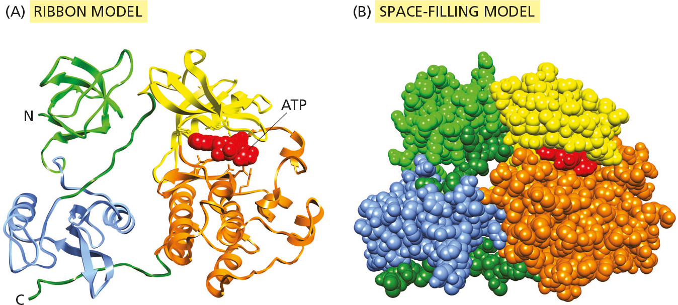
How many protein domains make up the Src protein kinase shown in the figure?
3
The relatively unstructured lengths of polypeptide chain that connect domains of protein can act as flexible hinges between domains.
true
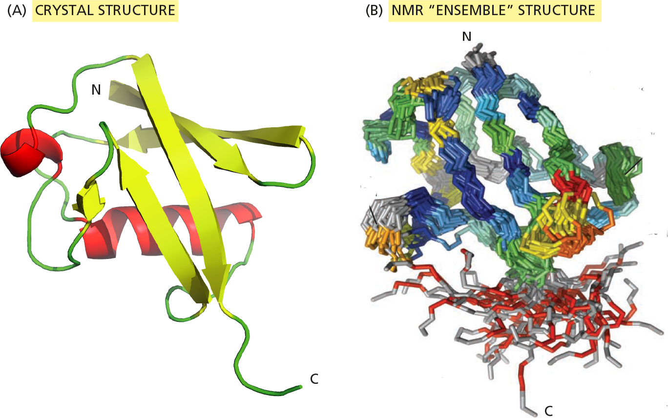
The crystal structure of ubiquitin at 0.18 nm resolution is shown as a ribbon diagram in the figure Part A, colored according to secondary structure. The NMR structure of ubiquitin, with red and yellow indicating high mobility regions of the protein, is shown in Part B.
Is the crystal structure or the NMR structure a more accurate representation of ubiquitin as it exists inside the cell? Why?
The NMR structure, because the atoms in cellular proteins are constantly moving.
A polypeptide chain of any sequence adopts a unique stable conformation that can be useful for the cell.
false

Often, the hard part of protein structure determination by x-ray diffraction is getting good crystals. In difficult cases, there are two common approaches for obtaining crystals: (1) using fragments of the protein and (2) trying homologous proteins from different species.
Examine the protein in the figure. Where would you cleave this protein to obtain fragments that might be expected to fold properly and perhaps form crystals?
point 4
Stiff, extended structures composed of a series of domains arranged in tandem are especially common in extracellular matrix molecules and in the extracellular portions of cell-surface receptor proteins.
Which one of the following types of protein module is more common in such proteins?
In-line modules with their N- and C-termini at opposite poles of the domain
The human genome encodes about 20,000 proteins. For about how many of those proteins do we still not have even the tiniest hint about their function?
10,000
The Cro repressor is a bacterial transcription regulator that binds to DNA to turn genes off. It is a symmetrical “head-to-head” dimer. Each of the two subunits of the dimer recognizes a particular short sequence of nucleotides in DNA.
If the sequence of nucleotides recognized by one subunit is represented as an arrow (→), so that the “head” of the arrow corresponds to DNA recognized by the “head” of the subunit, which one of the following arrangements of arrows represents the binding site for the Cro dimer?
→←
Actin is a major constituent of the cytoskeleton. Which one of the following statements correctly describes actin’s structure and location?
Actin forms helical filaments in the cell.
Proteins whose functions require that they span a long distance generally have a relatively simple, elongated three-dimensional structure.
true
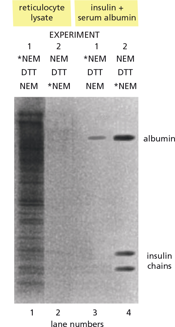
You are skeptical of the blanket statement that cysteines in intracellular proteins are not involved in disulfide bonds, while in extracellular proteins they are. To test this statement, you carry out the following experiment. As a source of intracellular protein, you use reticulocytes, which have no internal membranes and, thus, no proteins from the endoplasmic reticulum (ER) or other membrane-enclosed compartments. As examples of extracellular proteins, you use bovine serum albumin (BSA), which has 37 cysteines, and insulin, which has 6. You denature the soluble proteins from a reticulocyte lysate and the two extracellular proteins so that all cysteines are exposed.
To probe the status of cysteines, you treat the proteins with N-ethylmaleimide (NEM), which reacts covalently with the –SH groups of free cysteines but not with sulfur atoms in disulfide (S–S) bonds. In the first experiment, you treat the denatured proteins with radiolabeled NEM (*NEM), then break any disulfide bonds with dithiothreitol (DTT) and react a second time with unlabeled NEM (see the figure, lanes 1 and 3). In the second experiment, you do the reverse: you first treat the denatured proteins with unlabeled NEM, then break disulfide bonds with DTT and treat with radiolabeled *NEM (lanes 2 and 4). The proteins are separated according to size by electrophoresis on a polyacrylamide gel, which is then placed against x-ray film so that radioactive decay gives rise to the dark bands shown in the figure.
Proteins containing disulfide bonds would be expected to show up as dark bands in which lanes of the figure?
Lanes 2 and 4
Each one the following structures is capable of self-assembly from a solution of its component macromolecules EXCEPT:
cilium
Which of the following statements correctly describe how assembly factors aid the formation of complex biological structures?
I In some cases, assembly factors carry out a proteolytic cleavage that is an essential step in the assembly process.
II In some cases, assembly factors act as chaperones to ensure that subunits are properly folded before they assemble.
III In some cases, assembly factors provide a temporary scaffold for assembly that is not part of the final structure.
I and III
Why do you suppose that the most severe amyloid pathologies are neurodegenerative diseases?
The brain’s organized array of neurons is especially vulnerable.
Which one of the following statements about amyloid fibrils is true?
Many bacteria use amyloid fibrils to create a protective biofilm for survival.
Amino acid side chains in the interior of a protein play no role in the binding of a ligand to the protein.
false
Using evolutionary tracing, you have identified a few clusters of invariant amino acids in members of a protein family. Which one of the following statements about such clusters is correct?
They correspond to binding sites for one or more ligands.