PBL Finals Trimester 1 Combot
1/73
There's no tags or description
Looks like no tags are added yet.
Name | Mastery | Learn | Test | Matching | Spaced |
|---|
No study sessions yet.
74 Terms
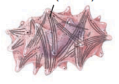
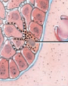
What are the black lines?
Intermediate filament (keratin) (in a keratinocyte cell)
What are the dots?
Melanin granules (in a melanocyte)
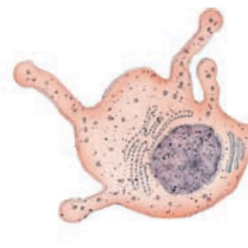
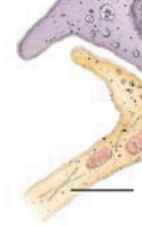
What is this?
langerhans cell
black line is pointing to?
Merkel (tactile) disc
black line is pointing to?
sensory neuron of a merkel cell

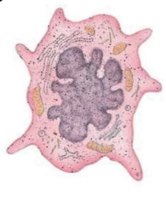
What type of cell is this
langerhans cell
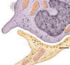
What type of cell is this? the one the black line is pointing to?
Melanocyte
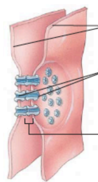
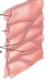
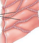
What is this?
gap junction
what is the line pointing to?
gap between cells
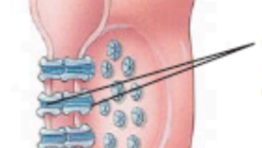
What is the lines pointing to? (photo of gap junction)
connexons (composed of connexins)
what are the top lines pointing to? (in a gap junction)
adjacent plasma membranes
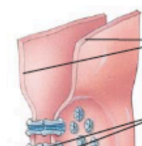
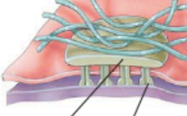
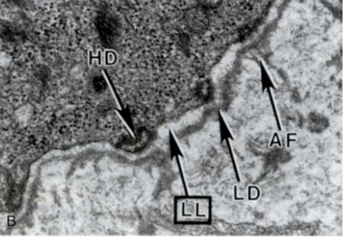
what is this overall photo of?
hemidesmosome
what is this blue part of a hemidesmosome?
intermediate filament (keratin)
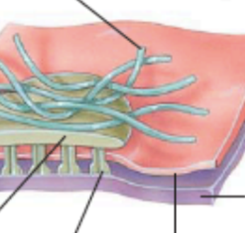
what is the pink layer and what is the purple layer (hemidesmosome)
pink = plasma membrane; purple = basement membrane
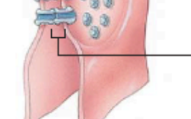
What are the two lines pointing to? (hemidesmosome)
top = plaque; bottom = transmembrane glycoprotein (integrin) in extracellular space
what is this?
tight junctions
what are the lines pointing to?
strands of transmembrane proteins
what is this?
adherens junction
what do the blue and green represent on this adherens junction?
blue = plaque; green = transmembrane glycoprotein (cadherin)
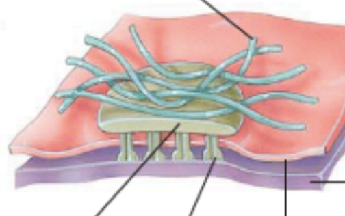
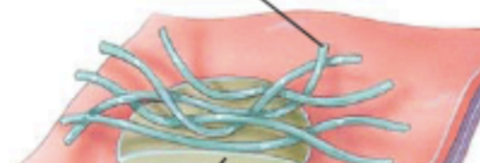
what is this photo of?
desmosome
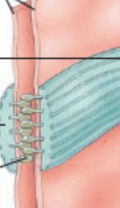
What are the blue lines?
intermediate filaments (keratin)
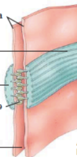
What is the thick blue line?
adhesion belt
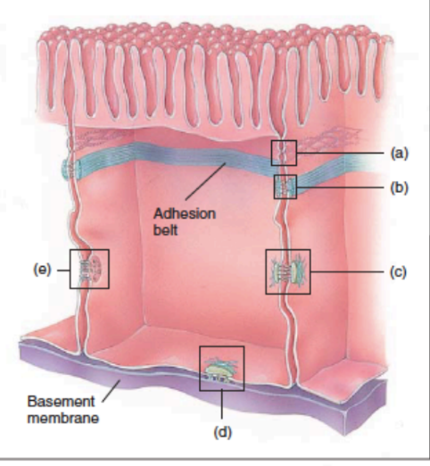
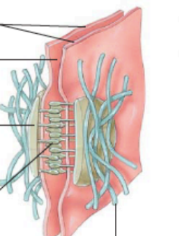
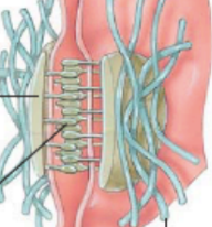
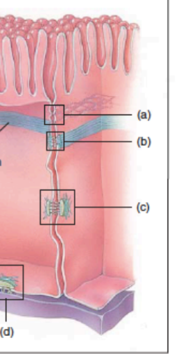
in this photo, what is a,b,c,d,e?
a = tight junction
b = adherens junction
c = desmosome
d = hemidesmosome
e = gap junction
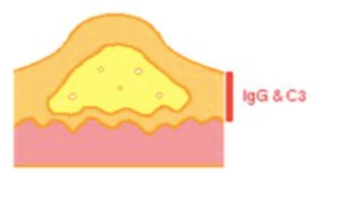
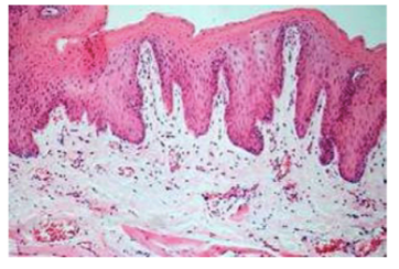
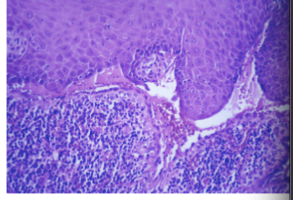
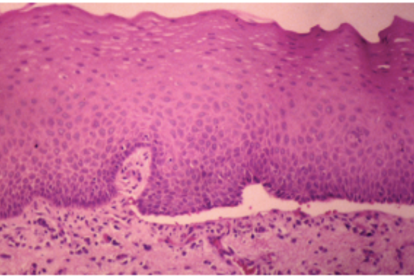
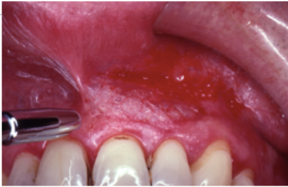
Which disease: blister within the epidermis; floor lined by basal csel acantholytic slcel ni blister fluid; intercellular igG &C3 by direct immunofluorescence
pemphigus vulgaris
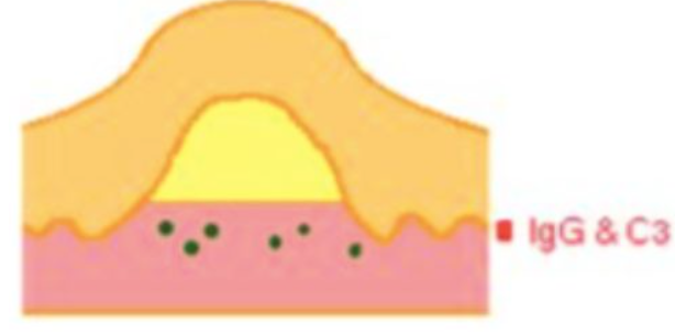
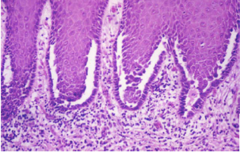
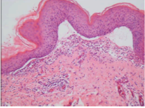
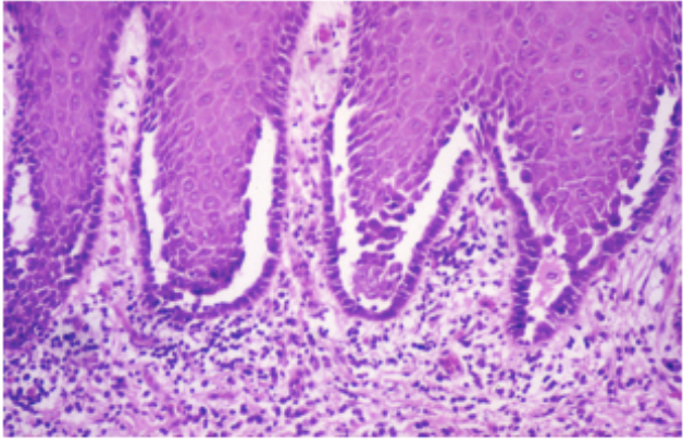
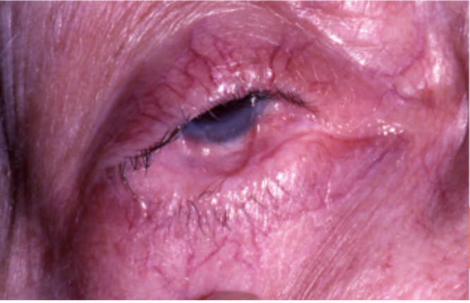
Which disease? subepidermal blister; eosinophil infiltrate underlying dermis basement membrane IgG &C 3 by direct immunofluorescence
pemphigoid
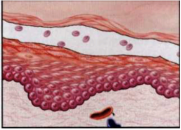
what kind of blister
Subcorneal (stratum corneum forms the roof of the bulla) (such as in PF)
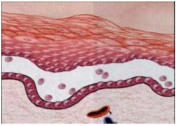
what kind of blister
suprabasal (such as in PV)
what kind of blister
subepidermal (such as in pemphigoid or dermatitis herpetiformis)
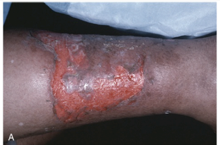
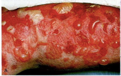
What disease is shown?
pemphigus vulgaris
what is this?
pemphigus vulgaris
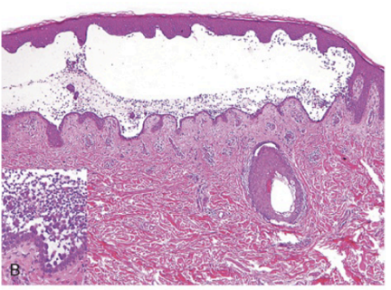
this is a histological slide of:
pemphigus vulgaris
PEMPHIGUS VULGARIS
pemphigus vulgaris
pemphigoid
pemphigoid
pemphigus vulgaris
pemphigoid
pemphigoid
pemphigus vulgaris
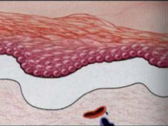
this shows
blister
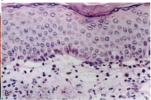
this shows
normal skin histology
Mycelex
Nystatin
prednisone
cyclophosphamide
pemphiGUS
pemphigoid
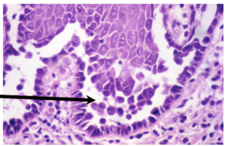
What cells?
Tzanck cells (multinucleated giant cells or macrophages) found in HSV, Varicella, Cytomegalovirus, and pemphigus
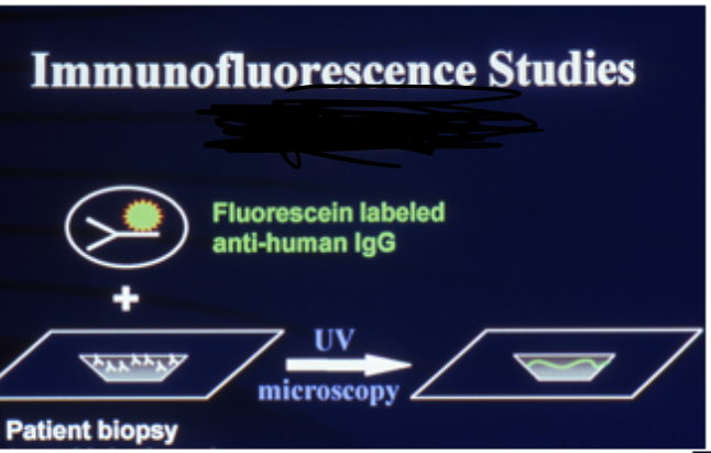
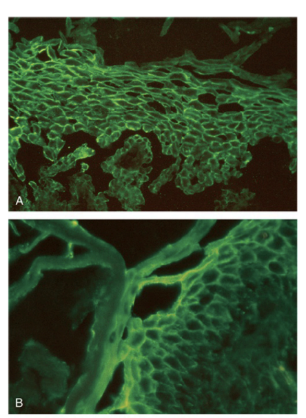
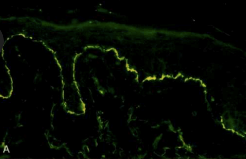
What type of immunofluorescence?
Direct (also be able to label other parts)
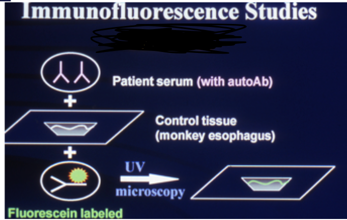
What type of immunofluorescence?
Indirect (also be able to label other parts)
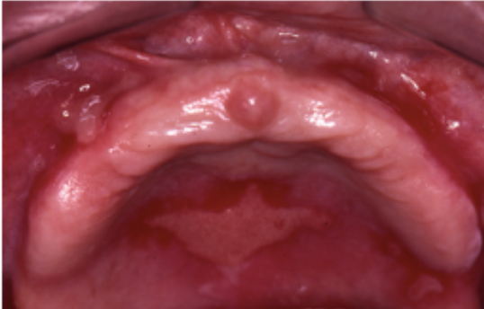
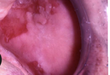
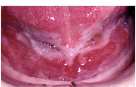
Cicatricial pemphigoid
Cicatricial pemphigoid
Cicatricial pemphigoid
Cicatricial pemphigoid
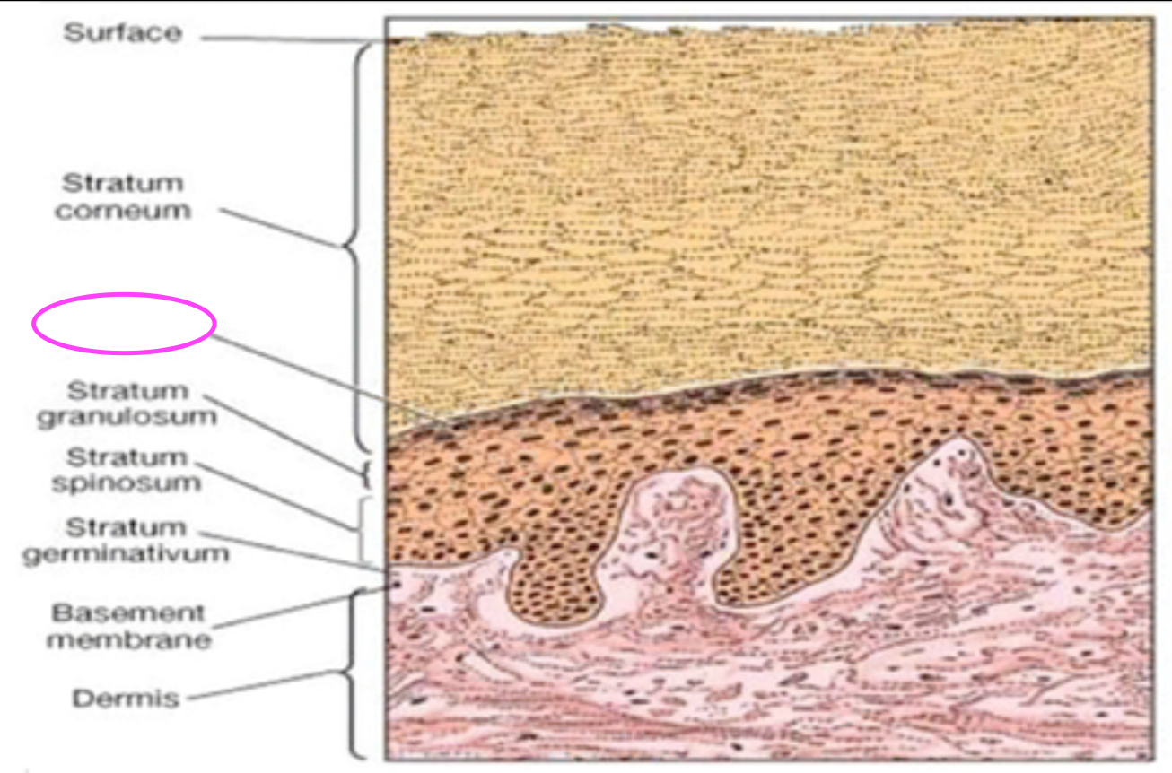
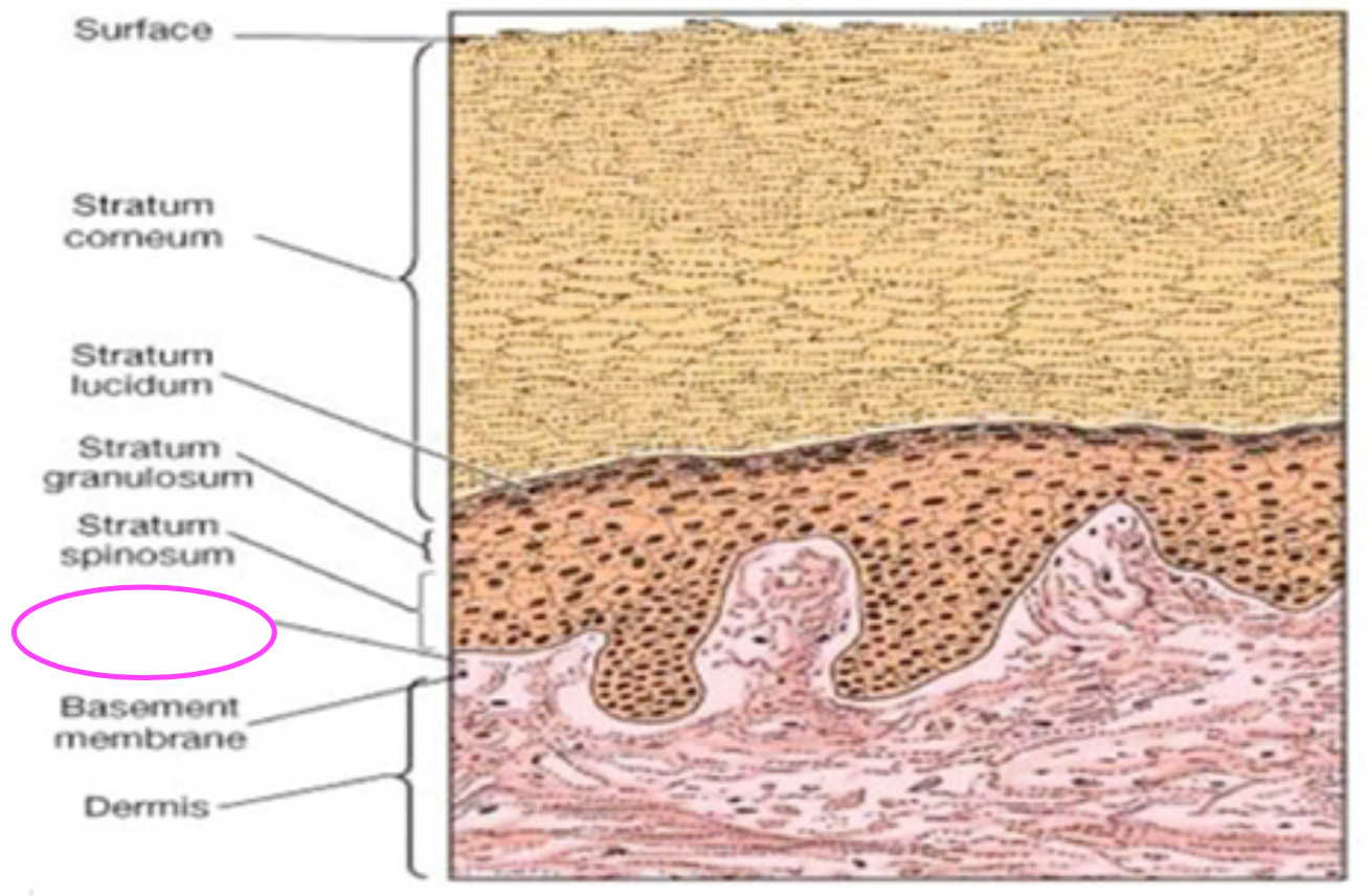
Fill in blank
Stratum lucidum
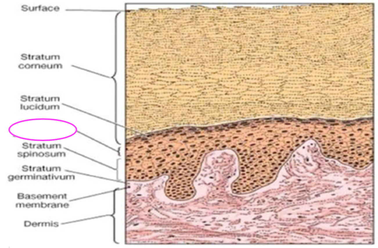
Stratum granulosum
Stratum germinativum
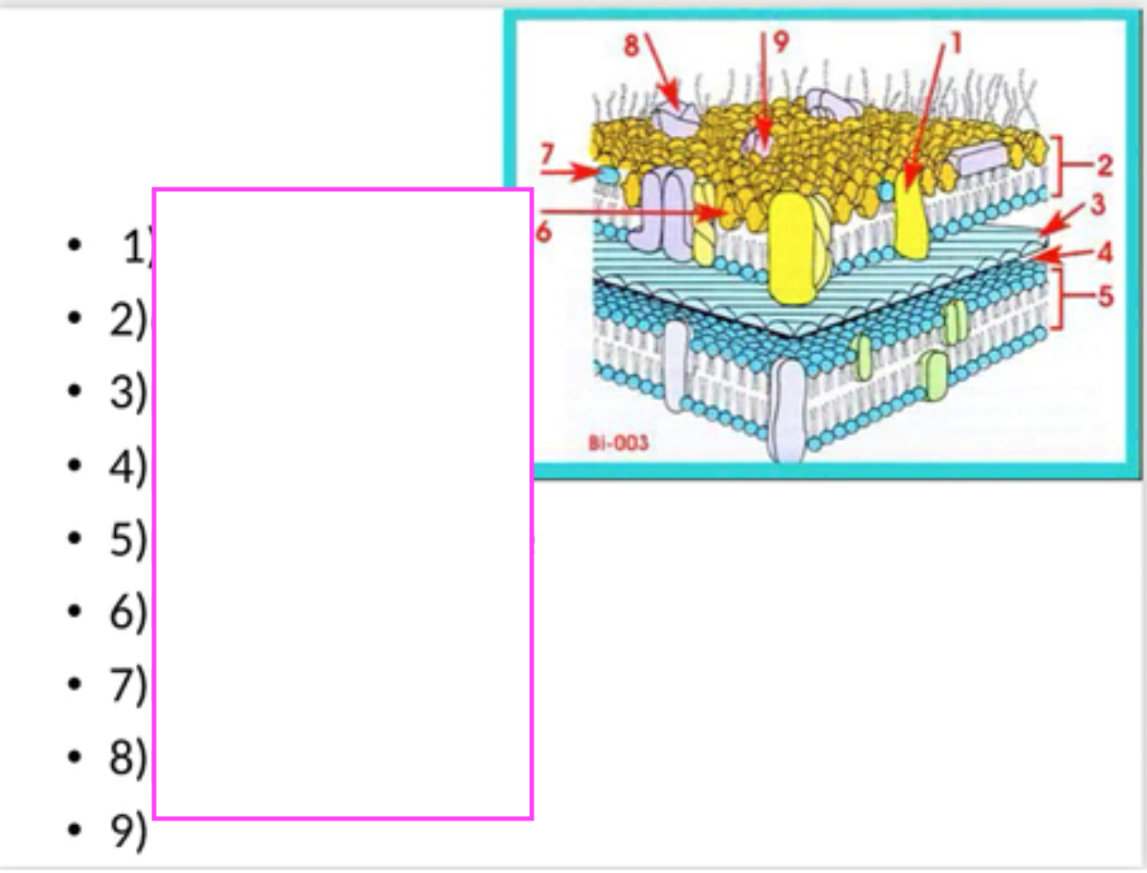
Fill in blanks #1-8
1: lipoprotein______ 2: outer membrane_____ 3: peptidoglycan_____ 4: periplasmic space_____5: plasma membrane _____6: LPS _____7: Lipid A_____ 8: porin
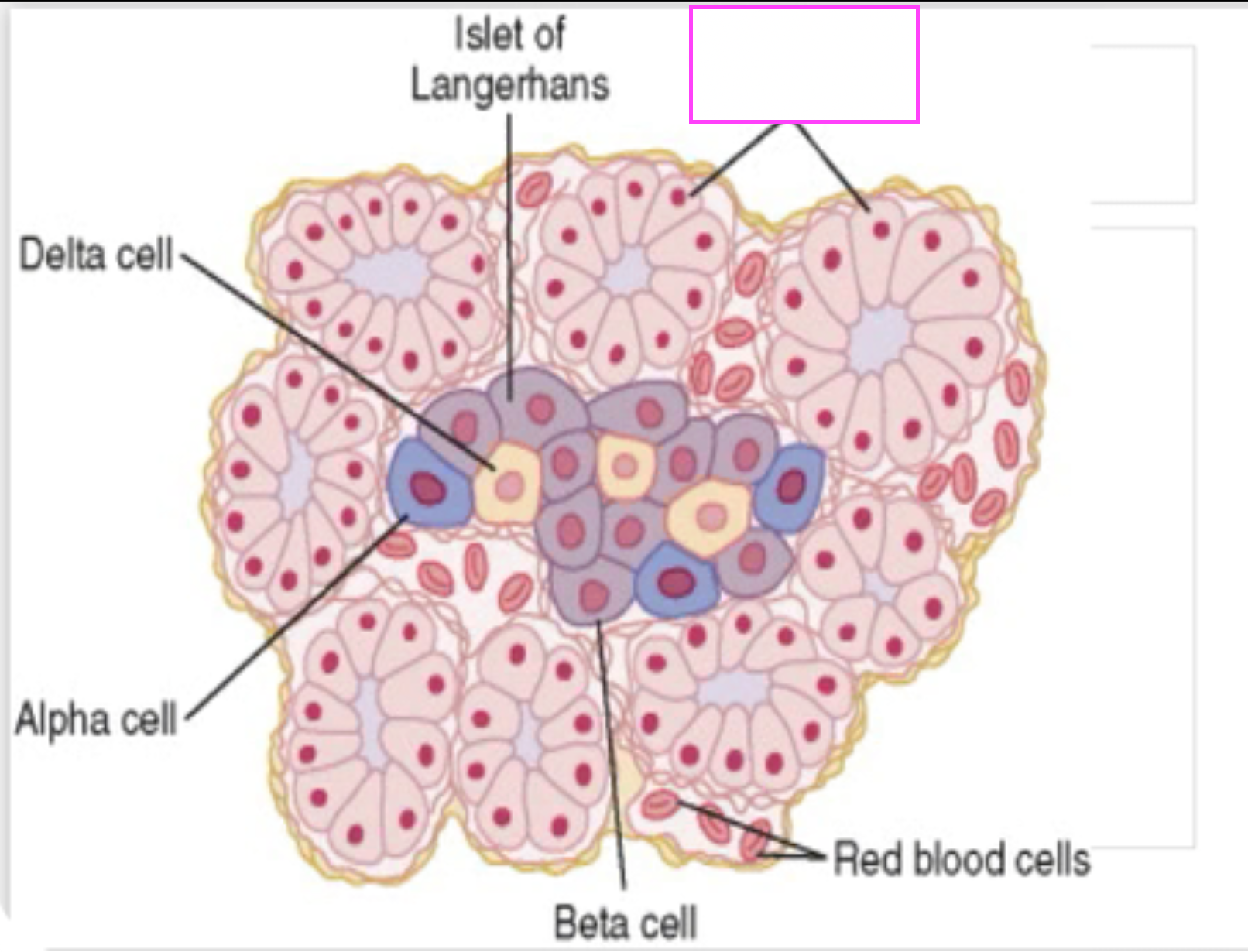
fill in blank
pancreatic acini
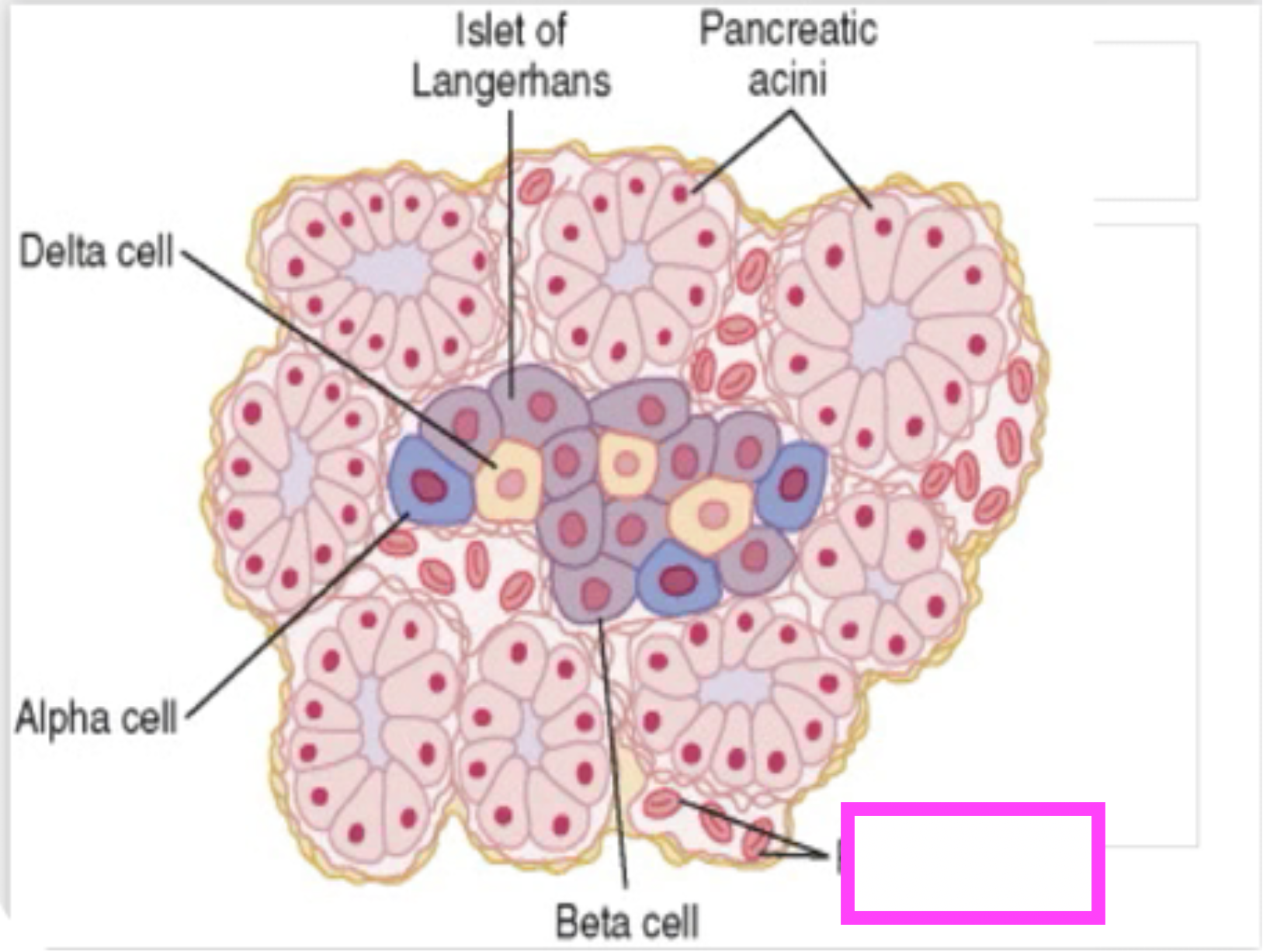
red blood cells
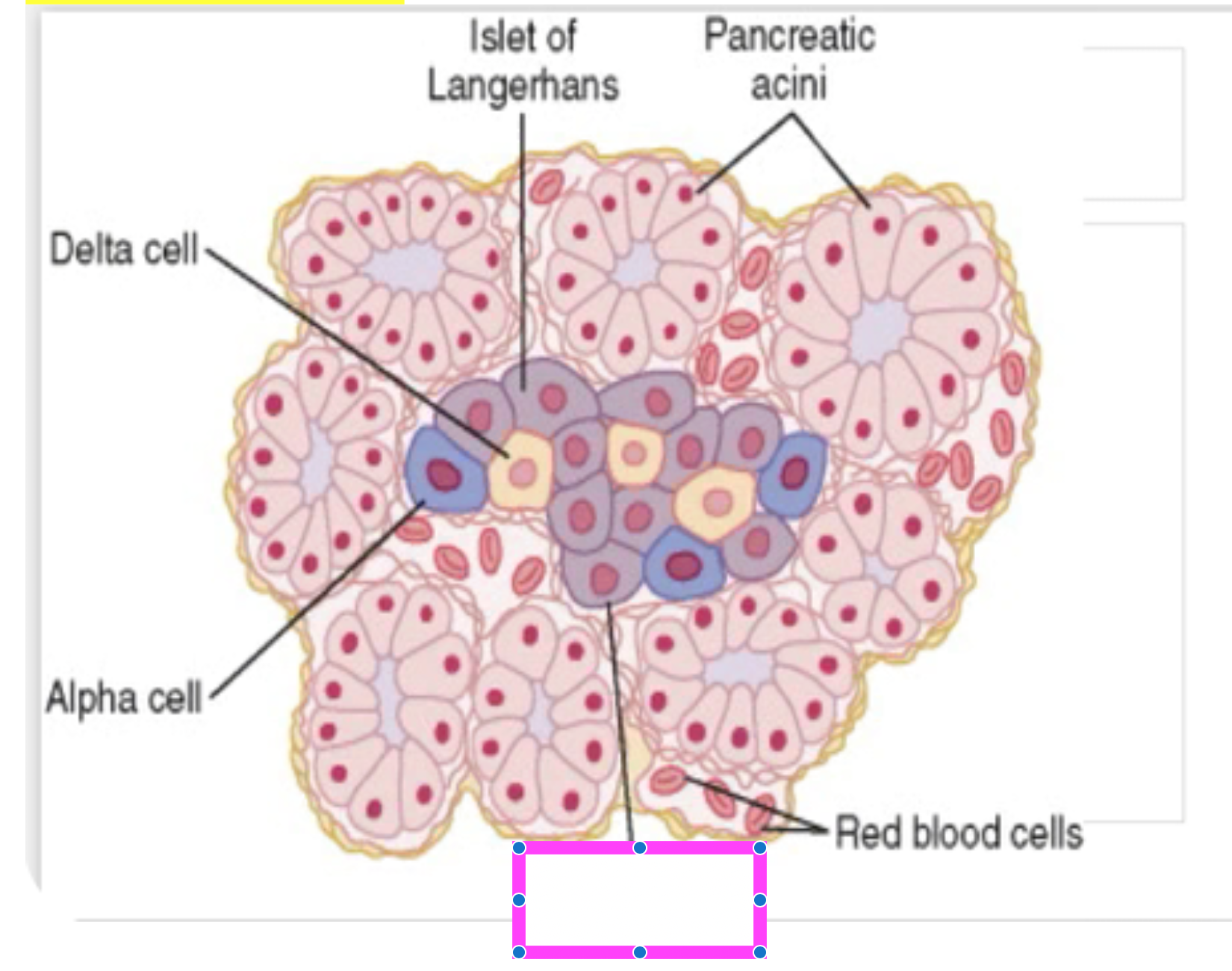
beta cells
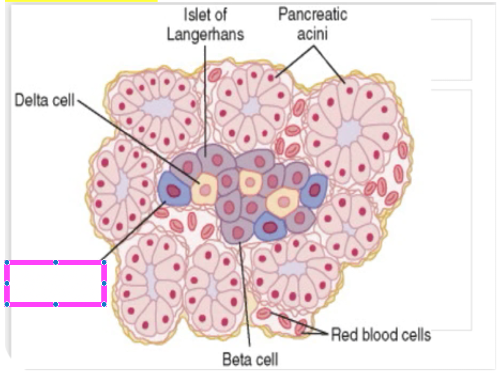
alpha cell
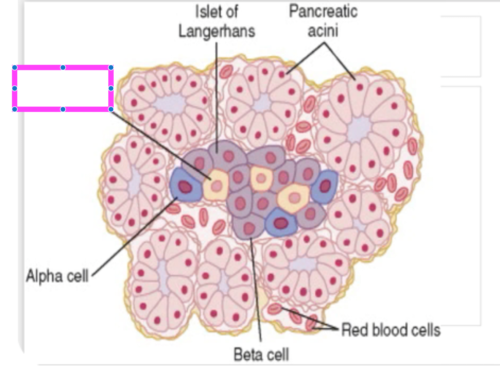
delta cell
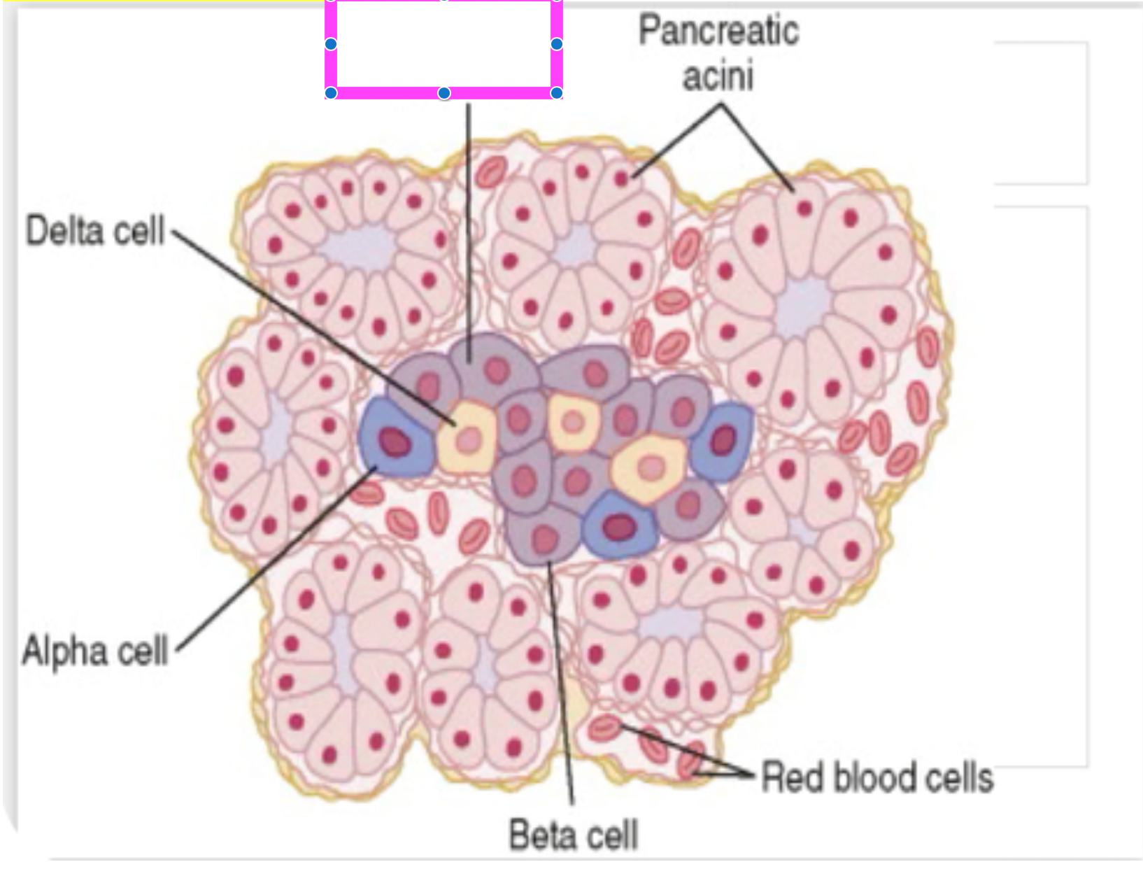
islet of langerhans
Which type of alveolar cells produce surfactant? (type I or type II)
Type II
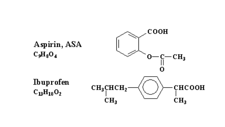
Which drug here contains a carboxylic acid group?
Both

Which drug here contains an ester group?
Aspirin
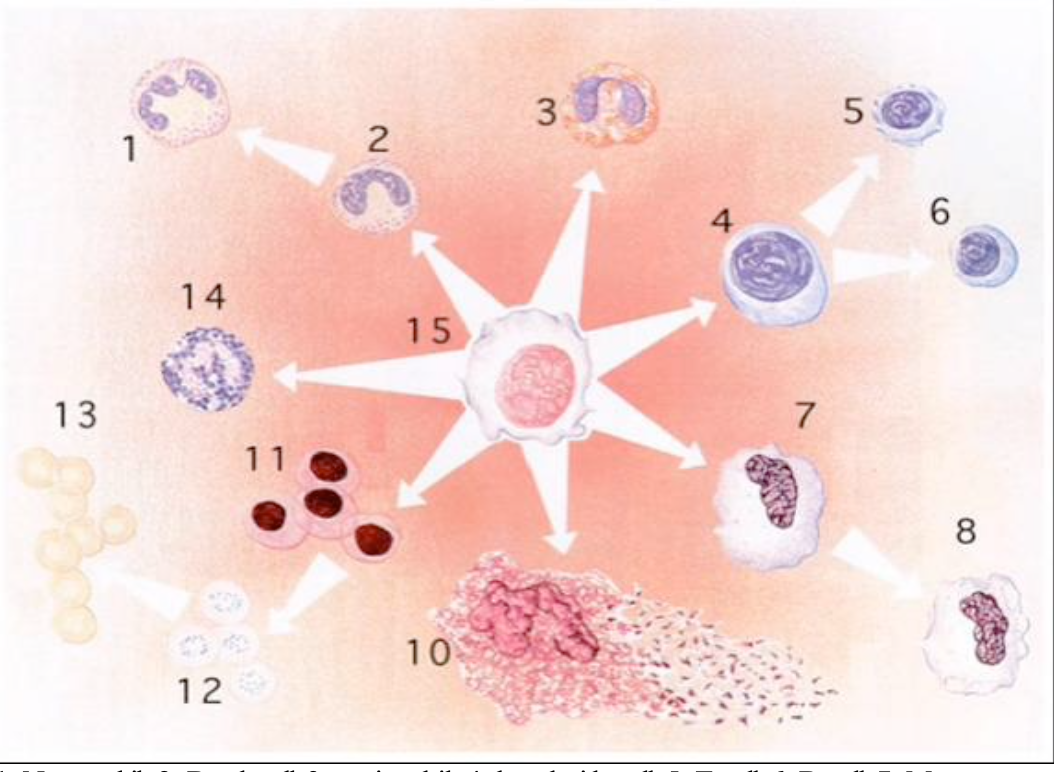
Label #1-15
1- neutrophil
2- band cell
3- eosinophil
4- lymphoid cell
5- T cell
6- B cell
7- Monocyte
8- Macrophage
9- none
10- megakaryocyte
11- proerythrocyte
12- reticulocyte
13- erythrocyte
14- basophil
15- pluripotent stem cell
T or F: RBC’s contain a nucleus
false

T or F: Monocytes can differentiate into macrophages
True

Platelets are derived from which of the following cells?
10- megakaryocytes
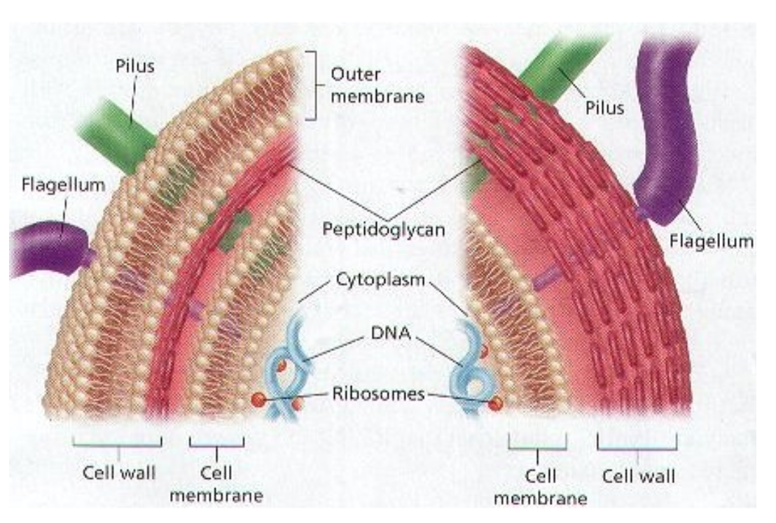
difference between left and right?
left = gram negative
right = gram positive
(note that gram positive bacteria have a thick peptidoglycan layer)
E coli is gram (+ or -)
gram negative