Bi 121 GRCC LAB exam 1
 Call Kai
 Learn
Learn Practice Test
 Spaced Repetition
 Match
Match Flashcards
 Knowt Play
1/93

There's no tags or description
Looks like no tags are added yet.
Name | Mastery | Learn | Test | Matching | Spaced | Call with Kai | Chat |
|---|
No analytics yet
Send a link to your students to track their progress
94 Terms
Diffusion
Movement of molecules from an area of higher concentration to an area of lower concentration. (form of passive transport)
selective permeability
A property of a plasma membrane that allows some substances to cross more easily than others.
passive transport
Requires NO energy, Movement of molecules from high to low concentration, Moves with the concentration gradient
concentration gradient
A difference in the concentration of a substance across a distance.
isotonic
denoting or relating to a solution having the same osmotic pressure as some other solution, especially one in a cell or a body fluid.
Hypertonic
Having a higher concentration of solute than another solution. (shrinks)
Hypotonic
Having a lower concentration of solute than another solution (swells)
Solute
A substance that is dissolved in a solution.
Osmosis
Diffusion of water through a selectively permeable membrane (passive transport)
active transport
Energy-requiring process that moves material across a cell membrane against a concentration difference
Crenation
This happens when a cell shrinks and shrivels; can result in cell death if severe.
-lysis
breakdown of a cell caused by damage to its plasma membrane
simple diffusion
movement of a solute from an area of high concentration to an area of low concentration
Superior Plane
everything above the transverse line (above the waist)

Inferior Plane
everything below the transverse line (below the waist). Opposite of superior plane.

Anterior (ventral)
front of the body

Posterior (dorsal)
back of body

Medial
Toward the midline of the body

Lateral
Away from the midline of the body

superficial
near the surface
Distal
farther from the origin of a body part or the point of attachment of a limb to the body trunk

deep (internal)
Away from the body surface; more internal
Proximal
Closer to the origin of the body part or the point of attachment of a limb to the body trunk

serous membrane
Membrane that lines a cavity without an opening to the outside of the body
thin layer of tissue that covers internal body cavities and secretes a fluid that keeps the membrane moist; also called serosa

parietal peritoneum
the outer layer of the peritoneum that lines the interior of the abdominal wall

visceral peritoneum
the inner layer of the peritoneum that surrounds the organs of the abdominal cavity

parietal pleura
lines the walls of the thoracic cavity

visceral pleura
the inner layer of pleura that surrounds each lung

parietal pericardium
outer layer of the pericardium
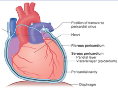
visceral pericardium (epicardium)
serous membrane covering the heart

sagittal plane
divides body into left and right

midsagittal plane
divides the body into equal left and right halves

frontal (coronal) plane
vertical plane dividing the body or structure into anterior and posterior portions
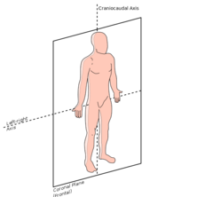
transverse plane
divides the body into superior and inferior parts

4 types of animal tissue
1. Epithelial
2. Connective
3. Muscle
4. Nervous

Biological Hierarchy
cells, tissues, organs, organ systems, organisms
epithelial tissue
Tissue that covers outside of the body and lines organs and cavities.
Cells fit closely together to form membranes, or sheets of cells
-Membranes always have one exposed surface or free edge, called the apical surface
-Cells are attached to and supported by an adhesive basement membrane
-Epithelial cells have no blood supply of their own; depend on diffusion of nutrients from the underlying connective tissue

simple squamous epithelium
Function: Allows passage of materials by diffusion and filtration in sites where protection is not important; secretes lubricating substances in serosae.
Location: Kidney glomeruli, air sacs of lungs, lining of heart, blood vessels, and lymphatic vessels; lining of ventral body cavity(serosae)
single layer of flattened cells
connective tissue
A body tissue that provides support for the body and connects all of its parts

types of epithelium cells
squamous, cuboidal, columnar
stratified squamous epithelium
Function: protects underlying tissues in areas subject to abrasion
Location: nonkeratinized type forms the moist lining of the esophagus, mouth, and vagina; keratinized type forms the epidermis of the skin, a dry membrane.

simple cuboidal epithelium
Function: secretion and absorption
Location: Kidney tubules; ducts and secretory portions of small glands, ovary surface.
single layer of flat cube like cells

simple columnar epithelium
Function: Absorption; secretion of mucus, enzymes, and other substances; ciliated type propels mucus (or reproductive cells) by ciliated action.
Location: nonciliated type lines most of the digestive tract (stomach to anal canal), gallbladder and excretory ducts of some glands; ciliated variety lines small bronchi, uterine tubes, and some regions of the uterus.
single layer of cells that are taller than they are wide

ciliated pseudostratified columnar epithelium
Ciliated pseudostratified columnar epithelia is the type of respiratory epithelium found in the linings of the trachea as well as the upper respiratory tract, which allows filtering and humidification of incoming air.

mucous (goblet) cell
Goblet cells are specialized for the synthesis and secretion of mucus. They acquired their name for their typical goblet, cup-like, appearance formed by the mucin granulae that fill up the cytoplasm

transitional epithelium
function: stretches readily and permits distension of urinary organ by contained urine
Location: lines the ureters, urinary bladder, and part of the urethra

apical (free) surface
the surface of the epithelial cell that faces the body surface, a body cavity, the lumen of an internal organ, or a tubular duct

basal lamina (basement membrane)
a layer of filaments and fibers that attach an epithelium to the underlying connective tissue

Keratinocytes
The most abundant epidermal cells, they function mainly to produce keratin.

connective tissue cells
fibroblasts, adipocytes, mast cells, white blood cells, macrophages, plasma cells
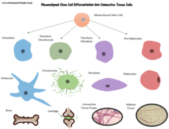
matrix
Connective tissues are made up of a matrix consisting of living cells and a non-living substance
Extracellular matrix provides support and anchorage for the shape of the cells, regulates and determines cells dynamic and behavior including cell survival, cell proliferation, cell polarity, cell differentiation, cell adhesion, and cell migration.
cells, fibers, and ground substance.

ground substance of connective tissue
Gelatinous or rubbery material found in between cells-protects by absorbing compressive forces.
Ground substance is primarily composed of water and large organic molecules, such as glycosaminoglycans (GAGs), proteoglycans, and glycoproteins

collagen fibers
a protein substance found in bone and cartilage, provides flexibility and strength

elastic fibers
Flexible and "stretchy" fibers that add elasticity to tissue

reticular fibers
Fibers made of collagen fibers that are very thin and branched. Forma tightly woven fabric that joins connective tissue to adjacent tissues.
acts as a supporting mesh in soft tissues such as liver, bone marrow, and the tissues and organs of the lymphatic system.

loose connective tissue
Tissue that binds epithelia to underlying tissues and holds organs in place. Contains collagenous, elastic, and recticular fibers.

dense connective tissue
dense regular, dense irregular, elastic; tendons and ligaments

special connective tissue
connective tissue that has a liquid or rigid extracellular matrix
areolar (loose) connective tissue
Binds skin to underlying organs; Forms delicate, thin layers between muscles; Fills in spaces underneath epithelia;
It is characterized by an abundance of ground substance, plus thin and relatively few fibres and cells The main cellular elements are fibroblasts and a smaller amount of adipocytes.
Located in the skin, areolar tissue binds the outer layers of the skin to the muscles lying underneath. They are also found in, around the mucous membranes, surrounding nerves, blood vessels and various other body organs. Its functions are as follows: Supports the internal organs.

adipose connective tissue
Function: provides reserve fuel; insulates against heat loss; supports and protects organs
Location: under skin, around kidneys and eyeballs, within abdomen, in breasts.

reticular connective tissue
Function: Fibers form a soft internal skeleton (stroma) that supports other cell types, including white blood cells, mast cells, and macrophages.
Location: Lymphoid organs (lymph nodes, bone marrow, and spleen).

dense connective tissue
Function: binds organs together
Location: tendons, ligaments, deeper layers of skin

hyaline cartilage connective tissue
Function: Supports and reinforces, provides a framework
Location: covers the ends of bones, costal cartilage in ribs, nose, rings in trachea, and larynx, forms most of the embryonic skeleton

elastic cartilage connective tissue
Function: Maintains shape and structure with great flexibility, supports and protects
Location: external ear, epiglottis (covers airway opening when swallowing)

Fibrocartilage Connective Tissue
Function: supports, protects, absorbs shock
Location: between bony parts of spinal column, parts of pelvic girdle and knee
Function: absorbs shock, supports, protects
Location: Intervertebral discs (between spinal vertebrae), pubic symphysis, meniscus of knee joint

layers of the integument
epidermis, dermis, hypodermis (subcutaneous layer)

melanocyte
cell in the basal layer that gives color to the skin

basal lamina (basement membrane)
a layer of filaments and fibers that attach an epithelium to the underlying connective tissue

sebaceous gland
oil-secreting gland in the dermis that is associated with hair follicles

sebum
oily substance secreted by sebaceous glands

sweat gland
The glands that secrete sweat, located in the dermal layer of the skin.

hair follicle
a small tubular cavity containing the root of a hair

shaft of hair follicle

arrector pili
a smooth muscle attached to hair follicles that causes "goose bumps" to appear on the skin when contracted

free edge of nail
the portion of the nail that grows out away from the body

nail body
visible part of the nail

nail bed
Portion of the living skin that supports the nail plate as it grows toward the free edge.

follicle-stimulating hormone (FSH)
regulates gamete production and hormonal activity of the gonads
pituitary gland

adrenocorticotropic hormone (ACTH)
regulates the endocrine activity of the adrenal cortex
pituitary gland

thyroid-stimulating hormone (TSH)
influences metabolism by controlling the growth and activity of the thyroid gland
pituitary gland
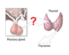
growth hormone
regulates muscle and bone growth
pituitary gland

prolactin
stimulates breast development and milk production
pituitary gland

oxytocin
stimulates contractions of uterus and milk ejection
pituitary gland

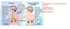
antidiuretic hormone (ADH)
reduces urinary output
pituitary gland
thyroid hormone (TH)
controls rate of metabolism
thyroid gland

calcitonin
decreased blood calcium levels by stimulating calcium depositing in the bones
thyroid gland

parathyroid hormone (PTH)
causes release of calcium from gland bone, thus increasing blood calcium levels
parathyroid gland

epinephrine and norepinephrine
act with sympathetic nervous system to produce the "flight/fight" response to stress
Adrenal gland

insulin
decreases blood sugar levels, helps glucose get into the body cells
pancreas

glucagon
increases blood sugar levels, stimulates liver to release glucose
pancreas

thymosins
affect production and differentiation of certain white blood cells (T lymphocytes
thymus

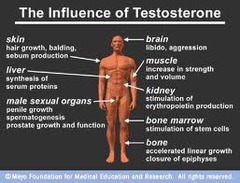
testosterone
stimulate formation of male reproductive organs and secondary sex characteristics, regulate the reproductive cycle.
testes
estrogen and progesterone
stimulate formation of female reproductive organs and secondary sex characteristics, regulate the reproductive cycle
ovaries

melatonin
Activates the reticular formation in the medulla oblongata which regulates circadian rhythms
Pineal gland
