Balance Wk1 bootcamp LG3
 Call Kai
 Learn
Learn Practice Test
 Spaced Repetition
 Match
Match Flashcards
 Knowt Play
1/99

There's no tags or description
Looks like no tags are added yet.
Name | Mastery | Learn | Test | Matching | Spaced | Call with Kai | Chat |
|---|
No analytics yet
Send a link to your students to track their progress
100 Terms
What is a signal sequence?
Answer: A string of hydrophobic amino acids on proteins destined for the endoplasmic reticulum.
Extra Information:
These proteins are destined for export or membrane integration.

What types of proteins contain an endoplasmic reticulum signal sequence according to the slide?
Answer: Proteins destined for export or membrane integration.
Extra Information:
Their signal sequence consists of a string of hydrophobic amino acids.

What does the signal sequence attract during protein synthesis?
Answer: The signal recognition particle.
Extra Information:
The signal recognition particle recognizes and binds the signal sequence.
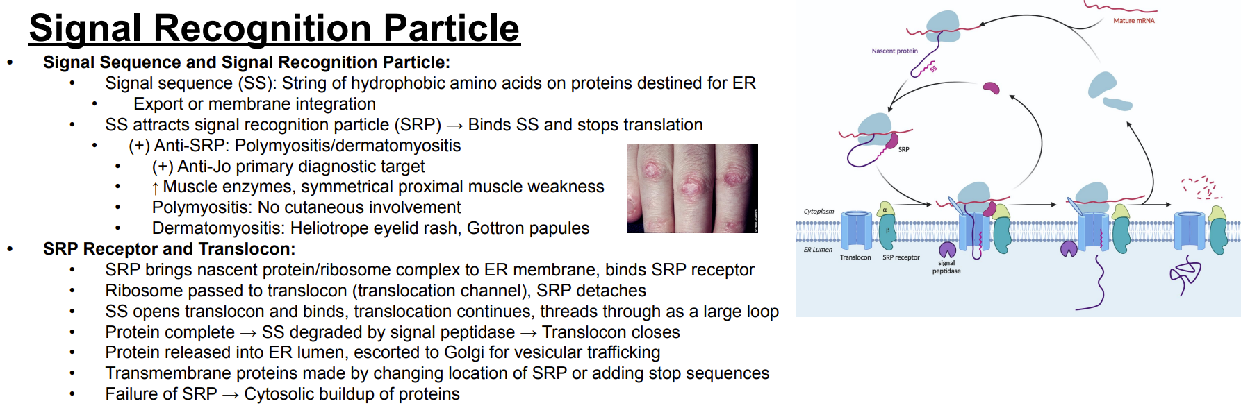
What happens when the signal recognition particle binds the signal sequence?
Answer: Translation stops.
Extra Information:
This temporary stop occurs while the nascent protein-ribosome complex is directed toward the endoplasmic reticulum.

Trace the first steps of endoplasmic reticulum targeting by the signal recognition particle.
Answer: Signal sequence emerges → signal recognition particle binds the signal sequence → translation stops → the signal recognition particle brings the nascent protein-ribosome complex to the endoplasmic reticulum membrane.
Extra Information:
The complex then interacts with the signal recognition particle receptor.

What does the signal recognition particle bind at the endoplasmic reticulum membrane?
Answer: The signal recognition particle receptor.
Extra Information:
This brings the nascent protein-ribosome complex to the endoplasmic reticulum membrane.
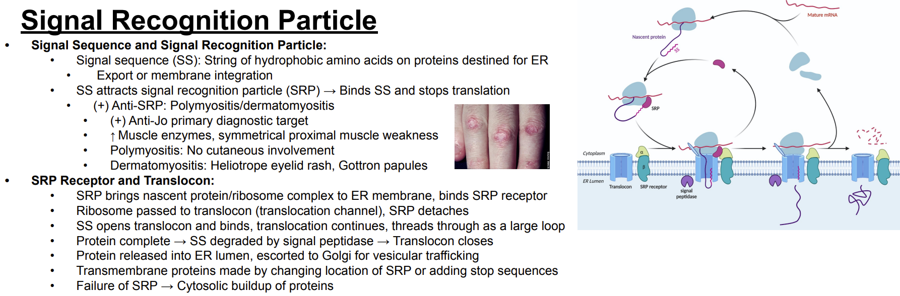
What happens after the signal recognition particle binds its receptor at the endoplasmic reticulum membrane?
Answer: The ribosome is passed to the translocon and the signal recognition particle detaches.
Extra Information:
The translocon is a translocation channel.

What is the translocon?
Answer: A translocation channel in the endoplasmic reticulum membrane.
Extra Information:
The ribosome is passed to the translocon after the signal recognition particle binds its receptor.

What opens the translocon?
Answer: The signal sequence.
Extra Information:
The signal sequence opens and binds the translocon.
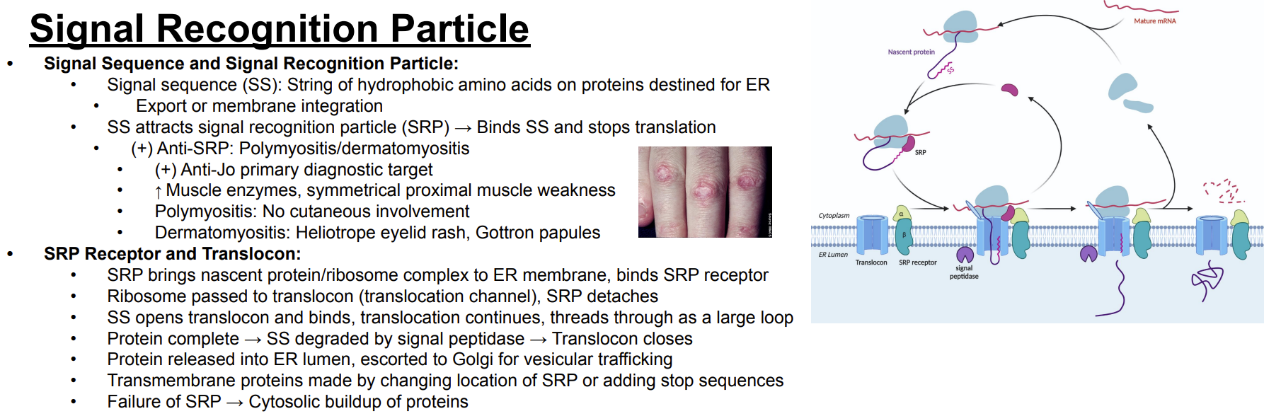
What happens to the growing protein after the signal sequence opens the translocon?
Answer: Translocation continues and the protein threads through the translocon as a large loop.
Extra Information:
The slide's diagram shows translation and translocation occurring at the endoplasmic reticulum membrane.

What happens to the signal sequence after the protein is completed?
Answer: The signal sequence is degraded by signal peptidase.
Extra Information:
The translocon then closes.
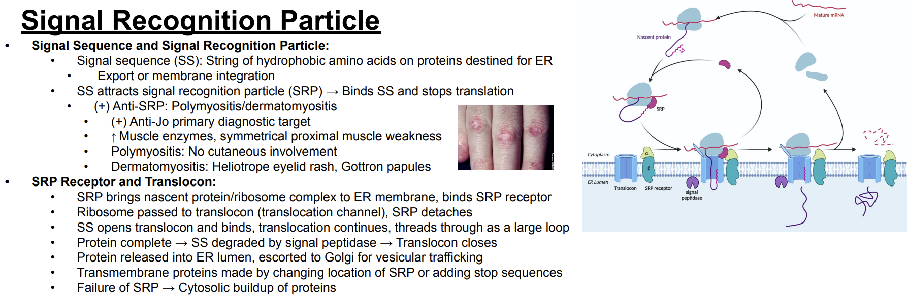
Which enzyme degrades the signal sequence after protein synthesis is complete?
Answer: Signal peptidase.
Extra Information:
Signal sequence degradation occurs before the completed protein is released into the endoplasmic reticulum lumen.
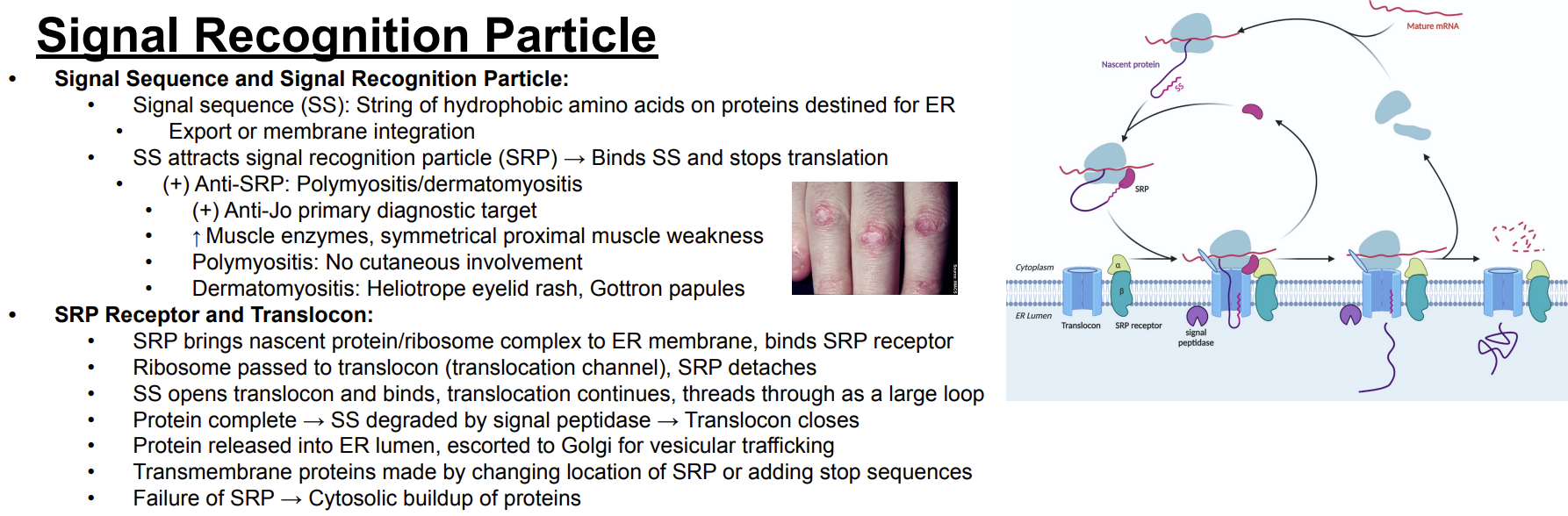
What happens to a completed protein after translocation into the endoplasmic reticulum?
Answer: The protein is released into the endoplasmic reticulum lumen and escorted to the Golgi for vesicular trafficking.
Extra Information:
This follows completion of translocation and degradation of the signal sequence.
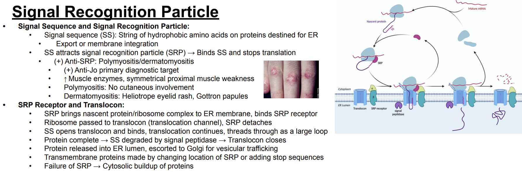
Trace the signal recognition particle pathway from signal sequence recognition to Golgi trafficking.
Answer: Signal sequence → signal recognition particle binds and stops translation → complex moves to the endoplasmic reticulum → signal recognition particle receptor → ribosome transfers to translocon → signal recognition particle detaches → signal sequence opens translocon → translocation continues → protein completes → signal peptidase degrades signal sequence → translocon closes → protein enters endoplasmic reticulum lumen → Golgi for vesicular trafficking.
Extra Information:
This integrates the complete pathway shown in the slide and diagram.
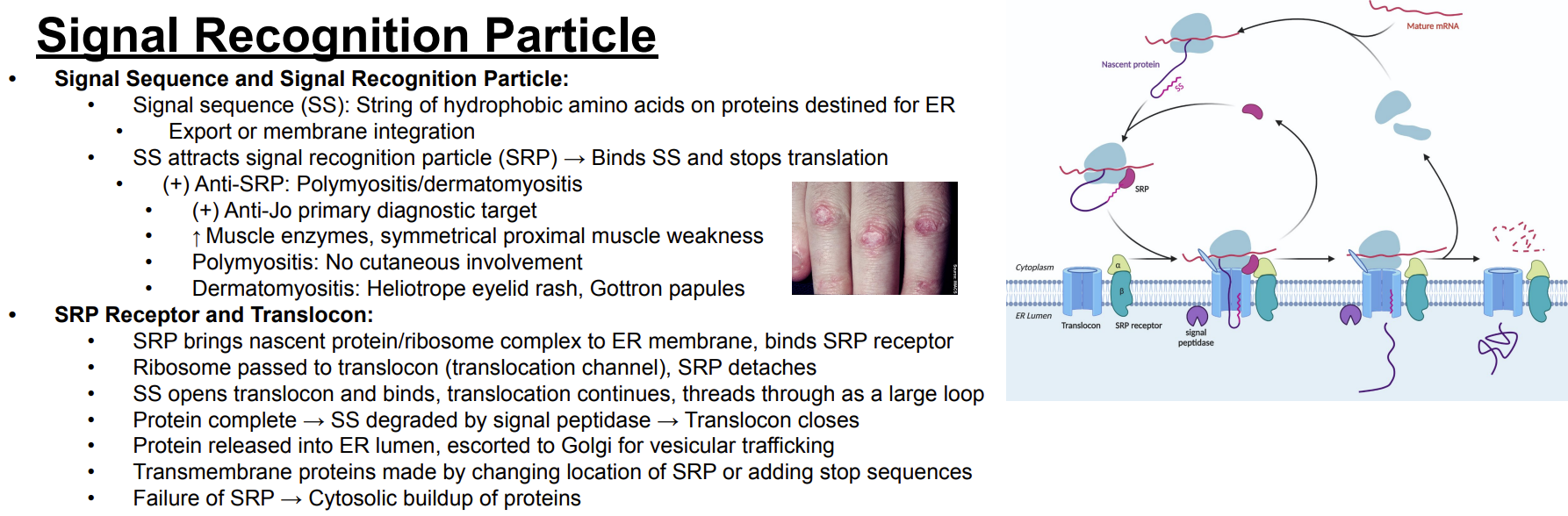
How are transmembrane proteins made according to the slide?
Answer: By changing the location of the signal recognition particle or adding stop sequences.
Extra Information:
These changes allow proteins to remain integrated into the membrane.

What happens when the signal recognition particle fails?
Answer: Proteins build up in the cytosol.
Extra Information:
Failure prevents normal targeting of these proteins to the endoplasmic reticulum.

Anti-signal recognition particle antibodies are associated with which diseases?
Answer: Polymyositis and dermatomyositis.
Extra Information:
The slide also identifies anti-Jo as a primary diagnostic target.

What muscle findings are associated with polymyositis and dermatomyositis on this slide?
Answer: Increased muscle enzymes and symmetrical proximal muscle weakness.
Extra Information:
Anti-signal recognition particle antibodies are associated with these conditions.

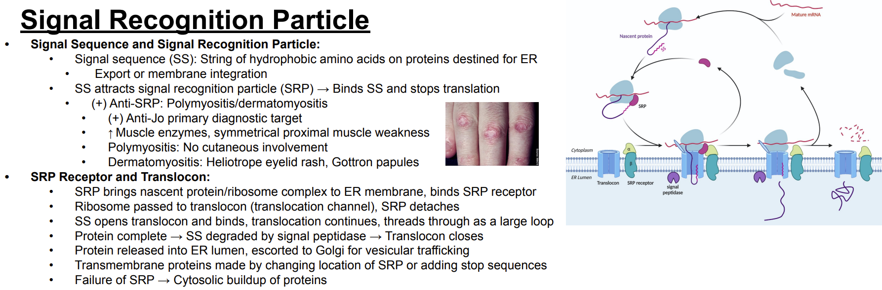
How do the cutaneous findings of polymyositis and dermatomyositis differ?
Answer: Polymyositis has no cutaneous involvement, while dermatomyositis has a heliotrope eyelid rash and Gottron papules.
Extra Information:
The slide includes an image demonstrating the characteristic papular skin findings.
What are the major features and functions of the rough endoplasmic reticulum?
Answer: It contains ribosomes, is located near the nucleus, performs protein synthesis, and carries out N-linked glycosylation.
Extra Information:
These features distinguish rough endoplasmic reticulum from smooth endoplasmic reticulum.

What are the major features and functions of the smooth endoplasmic reticulum?
Answer: It has no ribosomes, is located near the membrane, performs lipid synthesis, and contains glucose-6-phosphatase.
Extra Information:
These features distinguish smooth endoplasmic reticulum from rough endoplasmic reticulum.
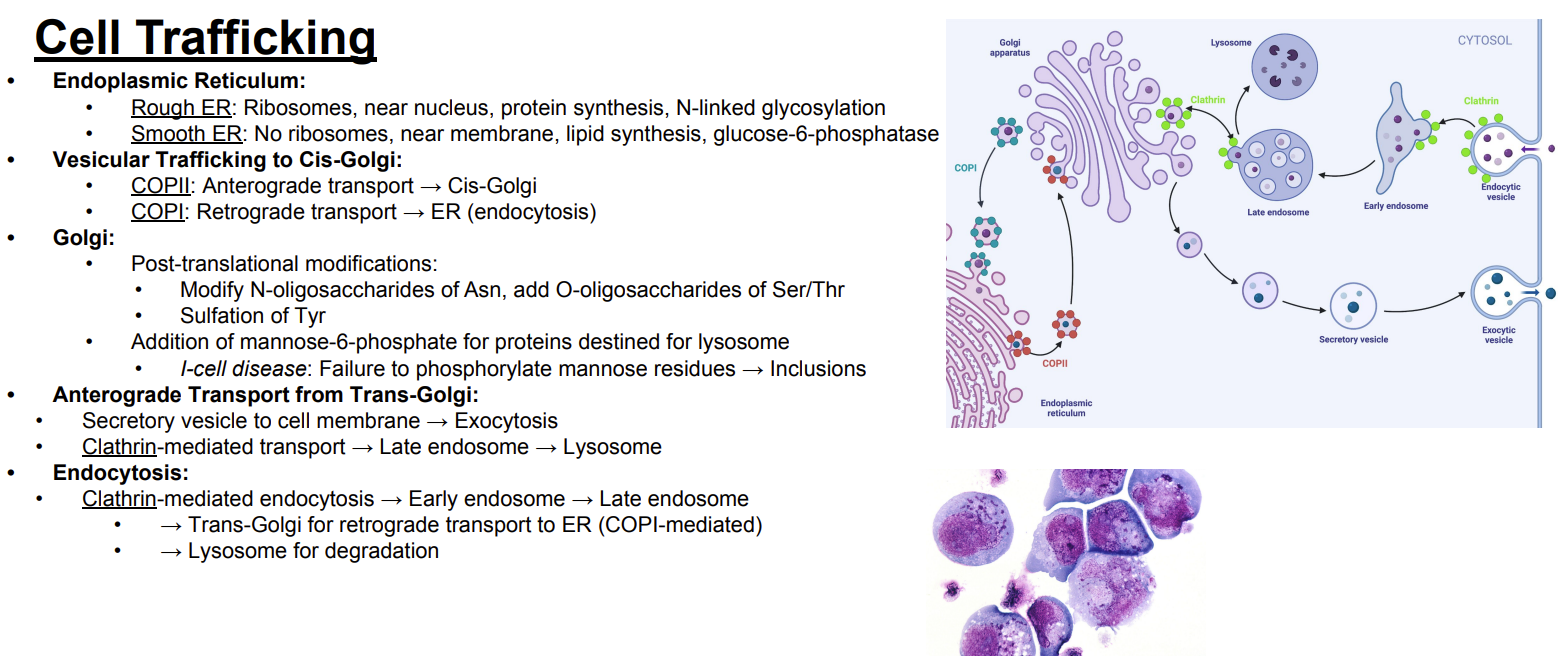
Compare the rough and smooth endoplasmic reticulum.
Answer:
rough endoplasmic reticulum has ribosomes, is near the nucleus, performs protein synthesis, and carries out N-linked glycosylation
smooth endoplasmic reticulum lacks ribosomes, is near the membrane, performs lipid synthesis, and contains glucose-6-phosphatase.
Extra Information:
This card integrates the major differences shown on the slide.

What does COPII mediate?
Answer: Anterograde transport from the endoplasmic reticulum to the cis-Golgi.
Extra Information:
Think: COPII → toward the Golgi.
2 steps forward 1 step back

What does COPI mediate?
Answer: Retrograde transport back to the endoplasmic reticulum.
Extra Information:
The slide labels this as retrograde transport.
2 steps forward 1 step back

Compare COPI and COPII vesicular trafficking.
Answer:
COPII = anterograde transport to the cis-Golgi
COPI = retrograde transport back to the endoplasmic reticulum.
Extra Information:
The slide's diagram shows these vesicles moving in opposite directions between the endoplasmic reticulum and Golgi.
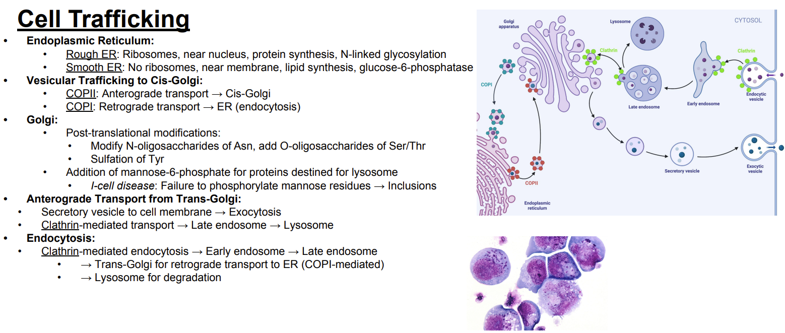
What post-translational carbohydrate modifications occur in the Golgi?
Answer: Modification of N-linked oligosaccharides on asparagine and addition of O-linked oligosaccharides to serine and threonine.
Extra Information:
These are post-translational modifications performed by the Golgi.

Which amino acid undergoes N-linked oligosaccharide modification in the Golgi?
Answer: Asparagine.
Extra Information:
The Golgi modifies N-linked oligosaccharides of asparagine.

Which amino acids receive O-linked oligosaccharides in the Golgi?
Answer: Serine and threonine.
Extra Information:
The Golgi adds O-linked oligosaccharides to these amino acids.
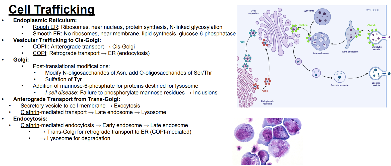
Which amino acid undergoes sulfation in the Golgi?
Answer: Tyrosine.
Extra Information:
Tyrosine sulfation is one of the Golgi's post-translational modifications.

What modification does the Golgi add to proteins destined for the lysosome?
Answer: Mannose-6-phosphate.
Extra Information:
Mannose-6-phosphate identifies proteins destined for the lysosome.

What defect occurs in I-cell disease according to the slide?
Answer: Failure to phosphorylate mannose residues.
Extra Information:
This disrupts the normal mannose-6-phosphate modification of proteins destined for lysosomes.

What cellular finding results from I-cell disease according to the slide?
Answer: Inclusions.
Extra Information:
I-cell disease results from failure to phosphorylate mannose residues.

Trace the secretory pathway from the trans-Golgi to the cell membrane.
Answer: Trans-Golgi → secretory vesicle → cell membrane → exocytosis.
Extra Information:
This is an anterograde transport pathway from the trans-Golgi.

How are proteins transported from the trans-Golgi to the lysosome?
Answer: Clathrin-mediated transport → late endosome → lysosome.
Extra Information:
The slide's diagram shows clathrin-coated trafficking toward the endosomal and lysosomal pathway.
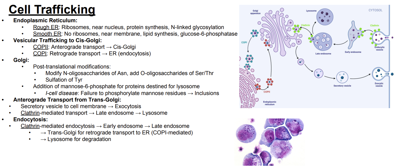
What is the pathway of clathrin-mediated endocytosis?
Answer: Clathrin-mediated endocytosis → early endosome → late endosome.
Extra Information:
Material entering the cell first reaches the early endosome.

What can happen to material after it reaches the late endosome?
Answer: It can be sent toward the trans-Golgi or to the lysosome for degradation.
Extra Information:
These are the destinations shown on the slide.

What happens to material sent from the endosomal pathway to the lysosome?
Answer: It undergoes degradation.
Extra Information:
The lysosome is the degradative destination shown in the trafficking pathway.

Trace the retrograde pathway from the endosomal system back to the endoplasmic reticulum.
Answer: Endocytosis → early endosome → late endosome → trans-Golgi → COPI-mediated retrograde transport → endoplasmic reticulum.
Extra Information:
COPI mediates the final retrograde transport back toward the endoplasmic reticulum.

Trace the major cell-trafficking pathways shown on the slide.
Answer: Endoplasmic reticulum → COPII → cis-Golgi → Golgi → COPI → endoplasmic reticulum → trans-Golgi → secretory vesicle → cell membrane → exocytosis → trans-Golgi → clathrin-mediated transport → late endosome → lysosome → endocytosis → early endosome → late endosome → lysosome or trans-Golgi.
Extra Information:
This card integrates the major trafficking routes shown in the diagram.

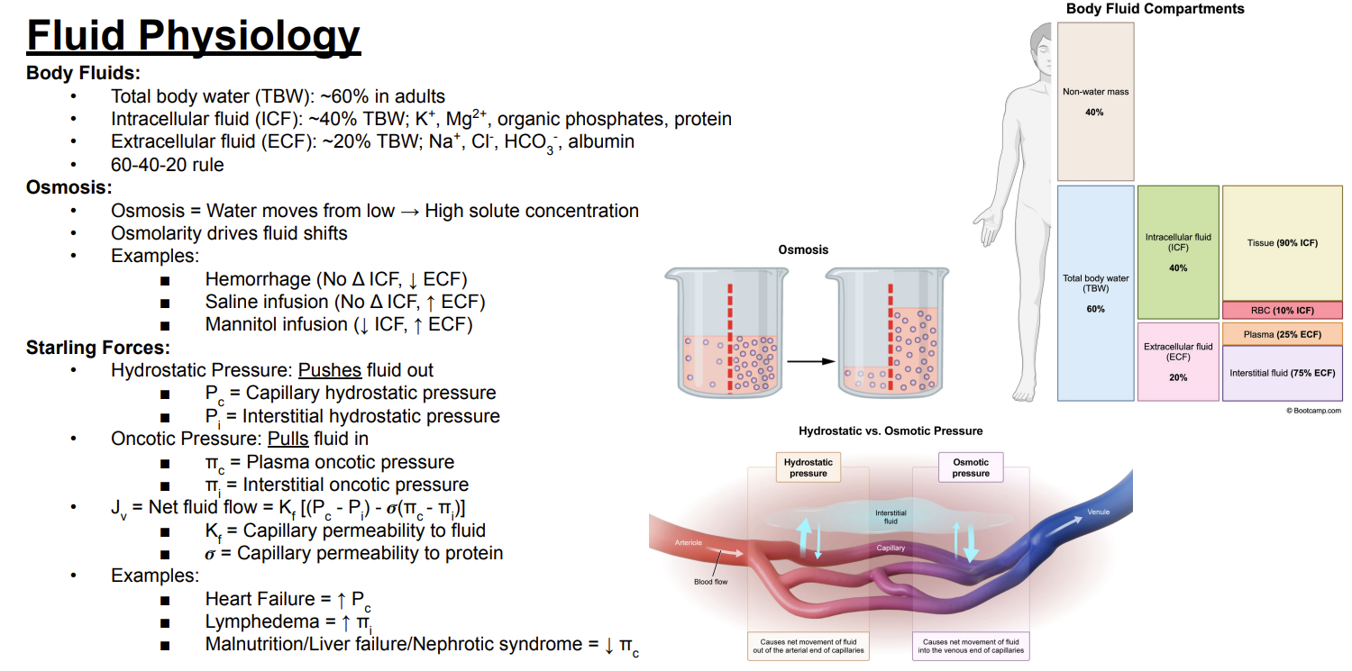
What is the 60-40-20 rule for body fluids?
Answer: Total body water is approximately 60% of body weight, intracellular fluid is approximately 40%, and extracellular fluid is approximately 20%.
Extra Information:
This is the 60-40-20 rule shown on the slide.

What percentage of body weight is total body water in adults?
Answer: Approximately 60%.
Extra Information:
This is the first number in the 60-40-20 rule.
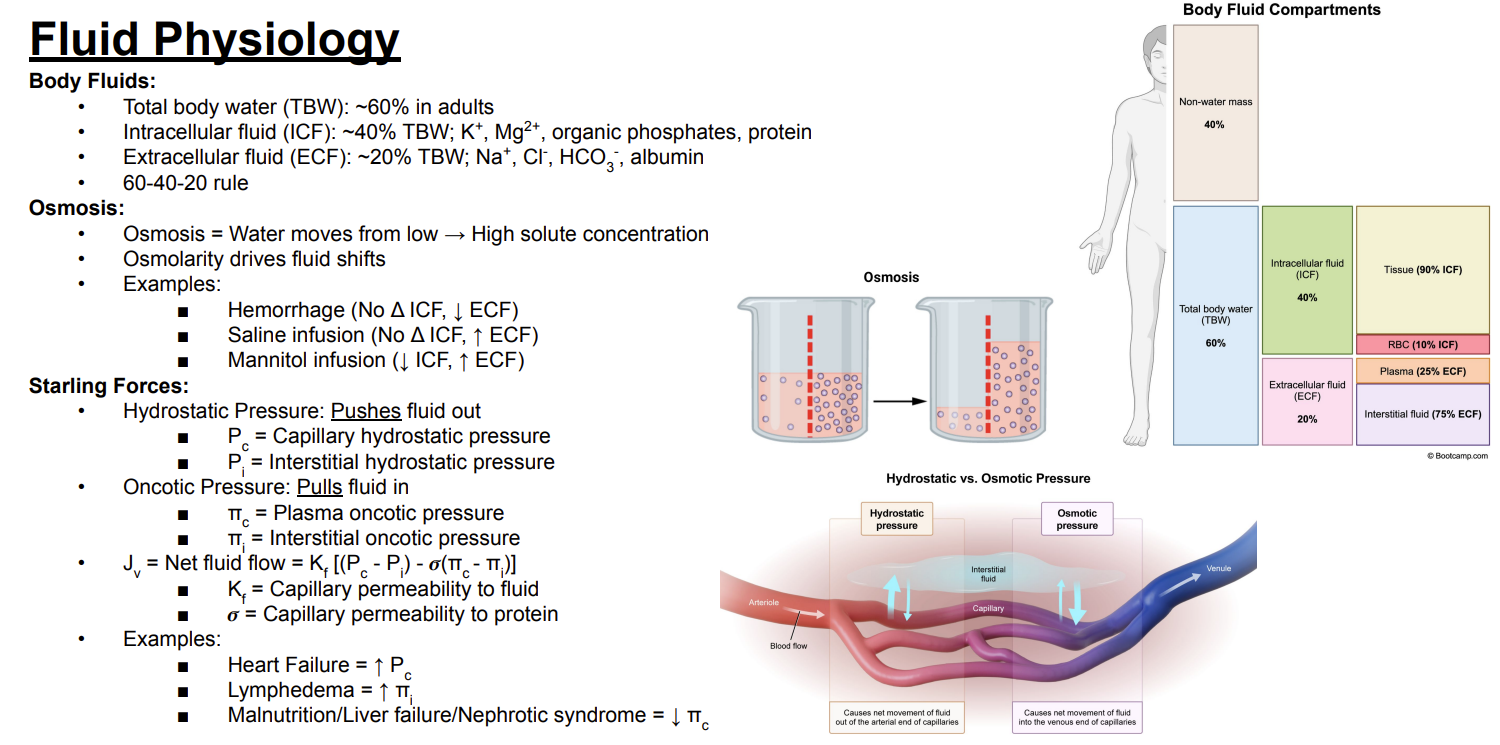
What percentage of body weight is intracellular fluid versus extracellular fluid?
Answer: Intracellular fluid is approximately 40%, while extracellular fluid is approximately 20%.
Extra Information:
Together with total body water at approximately 60%, this forms the 60-40-20 rule.

What are the major substances found in intracellular fluid according to the slide?
Answer: Potassium, magnesium, organic phosphates, and protein.
Extra Information:
These substances are associated with the intracellular compartment.
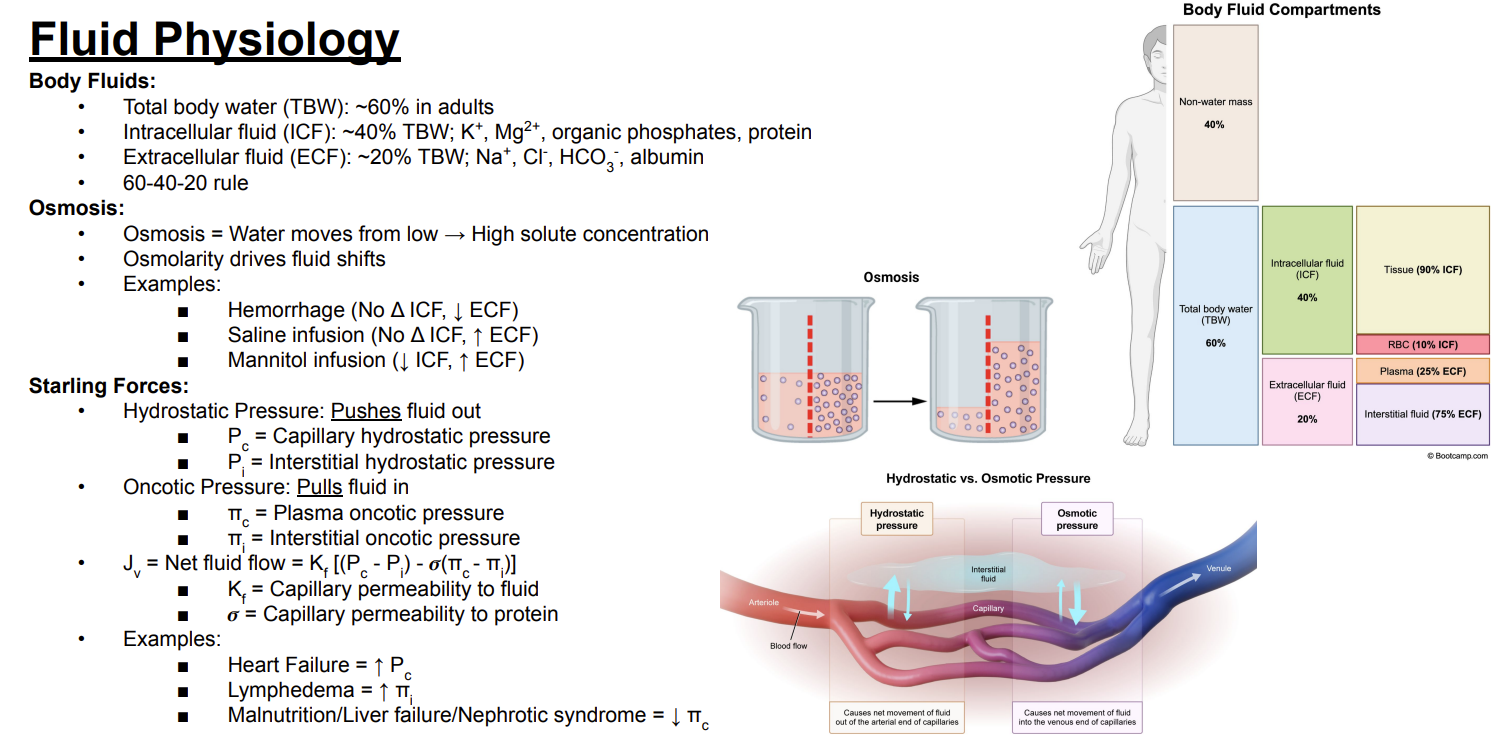
What are the major substances found in extracellular fluid according to the slide?
Answer: Sodium, chloride, bicarbonate, and albumin.
Extra Information:
These substances are associated with the extracellular compartment.

What is osmosis?
Answer: Movement of water from an area of low solute concentration toward an area of high solute concentration.
Extra Information:
Osmolarity drives fluid shifts.

What happens to intracellular and extracellular fluid volumes during hemorrhage?
Answer: Intracellular fluid does not change, while extracellular fluid decreases.
Extra Information:
Hemorrhage: no change in intracellular fluid and decreased extracellular fluid.

What happens to intracellular and extracellular fluid volumes after saline infusion?
Answer: Intracellular fluid does not change, while extracellular fluid increases.
Extra Information:
Saline infusion: no change in intracellular fluid and increased extracellular fluid.
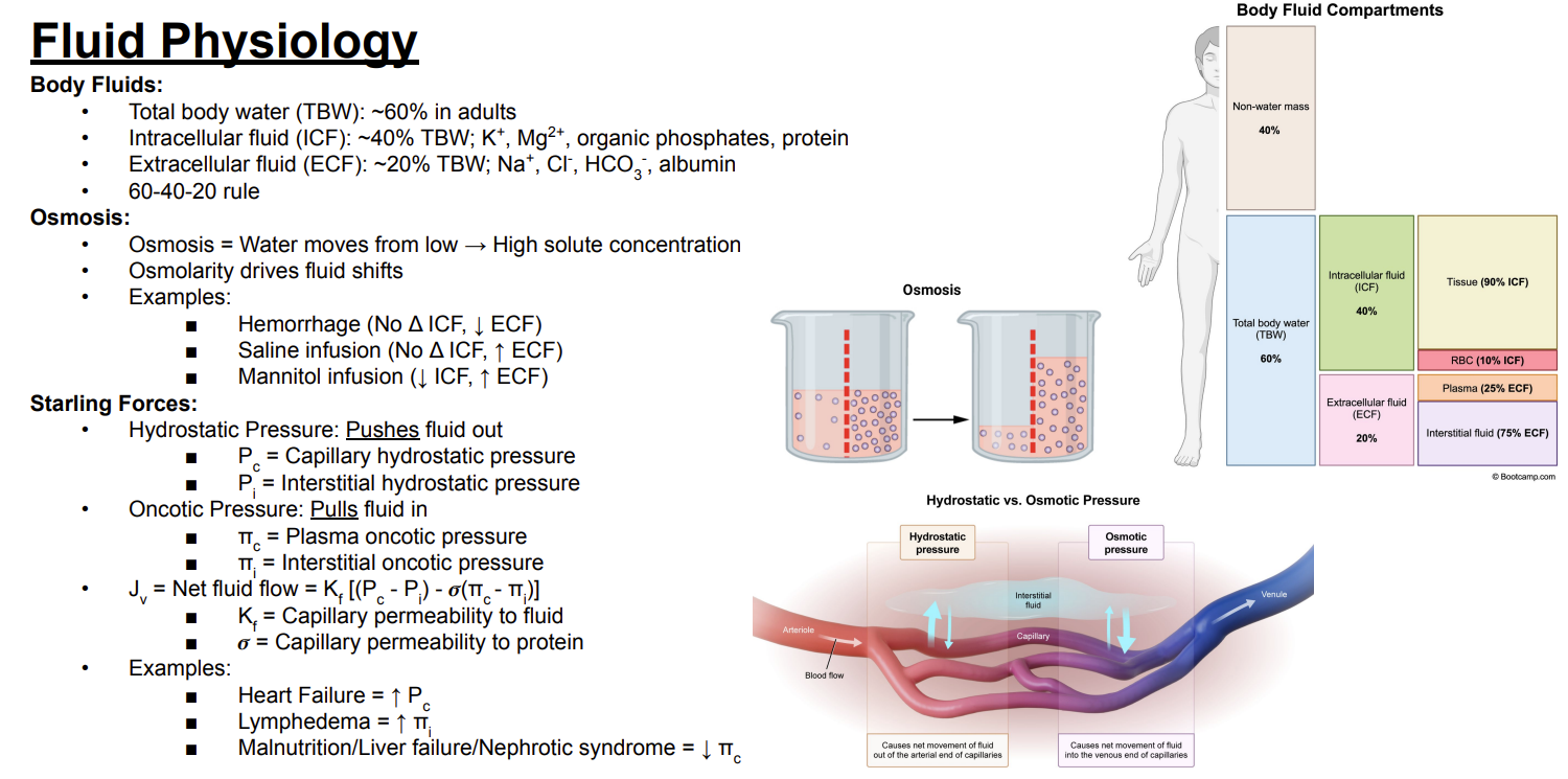
What happens to intracellular and extracellular fluid volumes after mannitol infusion?
Answer: Intracellular fluid decreases, while extracellular fluid increases.
Extra Information:
Osmolarity drives the fluid shift.

What is the basic difference between hydrostatic pressure and oncotic pressure?
Answer: Hydrostatic pressure pushes fluid out, while oncotic pressure pulls fluid in.
Extra Information:
Think: hydrostatic = push out & oncotic = pull in.

What do Pc and Pi represent in the Starling forces equation?
Answer: Pc is capillary hydrostatic pressure, and Pi is interstitial hydrostatic pressure.
Extra Information:
These are the hydrostatic pressure components of the equation.

What do πc and πi represent in the Starling forces equation?
Answer: πc is plasma oncotic pressure, and πi is interstitial oncotic pressure.
Extra Information:
These are the oncotic pressure components of the equation.
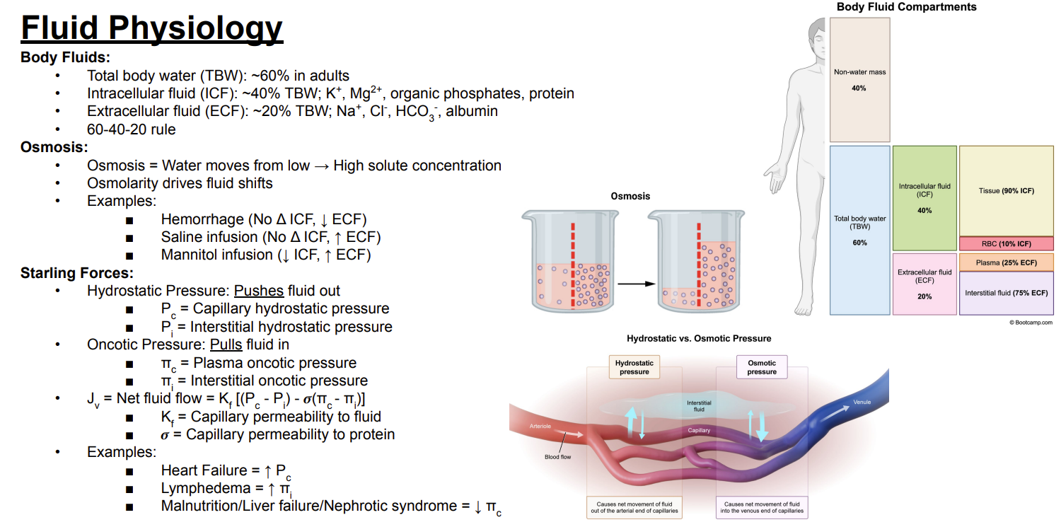
What is the Starling forces equation for net fluid flow?
Answer: Jv = Kf[(Pc − Pi) − σ(πc − πi)].
Extra Information:
Jv represents net fluid flow.
![<p>Answer: Jv = Kf[(Pc − Pi) − σ(πc − πi)].</p><p>Extra Information:</p><ul><li><p>Jv represents net fluid flow.</p></li></ul><p></p>](https://assets.knowt.com/user-attachments/d9565b69-646e-4d44-8be0-75eff022dcd4.png)
What does Kf represent in the Starling forces equation?
Answer: Capillary permeability to fluid.
Extra Information:
Kf is one component determining net fluid flow.

What does σ represent in the Starling forces equation?
Answer: Capillary permeability to protein.
Extra Information:
The slide includes σ in the oncotic component of the Starling equation.

Which Starling force is increased in heart failure?
Answer: Capillary hydrostatic pressure increases.
Extra Information:
Heart failure → increased Pc.
Increased hydrostatic pressure promotes fluid movement out.

Which Starling force is increased in lymphedema?
Answer: Interstitial oncotic pressure increases.
Extra Information:
Lymphedema → increased πi.
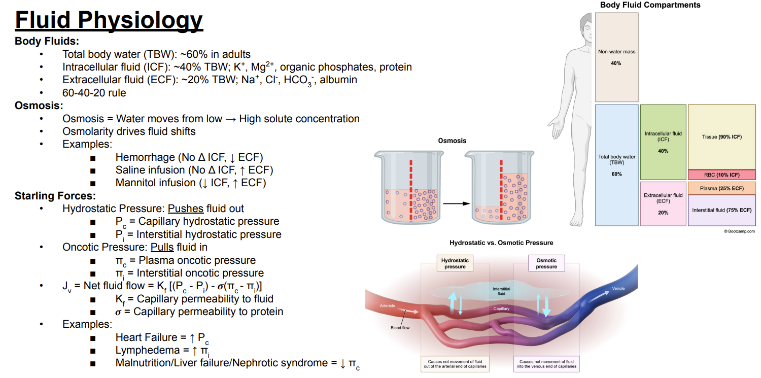
How do malnutrition, liver failure, and nephrotic syndrome affect Starling forces?
Answer: They decrease plasma oncotic pressure.
Extra Information:
Malnutrition, liver failure, and nephrotic syndrome → decreased πc.
Plasma oncotic pressure normally pulls fluid in.
What is the main function of the proteasome?
Answer: To degrade endogenous ubiquitinated proteins.
Extra Information:
The proteasome provides specific degradation of proteins tagged with ubiquitin.

What types of proteins are degraded by the ubiquitin-proteasome system according to the slide?
Answer: Misfolded proteins, proteins involved in cell-cycle regulation, and anti-apoptotic molecules.
Extra Information:
The proteasome primarily handles endogenous ubiquitinated proteins.

How does proteasomal degradation differ from lysosomal degradation according to the slide?
Answer: The proteasome degrades endogenous ubiquitinated proteins, while the lysosome degrades exogenous proteins.
Extra Information:
Ubiquitination specifically targets proteins toward the proteasome.

What is ubiquitin?
Answer: A regulatory protein tag added to proteins destined for the proteasome.
Extra Information:
The ubiquitination process is initiated using adenosine triphosphate.

To which amino acid residues is ubiquitin added during the conjugation cascade?
Answer: Lysine residues.
Extra Information:
Repeated addition of ubiquitin produces a polyubiquitin chain.

What are the three major enzymes in the ubiquitin conjugation cascade?
Answer: E1, E2, and E3.
Extra Information:
E1 = activation.
E2 = conjugation.
E3 = ligation.

What is the function of E1 in ubiquitination?
Answer: E1 activates ubiquitin and transfers it to E2.
Extra Information:
There is one E1 according to the slide.

What is the function of E2 in ubiquitination?
Answer: E2 conjugates and presents ubiquitin to E3.
Extra Information:
There are several E2 enzymes according to the slide.

What is the function of E3 in ubiquitination?
Answer: E3 facilitates attachment of ubiquitin to the specific target protein.
Extra Information:
E3 performs ligation.
There are many E3 enzymes, allowing target specificity.
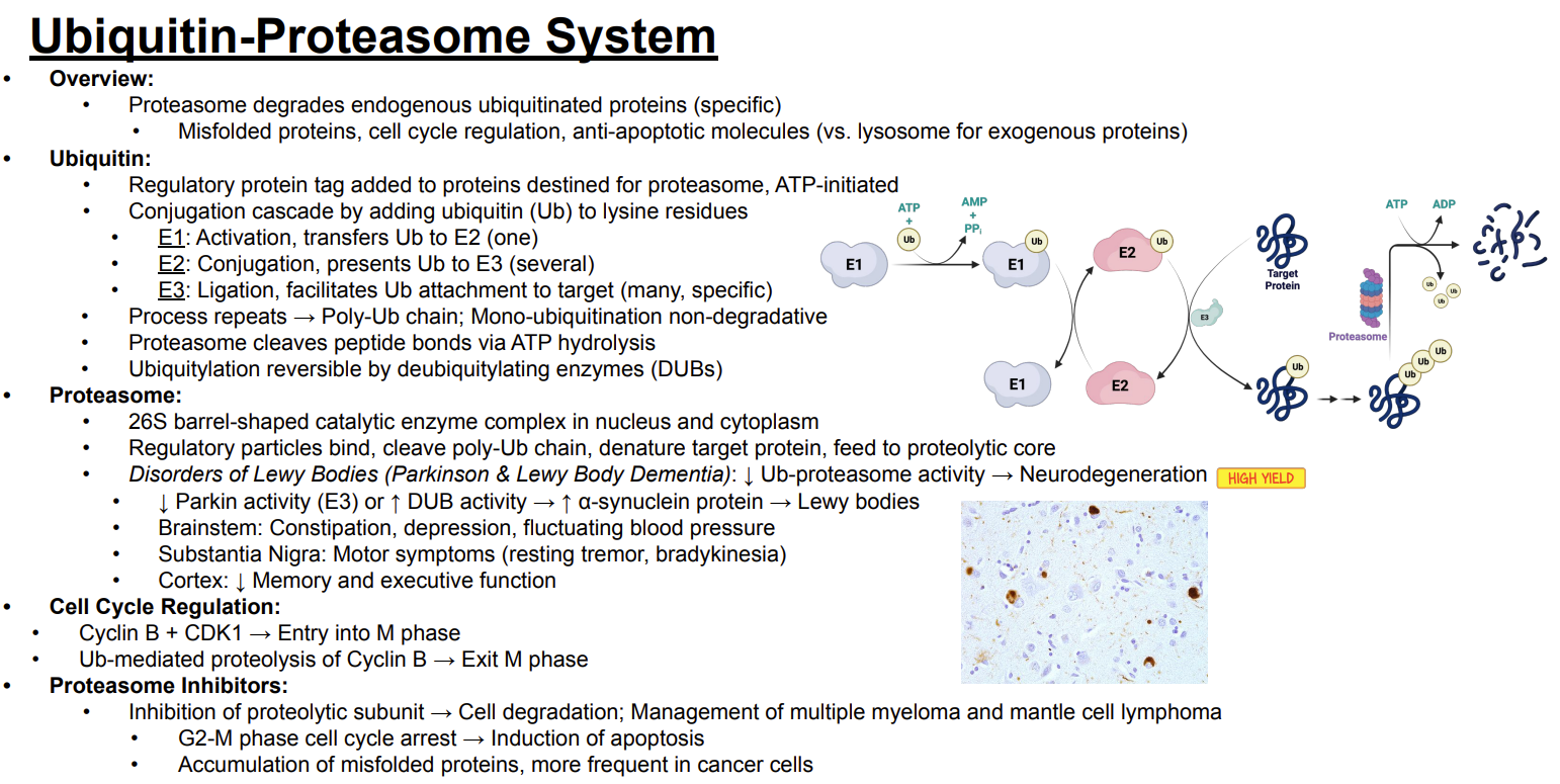
Trace the E1-E2-E3 ubiquitination pathway.
Answer: E1 activates ubiquitin and transfers it to E2 → E2 presents ubiquitin to E3 → E3 facilitates attachment of ubiquitin to the target protein.
Extra Information:
The process repeats to produce a polyubiquitin chain.

Which ubiquitination enzyme provides specificity for the target protein?
Answer: E3.
Extra Information:
There are many specific E3 ligases.

What happens when the ubiquitination process repeats on a target protein?
Answer: A polyubiquitin chain forms.
Extra Information:
The polyubiquitin chain allows the protein to be recognized by the proteasome.

Is monoubiquitination degradative according to the slide?
Answer: No. Monoubiquitination is non-degradative.
Extra Information:
Polyubiquitination is associated with proteasomal degradation on the slide.

How does the proteasome cleave proteins?
Answer: It cleaves peptide bonds using adenosine triphosphate hydrolysis.
Extra Information:
Proteasomal degradation is an energy-dependent process.
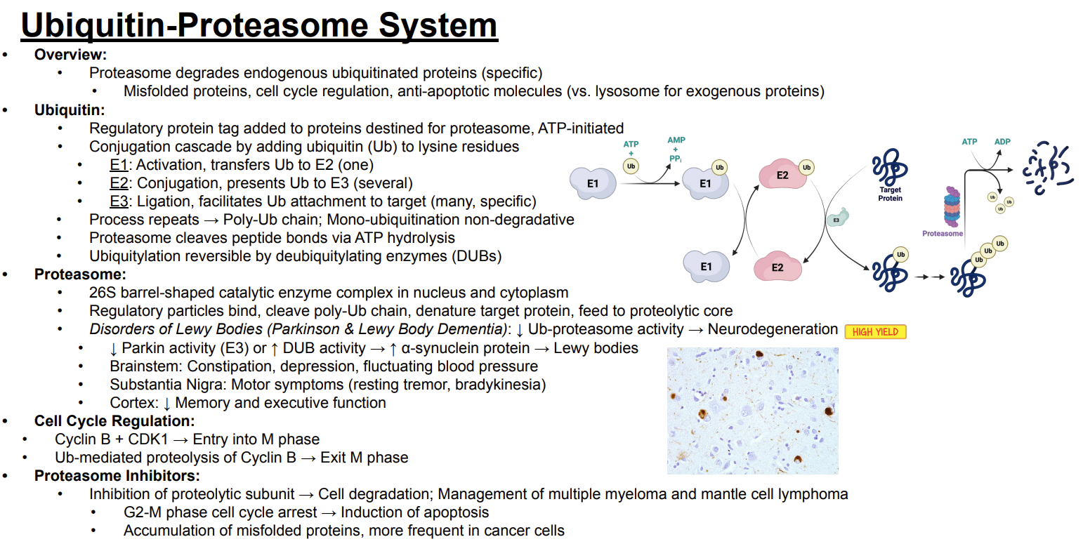
Is ubiquitination reversible?
Answer: Yes. Ubiquitination can be reversed by deubiquitylating enzymes.
Extra Information:
Deubiquitylating enzymes are abbreviated DUBs.
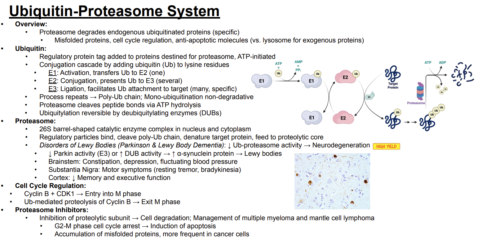
What is the 26S proteasome?
Answer: A barrel-shaped catalytic enzyme complex located in the nucleus and cytoplasm.
Extra Information:
It contains regulatory particles and a proteolytic core.

What do the regulatory particles of the proteasome do?
Answer: They bind the target, cleave the polyubiquitin chain, denature the target protein, and feed it into the proteolytic core.
Extra Information:
The proteolytic core then degrades the protein.

How is decreased ubiquitin-proteasome activity related to Lewy body disorders?
Answer: Decreased ubiquitin-proteasome activity leads to neurodegeneration.
Extra Information:
The slide associates this with Parkinson disease and Lewy body dementia.
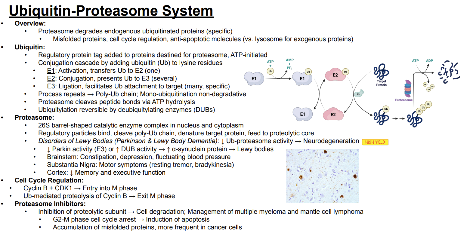
How can Parkin or deubiquitylating enzyme activity contribute to Lewy body formation?
Answer: Decreased Parkin activity or increased deubiquitylating enzyme activity → increased alpha-synuclein protein → Lewy bodies.
Extra Information:
Parkin is an E3 enzyme.
This pathway is marked as high yield on the slide.

What clinical findings are associated with Lewy body involvement of the brainstem, substantia nigra, and cortex?
Answer: Brainstem → constipation, depression, and fluctuating blood pressure → substantia nigra → resting tremor and bradykinesia → cortex → decreased memory and executive function.
Extra Information:
These findings are associated with Lewy body disorders on the slide.

How does the ubiquitin-proteasome system regulate progression through M phase?
Answer: Cyclin B plus cyclin-dependent kinase 1 causes entry into M phase, while ubiquitin-mediated proteolysis of Cyclin B causes exit from M phase.
Extra Information:
Proteasomal degradation therefore contributes to cell-cycle regulation.

What cell-cycle effect results from proteasome inhibition?
Answer: G2-M phase cell-cycle arrest followed by induction of apoptosis.
Extra Information:
Proteasome inhibition also causes accumulation of misfolded proteins.

How are proteasome inhibitors used clinically according to the slide?
Answer: They are used in the management of multiple myeloma and mantle cell lymphoma.
Extra Information:
They inhibit the proteolytic subunit.
This causes G2-M cell-cycle arrest, induction of apoptosis, and accumulation of misfolded proteins.
Misfolded protein accumulation is more frequent in cancer cells.

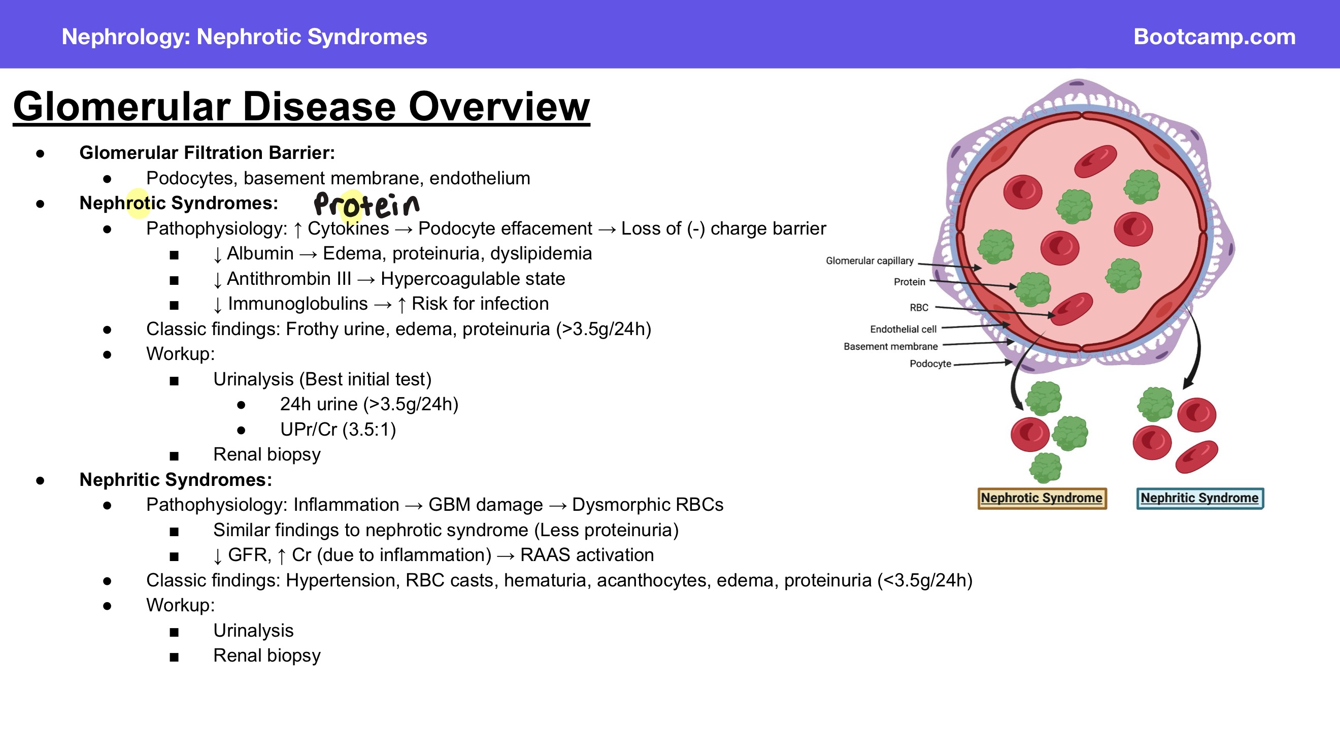
What are the three components of the glomerular filtration barrier?
Answer: Podocytes, basement membrane, and endothelium.
Extra Information:
These structures form the filtration barrier shown in the slide's glomerular capillary diagram.
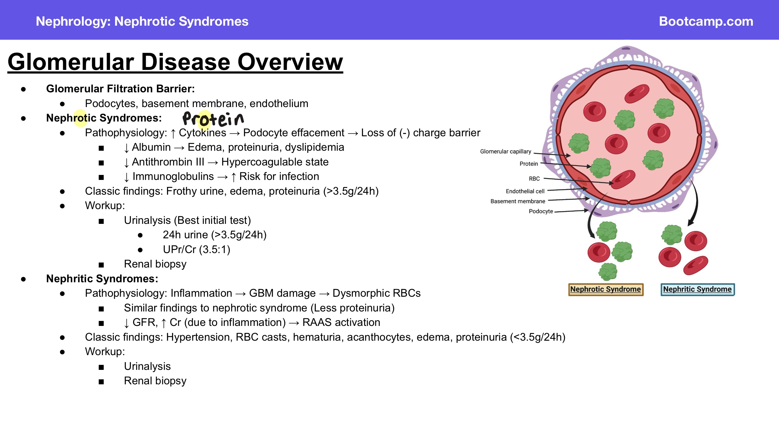
What is the pathophysiology of nephrotic syndrome according to the slide?
Answer: Increased cytokines → podocyte effacement → loss of the negative charge barrier.
Extra Information:
- Loss of the filtration barrier allows increased protein loss in the urine.

What happens to albumin in nephrotic syndrome?
Answer: Albumin decreases.
Extra Information:
- Loss of albumin is associated with edema, proteinuria, and dyslipidemia.
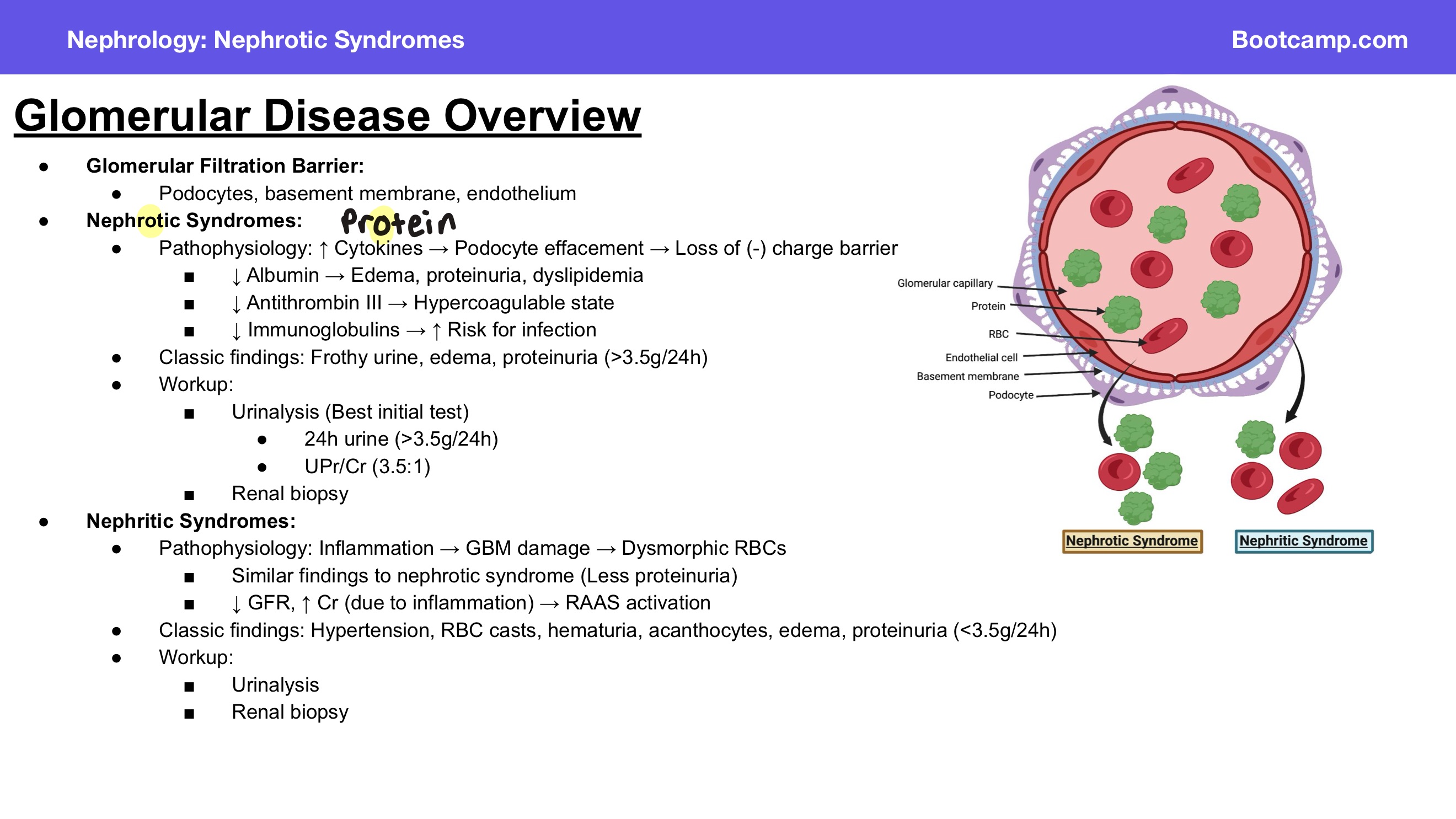
How does decreased albumin contribute to the findings of nephrotic syndrome?
Answer: Decreased albumin is associated with edema, proteinuria, and dyslipidemia.
Extra Information:
Albumin loss is one of the major protein abnormalities shown on the slide.

What happens to antithrombin III in nephrotic syndrome, and what is the consequence?
Answer: Antithrombin III decreases → hypercoagulable state.
Extra Information:
- Loss of antithrombin III is one of the consequences of nephrotic syndrome.

Why does nephrotic syndrome cause a hypercoagulable state?
Answer: Decreased antithrombin III.
Extra Information:
- This is one of the important consequences of urinary protein loss shown on the slide.
What happens to immunoglobulins in nephrotic syndrome, and what is the consequence?
Answer: Immunoglobulins decrease → increased risk for infection.
Extra Information:
- Loss of immunoglobulins increases susceptibility to infection.

What are the classic findings of nephrotic syndrome?
Answer: Frothy urine, edema, and proteinuria greater than 3.5 g/24 hours.
Extra Information:
- Heavy proteinuria is a defining finding on the slide.

How much proteinuria is associated with nephrotic syndrome?
Answer: Greater than 3.5 g/24 hours.
Extra Information:
- The slide also lists a urine protein-to-creatinine ratio of 3.5:1.

What is the best initial test in the workup of nephrotic syndrome?
Answer: Urinalysis.
Extra Information:
- Additional evaluation includes quantification of urinary protein and renal biopsy.

What urine protein findings support nephrotic syndrome?
Answer: Greater than 3.5 g of protein in a 24-hour urine collection or a urine protein-to-creatinine ratio of 3.5:1.
Extra Information:
- These values reflect heavy proteinuria.

What tests are included in the workup of nephrotic syndrome?
Answer: Urinalysis, urinary protein measurement, and renal biopsy.
Extra Information:
- Urinalysis is the best initial test according to the slide.

What is the pathophysiology of nephritic syndrome?
Answer: Inflammation → glomerular basement membrane damage → dysmorphic red blood cells.
Extra Information:
- The inflammatory damage allows abnormal red blood cells to appear in the urine.

Why are dysmorphic red blood cells seen in nephritic syndrome?
Answer: Inflammation damages the glomerular basement membrane.
Extra Information:
Dysmorphic red blood cells are a consequence of glomerular injury.

How does glomerular filtration rate change in nephritic syndrome?
Answer: Glomerular filtration rate decreases.
Extra Information:
- The decrease occurs due to inflammation.
- Creatinine increases and the renin-angiotensin-aldosterone system is activated.

Trace the effect of inflammation on kidney function in nephritic syndrome.
Answer: Inflammation → decreased glomerular filtration rate → increased creatinine → renin-angiotensin-aldosterone system activation.
Extra Information:
- This pathway is specifically shown on the slide.

What are the classic findings of nephritic syndrome?
Answer: Hypertension, red blood cell casts, hematuria, acanthocytes, edema, and proteinuria less than 3.5 g/24 hours.
Extra Information:
- Nephritic syndrome has less proteinuria than nephrotic syndrome.

How much proteinuria is associated with nephritic syndrome?
Answer: Less than 3.5 g/24 hours.
Extra Information:
- Compare with nephrotic syndrome, which has proteinuria greater than 3.5 g/24 hours.

What tests are included in the workup of nephritic syndrome?
Answer: Urinalysis and renal biopsy.
Extra Information:
- These are the two tests listed on the slide.

Compare the classic findings of nephrotic and nephritic syndromes.
Answer:
Nephrotic syndrome = frothy urine, edema, and proteinuria greater than 3.5 g/24 hours
nephritic syndrome = hypertension, red blood cell casts, hematuria, acanthocytes, edema, and proteinuria less than 3.5 g/24 hours.
Extra Information:
Nephrotic syndrome centers on podocyte effacement and loss of the negative charge barrier.
Nephritic syndrome centers on inflammation and glomerular basement membrane damage.
