Neurological disorders, seizures and epilepsy
 Call Kai
 Learn
Learn Practice Test
 Spaced Repetition
 Match
Match Flashcards
 Knowt Play
1/81

There's no tags or description
Looks like no tags are added yet.
Name | Mastery | Learn | Test | Matching | Spaced | Call with Kai | Chat |
|---|
No analytics yet
Send a link to your students to track their progress
82 Terms
direct injection of local anesthetic agents into the fetal scalp (associated with transient bradycardia and fixed dilated pupils), severe anoxia, or congenital brain malformation
seizures noted in the delivery room are often caused by
infection or drug withdrawal
seizure in the first 5 days of life is most likely a result of
benign familial neonatal seizures
these seizures have onset on days 3-7 and resolve by 1-4 months, and are due to mutations in KCNQ2 and KCNQ3 potassium channel genes.
subarachnonid hemorrhage
In an infant who appears well, a sudden onset on days 1-3 of life of seizures that are of short duration and that do not recur may be the result of
local cerebral infarction
focal seizures are often the result of
define a simple febrile seizure
more than 15 min
focal or
more than 2 episodes in 24 hours
define a complex febrile seiizure
myoclonic seizures
-very brief LOC leading to the person's arms/legs jerkingg sharply and uncontrollably
tonic seizures
-instant LOC; person goes stiff and falls to the ground
tonic-clonic seizure
-instant LOC
-person will go stiff and then move into jerky movements
-may experience incontinence
absence seizures
-brief loss of awareness accompanied by eyelid flutter or simple automatisms
-usually 4-6 years old
-characteristic electroencephalographic (EEG) patterns consist of generalized 3-Hz spike-and-wave activity
hyperventilation
an absence seizure can often be provoked by
focal simple seizure
No LOC, experince altered feelings or sensation, symptoms depend on the area of the brain affected, usually 20-60 seconds, returns to the pre-seizure state and remembers of the event
focal complex seizure
previously termed complex partial seizures ) can have similar sensorimotor signs but also have associated alteration of consciousness. Although the child may not be completely unresponsive, subtle slowing or alteration of mental status (dyscognitive features) may occur
west syndrome (infantile spasm)
Brief contractions of the neck, trunk, and arm muscles, followed by a phase of sustained muscle contraction lasting less than 2 seconds. Spasms occur most frequently when the child is awakening or going to sleep. Each jerk is followed by a brief period of relaxation with repeated spasms in clusters of variable duration.
-infantile spasms, developmental regression, and dramatically abnormal EEG pattern (hypsarrhythmia)
triad of west syndrome
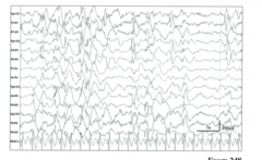
Hypsarrhythmia
a pattern of chaotic high-voltage slow waves, spikes, and polyspikes
-seen in west syndrome
3-8 months
peak age of onset of infantilie spasms
adrenocorticotropic hormone, high-dose oral corticosteroids, and vigabatrin
first line treatment for infantile spasms
Benign epilepsy with centrotemporal spikes (benign rolandic epilepsy)
Autosomal dominant genetic disorder linked to abnormal neuronal potassium channels. Otherwise well newborns present with focal seizures toward the end of the first week of life, leading to the colloquial term fifth-day fits . Response to treatment is generally excellent, and the long-term outcome is typically favorable
lennox-gastaut syndrome
Severe epilepsy syndrome with variable age of onset although most children present before age 5 years. Frequent, multiple seizure types including atonic, focal, atypical absence, and generalized tonic, clonic, or tonic-clonic semiologies characterize the disorder. Many children have underlying brain injury, malformations, or genetic etiologies. The seizures are typically difficult to control, and most patients have significant intellectual disability.
Absence seizures typically begin in the early school years and usually resolve by late childhood or adolescence. If absence does not remit, nearly half will go on to develop juvenile myoclonic epilepsy
prognosis of childhood absence epilepsy
Ethosuxamide
1st choice med for absence epilepsy
juvenile myoclonic epilepsy
Most common generalized epilepsy among adolescents and young adults.
-usually lifelong
juvenile myoclonic epiplepssy
Onset is typically in early adolescence with myoclonic jerks (exacerbated in the morning, often causing the patient to drop objects), generalized tonic-clonic seizures, and absence seizures. Seizures usually resolve promptly with anti seizure medication
-valproic acid
-levetiracetam lower risk (preferred in girls bc of less teratogenicity)
classic treatment of juvenile myoclonic epilepsy
(1) ongoing seizure activity for 5 or more minutes or (2) repetitive seizures without recovery of consciousness
define a neurologic emergency
Initial management is with a benzodiazepine and should be administered if the seizure persists longer than 5 minutes. Lorazepam, diazepam, and midazolam are all effective agents and should be given as an adequate single dose (rather than successive smaller doses), which can be repeated one time. IV administration is preferred. When IV access is not available, benzodiazepines may be administered via rectal, intranasal, buccal, or intramuscular routes.
initial management of status epilepticus
Lorazepam, diazepam, and midazolam
preferred benzos in acute seizure tx
-newborns and infants: phenobarbital
-older children: IV fosphenytoin, valproic acid, or levetiracetam
tx of ongoing seizures after two doses of benzodiazepine
cerebral palsy
A group of non-progressive, but often changing, motor impairment syndromes secondary to anomalies or lesions of the brain arising before or after birth
diagnosed in the first 18 months of life when they fail to attain motor milestones or show abnormalities such as asymmetric gross motor function, hypertonia, or hypotonia
when are most pts with CP diagnosed?
spastic cerebral palsy
-most common form of CP
-results from injury to the UMN of the pyramidal tract
-characterized by abnormal movement pattern, increased tone, or pathologic reflexes
dyskinetic cerebral palsy
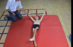
CP type dominated by abnormal patterns of movement and involuntary, uncontrolled recurring movements
ataxic cerebral palsy
-results from cerebellar injury and features abnormal posturue or movement and loss of orderly muscle coordination or both
-type of CP
acute disseminating encephalomyelitis
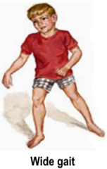
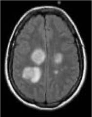
Acute, inflammatory demyelinating disorder that may affect infants and children after a febrile illness. Children may present with a variety of symptoms and signs including monocular vision loss (e.g., acute optic neuritis), hemiparesis, ataxia, seizures, headache, weakness, or dysphagia.
Intestinal infection by Clostridium botulinum , which produces a neurotoxin that blocks presynaptic cholinergic transmission. Young age and the absence of competitive bowel flora predispose infants to this disease. Infants may ingest dust, soil, or food (e.g., honey, poorly canned foods) contaminated with spores
describe the pathophys of botulism in children
Infants typically present with constipation and poor feeding. Hypotonia and weakness develop progressively, along with cranial nerve dysfunction manifested by decreased gag reflex, diminished eye movements, decreased pupillary contraction, and ptosis.
presentation of botulinism in infants
duchenne muscular dystrophy
X-linked disorder (Xp21) that arises from a mutation in the dystrophin gene
Duchenne muscular dystrophy
-at about 2-3 years old, male develops an awkward gait and inability to run
-x of mild delay in motor milestones
-pseudohypertrophy of calves
by 12, most boys are not walking and use a wheel chair full time
prognosis of DMD
Studies have shown that chronic oral steroid therapy delays the motor disability and improves longevity by preventing cardiac and pulmonary decline, even after a boy stops ambulating.
potential pharm management of DMD
acute inflammatory demyelinating polyradiculoneuropathy
most common variant of guillan=barre syndrome
acute inflammatory demyelinating polyradiculoneuropathy
-GBS variant
classically occurs about 10 days after a respiratory or gastrointestinal infection (e.g., Mycoplasma pneumoniae or Campylobacter jejuni ). It is the most common cause of acute flaccid paralysis in children.
Guillain-Barre Syndrome (GBS)
Areflexia, flaccidity, and symmetric ascending weakness. Progression can occur rapidly, in hours, or more indolently over weeks. Typically, symptoms start with numbness or paresthesia in the hands and feet, then a heavy, weak feeling in the legs. Weakness ascends to involve the arms, trunk, and bulbar muscles (tongue, pharynx, larynx). Deep tendon reflexes are absent even when strength is relatively preserved.
sometimes normal early in the illness but classically shows elevated protein levels without significant pleocytosis (albuminocytologic dissociation)
LP findings of GBS
Most patients are treated initially with intravenous immunoglobulin (IVIG). Plasma exchange is an alternative option. Physical, occupational, and speech therapies are mainstays of treatment.
treatment of GBS
fluctuating skeletal muscle weakness
cardinaal feature of juvenile myasthenia gravis
transient myasthenic syndrome develops in the first hours to days after birth in neonates born to mothers with myasthenia gravis related to maternal anti-AChR antibodies
Signs include weak facial movements, poor feeding, hypotonia, respiratory difficulty, and variable extremity weakness
-presentation of babies born to myasthenia gravis mothers
require cholinesterase inhibitors and supportive care for a few days to weeks until the weakness remits.
treatment of transient myastheniaa gravis in neonates
some types can respond to pyridostigmine or other drugs that improve neuromuscular junction function, other types can be exacerbated by pyridostigmine
treatment of congenital myasthenic syndromes
spinal muscular atrophy
defined by progressive degeneration of anterior horn cells
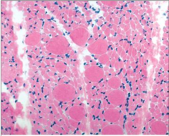
severe hypotonia, generalized weakness, and facial involvement. Infants have normal cognitive, social, and language skills and sensation. Fasciculations are best identified by inspecting the tongue when the child is asleep. Deep tendon reflexes are absent. With disease progression, breathing patterns change. Weak intercostal muscles and a relatively stronger diaphragm result in a collapsed chest wall (bell-shaped chest) and prominent abdominal movements with rapid, shallow breathing.
features of spinal muscular atrophy type one
-deficient enzyme: galactocerebrosidase
-accumulated substrate: galactocerebroside
deficient enzyme and accumulated substrate of Krabbe disease
Krabbe disease
Caused by inability to produce one lysosomal enzyme
Person cannot produce myelin for nerve cells, which leads to severe damage to the nervous system
Krabbe disease
autosomal recessive lysosomal storage disease
-Peripheral neuropathy, destruction of oligodendrocytes, developmental delay, optic atrophy, globoid cells
Hunter syndrome
Mild hurler + aggressive behavior, no corneal clouding
Deficient enzyme → iduronate-2-sulfatase
Accumulated substrate → heparan sulfate, dermatan sulfate
deficient enzyme and accumuluated substrate in Hunter syndrome
Hunter syndrome
X-linked recessive
similar to Hurler, deficient L iduronosulfate sulfatase, accumulations of heparin sulfate and dermatan sulfate, hepatospenomegaly, micrognathia, retinal degeneration, joint stiffness, mental retardation, cardiac lesions p59
Hurler syndrome
autosomal recessive - corneal clouding, course facial features, joint stiffness, mental retardation, hepatosplenomegaly
Deficient enzyme → alpha-L-iduronidase
Accumulated substrate → heparan sulfate, dermatan sulfate
deficient enzyme and accumulated substrate in hurler syndrome
Rett syndrome
-Neurodevelopmental disorder that classically affects girls
-Development appears normal during the first 6-18 months of life, but this is followed by developmental regression, loss of purposeful hand movements, loss of verbal communication skills, gait apraxia, and stereotypic repetitive hand movements that resemble washing, wringing, or clapping of the hands. Girls also develop acquired microcephaly.
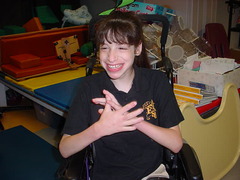
The etiology is a mutation on an X chromosome gene coding for methyl-CpG-binding protein 2 (MECP2) transcription factor
etiology of Rett syndrome
Tay-Sachs disease
-autosomal recessive
Progressive neurodegeneration, developmental delay, hyperreflexia, hyperacusis, "cherry red" spot on macula (lipid accumulation in ganglion cell layer), lysosomes with onion skin, no hepatosplenomegaly (vs niemann-pick
Deficient enzyme → hexosaminidase A
Accumulated substrate → GM2 ganglioside
deficient enzyme and accumulated substrate in Tay-sachs
-autosomal dominant
-mutations of NF1 gene which codes for tumor suppressor gene, neurofibromin
genetics associated with neurofibromatosis type 1
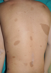
-cafe au lait spots
-cutaneous neuroffibromas
-iris hamartomas
cardinal features of neurofibromatosis type 1
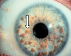
neurofibromatosis type 1
The presence of six or more café-au-lait spots larger than 5 mm in a prepubescent child suggests the diagnosis.
-learning disability, scoliosis, seizures, moyamoya
-optic nerve gliomas, astrocytomas, malignant peripheral nerve tumors
common complications associated wit NF type 1
bilateral acoustic schwannomas, schwannomas of other cranial and spinal nerves, meningiomas, and glioma
NF type 2 preddispoess the pt to these kindd of tumors
Lisch nodules, café-au-lait spots, and axillary freckling (seen in NF1) are not features of NF2.
main difference between NF type 1 and NF type 2
Sporadic (not inherited) and is caused by a somatic mosaic mutation of the GNAQ gene
genetics associate with Sturge-Weber syndrome
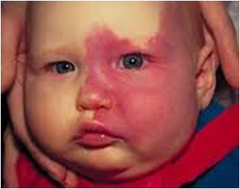
-angiomas of the leptomeninges in association with ipsilateral port-wine stain involving the opthalmic division of the trigeminal nerve
main features of Sturge-Weber ssyndrome
Sturge-Weber syndrome
Skin and meningeal angiomatous lesions. Port-wine nevus: skin angioma in ophthalmic division of trigeminal nerve. Pial angiomas may result in chronic ischemia, gliosis, and gyral cortical calcifications. Enlargement of deep and subependymal veins may mimic arteriovenous malformations.
Tuberous sclerosis
Autosomal dominant disorder, is characterized by hamartomas in many organs, especially the brain, eyes, skin, kidneys, and heart.
May have retinal lesions (retinal hamartomas, white depigmented patches) and brain lesions (cortical tubers, subependymal nodules, hydrocephalus). Tubers in the cerebral cortex are areas of dysplasia that, in combination with other microscopic areas of abnormal development, are responsible for the symptoms of intellectual disability and epilepsy.
main features of tuberous sclerosis
tuberous sclerosis
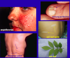
Skin lesions: Adenoma sebaceum. Ash-leaf spots. Brain lesions: Subependymal hamartomas. Cortical tubers. Subependymal giant cell astrocytomas located at foramina of Monro (May lead to hydrocephalus)
Subependymal giant cell astrocytoma
Seen in 10% of patients with tuberous sclerosis (look for subependymal and cortical hamartomas). WHO grade I. Commonly calcifies. and slow-growing, with calcification a common feature. Almost always produces some degree of hydrocephalus.
Tuberous sclerosis
one of the most common causes of infantile spasms; in this context, the infantile spasms often respond to treatment with vigabatrin
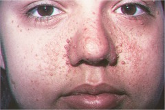
facial angiofibromas
Multiple smooth-surfaced papules most often found on the nose or nasolabial fold in tuberous sclerosis
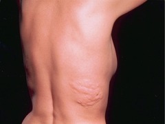
shagreen patches
Areas of rough, leathery, orange peel-like skin typically seen on the lower back or nape of the neck in tuberous sclerosis
-ash leaf spots
-facial angiofibromas
-shagreen patches
main extracerebral manifestations of tuberous sclerosis