8 Topography of the carnivore's abdomen
 Call Kai
 Learn
Learn Practice Test
 Spaced Repetition
 Match
Match Flashcards
 Knowt Play
1/80

Earn XP
Description and Tags
W3 Elnady
Name | Mastery | Learn | Test | Matching | Spaced | Call with Kai | Chat |
|---|
No analytics yet
Send a link to your students to track their progress
81 Terms
What are the transverse abdominal regions?
1. cranial- at caudal borders of the costal arches just cranial to the umbilicus
2. caudal- cranial to the wing of each ilium

What are the 9 abdominal subregions?
1. cranial abdominal region (epigastric and right and left hypochondriac)
2. middle abdominal region (umbilical and right and left lateral/flank; dorsal part of paralumbar fossa)
3. caudal abdominal region (pubic and R/L inguinal)
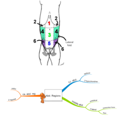
What is the lateral (flank) fold?
the fold of skin that attaches the caudal abdomen to the cranial thigh
what is the paralumbar fossa?
the upper part of the lateral abdominal region

where is the paralumbar fossa
it is the triangular depression just cranial to the hip and ilium, it is caudal to the costal arch (last rib) and ventral to the lumbar vertebrae
it is bound cranially by the last rib, dorsally by the transverse processes, and caudally by the internal abdominal oblique
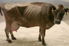
Why is the paralumbar fossa important?
it is used in large animals to gain access to the rumen

what is the flank?
the ventrolateral part of the abdominal wall between the thoracic cage and bones of the pelvis
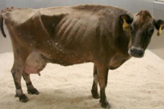
What are the boundaries of the abdominal cavity?
it is bound cranially by the diaphragm, caudally by the pelvic inlet, dorsally by the lumbar/sacral vertebrae, diaphragmatic pleura, and hypaxial muscles, and laterally/ventrally by the abdominal muscles

what are the layers of the abdominal wall?
1. superficial fascia (encloses cutaneous trunci)
2. deep fascia (thoracolumbar fascia, tunica flava abdominis (large animals))
3. muscles
4. internal fascia
5. parietal peritoneum
What is special about the superficial fasci?
it is only present in carnivores, it is neither attached to the spinous processes nor to the linea alba. so the wet dog can move their trunk skin extensively to dry themselves
what is the arrangement of the abdominal wall muscles?
The EAO fibers are oriented caudoventrally
The IAO fibers are oriented cranioventrally
Transverse abdominis fibers are oriented dorsally

what is the insertion of the abdominal wall muscles?
linea alba and prepubic tendon
What are the actions of the abdominal muscles?
1. abdominal press
2. bilateral (both sides contract) = flex trunk ventrally
3. unilateral (one side contracts) = rotate the trunk laterally
what nerves supply the abdominal muscles?
lateral branches of the last several intercostal nn.
first three lumbar nn.
why are the arrangements of the muscles important?
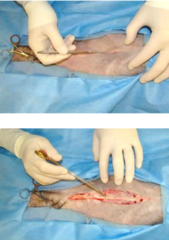
during flank incisions (as in spays/neuter or caesarian sections) the surgeon separates the muscles along the direction of their fibers instead of cutting though them (so there is less bleeding)

what does the internal fascia (sub-serous fascia) do?
it attaches the parietal layer of the pleura/peritoneum to the body wall
ex. endothoracic fascia and endo-abdominal fascia
What does the endothoracic fascia do?
lines the thoracic cavity
What does the endo-abdominal fascia do? (sub serous fascia)
it lines the abdominal cavity and extends into the pelvic cavity (endopelvic fascia)
ex. transversalis fascia is over the transversus abdominis m.

what is the linea alba?
an elongate ventral midline tendinous structure from the xiphoid cartilage to the pubis. it contains the umbilicus.
it is a common insertion for abdominal mm.
the Linea alba is easier to locate ______________________ because it becomes thinner near the pubis
near the umbilicus

Should you make an incision on the linea alba or a paramedian cut?
There are pros and cons
If you open the Linea alba there is no bleeding however it will take time to heal.
There is less sensory innervation (less post-operative pain)
there is strong closure
- if you do a paramedian incision, you need a stronger suture and it is more painful, but it heals faster.
the linea alba serves as the attachment site for ___________________
1. ventrolateral abdominal mm.
2. falciform ligament
3. median ligament of the bladder
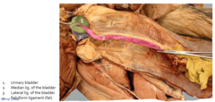
where is the umbilicus/ belly button?
it is a closed scar in the ventral midline (@ the transverse plane across the last ribs)
it is in the cranial part of the umbilical region
it is a landmark in abdominal surgical incisions
_____________ hair surrounds the umbilicus grows in whirl
vortex
what is an umbilical hernia?
escape of abdominal contents through the umbilical ring (if not closed)

Where is the liver in the abdomin?
it is against the diaphragm
it is in the right and left hypochondriac and xiphoid regions
chiefly on the right side

where do you perform a needle biopsy of the liver?
Right side; 7th ICS
Left side: caudodorsal to xiphoid

What are the two types of stomachs?
1. simple = monocular = one chamber (human, cat, horse, pig)
2. complex = multilocular (ruminants)

What are the two types of simple stomachs?
1. simple glandular = one type of mucous membrane (man, dog, cat)
2. simple/composite = one area glandular and the other non-glandular (horse and pig)
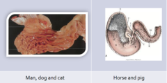
true/false- normally the stomach is non-palpable
true- it is usually hidden by the ribs

true/false- the stomach is freely moveable
TRUE- BUT the cardiac and pyloric extremities are relatively fixed

What is the shape of the stomach in the dog?
It is U-shaped and the pylorus is right of the mid-line

What is the shape of the feline stomach?
angulated (J-shaped)
pyloric lies on or near the midline

can the empty stomach be palpated?
NO- it is in the intrathoracic part of the abdominal cavity
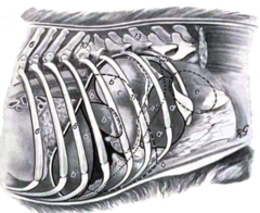
true/false- the stomach is attached to the spleen
yes, whenever the stomach moves the spleen will move with it because it is attached

when the stomach is moderately full where is it located?
under the last four ribs; it contacts the abdominal floor
part of the spleen protrudes over the costal arch

When the stomach is full where is it?
it is reaching a level of L2,3,4 and pelvic brim. it reaches from the right to the left body walls.
it displaces the spleen and left kidney caudally

can the full stomach be palpated?
YES- it has extensive contact with the abdominal floor and easily palpated
How can you measure the length of a stomach tube?
before a stomach tube is passed, the length of the tube to be passed is marked by measuring from the nose to the last rib

what are the parts of the small intestine?
1. duodenum
2. jejunum
3. ileum

What are the parts of the large intestine?
1. cecum
2. colon
3. rectum and anal canal
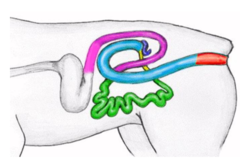
where is the cranial duodenum?
- it is fixed to the liver, but can move up and down with pyloric end
- may be cr. or ca. to pancreas (depends on position of the stomach)

the lesser curvature is _____________
cranial

the greater curvature is ______________
caudal

true/false- the cranial part of the duodenum is fairly mobile, the remainder of the duodenum is relatively fixed in position
TRUE

The descending duodenum is on the ____________ side
RIGHT

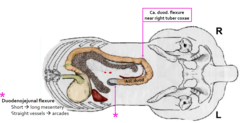
where is the caudal duodenal flexure located?
it is near the right tuber coxae caudal to the right kidney

the descending duodenum used as a landmark to find the _______________________
right lobe of the pancreas

the mesoduodenum contains the ______________________
right lobe of the pancreas

where is the ascending duodenum?
it extends cranially, in the median plane
it almost reaches the greater curvature of the stomach
what happens at the duodenojejunal flexure?
there is a change from short mesentery to the long mesentery
the mesoduodenum of the ascending duodenum is attached to the ______________________
descending mesocolon
it is called the duodenocolic fold
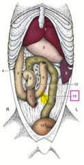
what is the clinical importance of the duodenocolic fold?
it is a landmark used during "running of the bowel" during celiotomy
what are the parts of the jejunum?
the lesser (Mesenteric) and greater (antimesenteric) curvatures
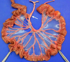
the mesojejunum is attached to the ___________________
short root of the mesentery

how can you identify the ileum from the jejunum?
-the ileum has mesenteric and anti-mesenteric blood vessels on both sides
-it has short mesentery and the ileocecal fold
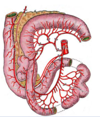
where is the ileum?
-it ends dorsally on the right side of the body (right to ascending duodenum)
-it opens into the ascending colon (ileo-colic junction)
the cat has the ________________ junction
ileo-ceco-colic junction
what is the most common part of the small intestine that is involved in intussusception?
ileum

what are the parts of the pancreas?
1. left lobe of the pancreas
2. body
3. right lobe of the pancreas

the pancreas extends almost _____________________ across the ventral aspect of the vertebral column
transversely

what is the cecum?
it is the diverticulum of the colon

where is the cecum located?
- in the right side of the cranial abdomen
- close to the dorsal body wall (L2-L4): dorsal to the jejunum

how does the cecum of the dog differ from the cecum of the cat?
in the cat the cecum smoothly joins the ascending colon with no demarcation
the dog has a larger cecum since it is more herbivorous. it is S shaped
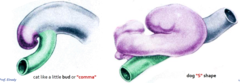
true/false- the cecum and the colon are pushed against the roof of the abdomen by the small intestine
TRUE
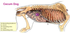
what are the parts of the colon?
1. ascending
2. transverse
3. descending
4. mesocolon

where is the ascending colon located?
9
in right dorsal portion of abdominal cavity
right to cranial mesenteric artery
right colic flexure

where is the transverse colon?
10
cranial to the root of the mesentery and cranial to cranial mesenteric artery
left colic flexure

where is the descending colon?
11
left to the midline and cranial mesenteric artery
At the pelvic inlet it continues as the rectum

what is present in the lumbar area "gutter"?
-adrenal glands
-kidneys
-ureters
-uterus/ovaries

in order to examine the right lumbar gutter, what organ must you retract?
the descending duodenum. use the mesoduodenum to retract the intestine to the left (toward the midline)
to examine the left lumbar gutter, what organ must you retract?
the descending colon
use the descending mesocolon to retract the abdominal contents to the right (toward the midline)
what are the parts of the adrenal gland?
cortex and medulla
Does the cortex or the medulla or the adrenal gland have nerve supply?
the medulla has abundant nerve supply. The cortex lacks nerve supply
The medulla of the adrenal gland has is innervated by _______________________
sympathetic postganglionics
true/false- the adrenal medulla is essential for life
FALSE- the dog can live if the adrenal medulla is bilaterally removed. BUT the cortex is essential for life
how are the adrenal glands supplied with blood?
-they lie close to their rich source of arterial supply and venous drainage

where are the kindeys located?
in the sublumbar region. They are retroperitoneal and lie on either side of the aorta and caudal vena cava
true/false- the palpation of both kidneys is easier in dogs than cats
FALSE- in the dog it is hard to palpate the right kidney because it is more cranial. The left is less firmly attached to the sublumbar area than the right
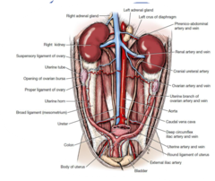
what is something that needs to be kept in mind about the bladder when performing surgery?
in midline celiotomy, the bladder is in danger of being accidentally incised if urine has not been expelled prior to surgery.
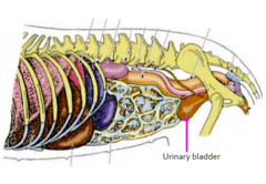
where can the bladder be palpated?
in the ventral caudal abdomen
