Textbook 9-12
1/288
There's no tags or description
Looks like no tags are added yet.
Name | Mastery | Learn | Test | Matching | Spaced |
|---|
No study sessions yet.
289 Terms
What is the primary function of hearing, and how does it reflect both shared sensory principles and unique evolutionary adaptations?
It involves detecting and interpreting sound using principles common to all senses, while also showcasing distinct biological solutions that evolved at different times to address specific environmental challenges.
Amplitude
In reference to vestibular sensation, the size (increase or decrease) of a head movement (with angular velocity, linear acceleration, tilt, etc.).
intensity
In reference to sound, the magnitude of displacement (increase or decrease) of a pressure wave. Amplitude is perceived as loudness.
Frequency
In reference to sound, the number of times per second that a pattern of pressure change repeats. Frequency is perceived as pitch.
Hertz (Hz)
A unit of measure for frequency; 1 hertz equals 1 cycle per second.
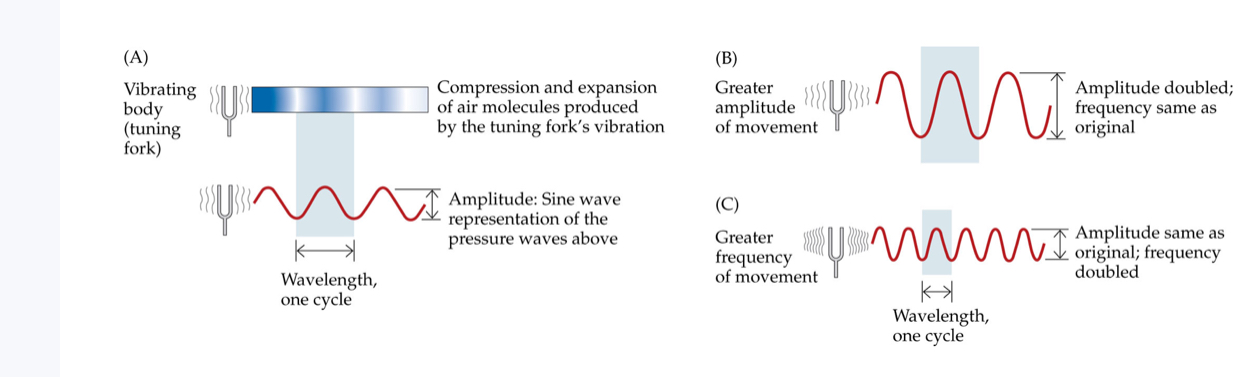
Amplitude and frequency
(A) Sound waves are described by the frequency and amplitude of pressure fluctuations. Changes in amplitude (B) and frequency (C) are shown for sine waves, the simplest kind of sound wave
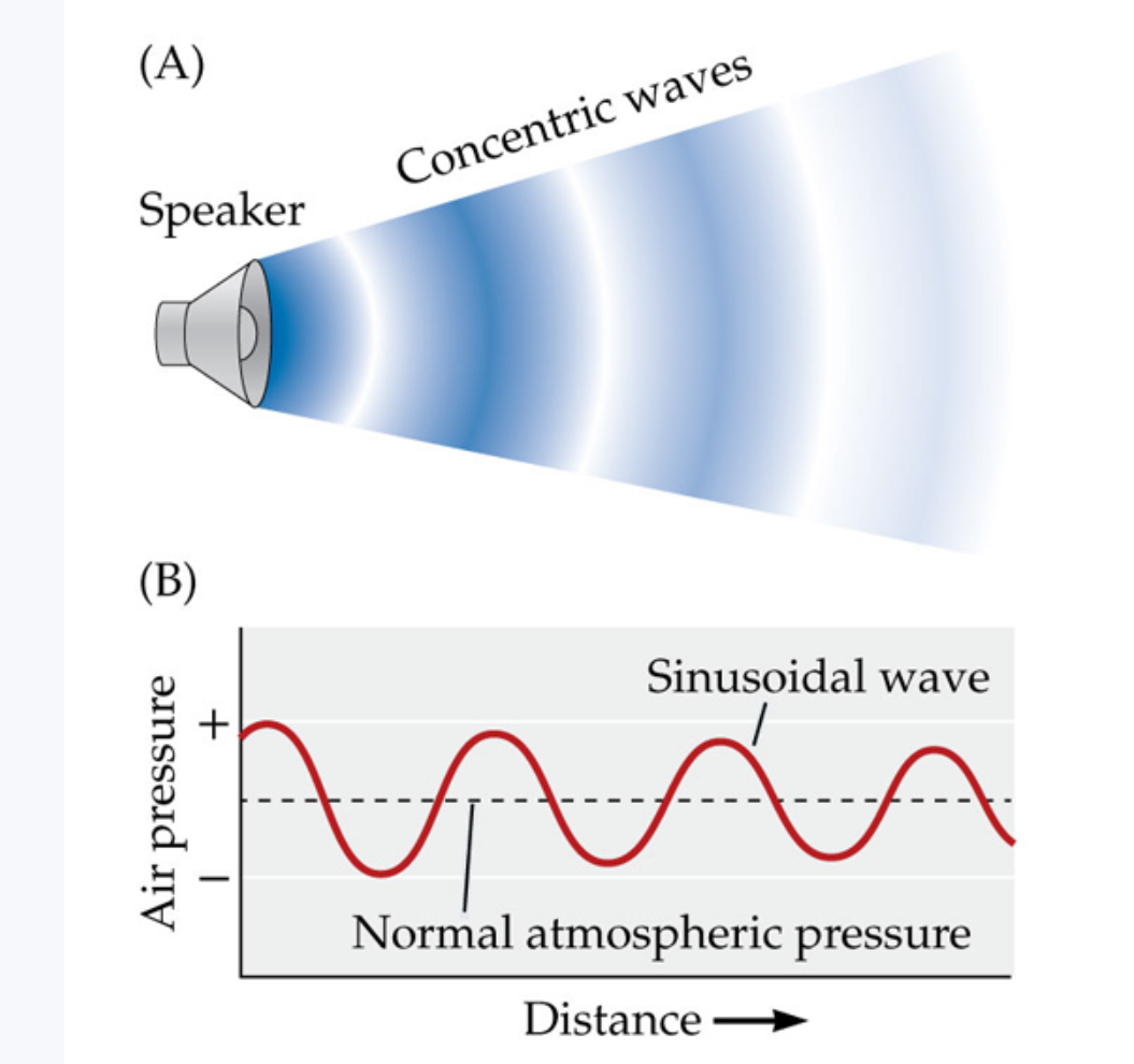
Sounds moving away from sources
The pattern of pressure fluctuations of a sound, distances peak to peak, stays the same as the sound wave moves away from the source (A), but the amount of pressure change, the height of peaks relative to the depth of valleys, decreases with increasing distance (B)
Loudness
The psychological aspect of sound related to perceived intensity (amplitude).
Pitch
The psychological aspect of sound related mainly to the perceived frequency.
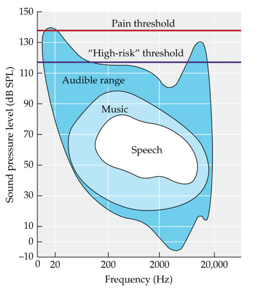
Range of human hearing
Humans can hear frequencies that range from about 20 to 20,000 Hz across a very wide range of intensities, or sound pressure levels (dB SPL, where dB = decibels)
Decibels (dB)
A unit of measure for the physical intensity of sound. Decibels define the difference between two sounds as the ratio between two sound pressures. Each 10:1 sound pressure ratio equals 20 dB, and a 100:1 ratio equals 40 dB.
Pure tone / sine wave
The single waveform for which variation as a function of time is a sine function. In hearing research, this is sometimes referred to as a pure tone.
Spectrum
A representation of the relative energy (intensity) present at each frequency.
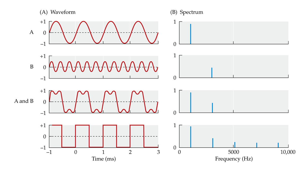
Waveforms and spectra
A spectrum displays the amplitude for each frequency present in a sound wave. Each signal is shown as a waveform (A) and as a spectrum (B).
Harmonic spectra
The spectrum of a complex sound in which energy is at integer multiples of the fundamental frequency.
fundamental frequency
The lowest-frequency component of a complex periodic sound.
Spectral shapes and timbre
Harmonic sounds with the same fundamental frequency can sound different because amplitudes of individual frequency components are different, resulting in different spectral shapes. For example, different musical instruments playing the same note (the same fundamental frequency, abbreviated f) sound different. C4 = middle C.
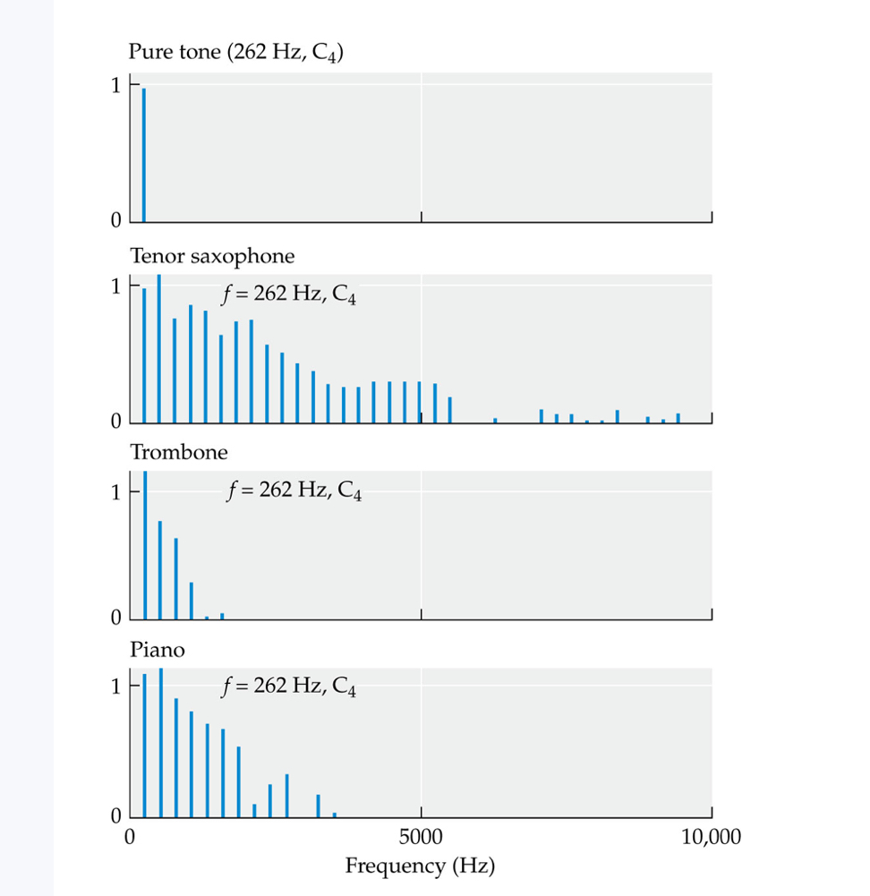
Timbre
The psychological sensation by which a listener can judge that two sounds with the same loudness and pitch are dissimilar. Timbre quality is conveyed by harmonics and other high frequencies.
How are sound waves produced, and what medium typically carries them for humans?
They are created when an object vibrates, causing nearby molecules—usually air—to vibrate as well. This produces pressure fluctuations in the medium that travel as waves away from the source.
What happens to the pattern and strength of a sound wave as it moves away from its source?
While the pattern of pressure fluctuations remains the same, the magnitude of those fluctuations decreases because the initial pressure change disperses over a larger area.
How does the speed of sound vary between different media, and how does it compare to the speed of light?
It moves faster in denser substances, traveling about 340 meters per second in air and around 1500 meters per second in water. Light moves through air nearly a million times faster than sound, explaining why lightning is seen before thunder is heard.
What aspect of a sound wave corresponds to loudness, and how is it defined?
The amplitude, or intensity, represents the magnitude of pressure changes in a sound wave and determines how loud it sounds.
What aspect of a sound wave corresponds to pitch, and how is it defined?
The frequency, which measures how quickly the pressure fluctuates, determines the pitch. Higher frequencies produce higher pitches, and lower frequencies produce lower pitches.
How is the frequency of a sound wave measured?
It is measured in hertz (Hz), where 1 Hz equals one cycle of pressure fluctuation per second.
What frequency range can young, healthy human ears typically detect?
Approximately 20 to 20,000 Hz.
How does animal size generally relate to the range of frequencies it can hear?
Larger animals tend to detect lower frequencies, while smaller animals are better at detecting higher frequencies.
Why are decibels (dB) used to measure sound amplitude, and what kind of scale is it?
They allow a compact description of a huge range of intensities using a logarithmic, compressed scale where each 10:1 sound pressure ratio equals 20 dB.
What is an important consideration when interpreting decibel differences between sounds?
Small differences in decibels can represent very large physical differences in sound pressure. For example, a 44 dB difference can mean a sound is over 150 times more intense.
What is a pure tone, and how is it related to sine waves?
consists of a single sine wave, representing the simplest type of sound wave with a single frequency and amplitude.
How can complex sounds be described in terms of their frequency components?
By using a spectrum that displays the amount of energy or amplitude present at each frequency within the sound.
What is a harmonic spectrum, and what typically produces it?
It consists of multiple frequency components that are integer multiples of a fundamental frequency, often produced by vibrating sources like vocal cords or musical instruments.
What is the fundamental frequency in a harmonic sound?
It is the lowest-frequency component of the sound and serves as the basis for determining the other harmonics.
What is timbre, and what does it depend on?
It refers to the perceptual quality that distinguishes different sounds with the same fundamental frequency. It depends partly on the shape of the sound's spectrum—the pattern of amplitudes for each harmonic.
Pinna
The outer, funnel-like part of the ear.
Ear canal
The canal that conducts sound vibrations from the pinna to the tympanic membrane and prevents damage to the tympanic membrane.
Outer ear
The external sound-gathering portion of the ear, consisting of the pinna and the ear canal.
tympanic membrane
The eardrum; a thin sheet of skin at the end of the outer ear canal. The tympanic membrane vibrates in response to sound.
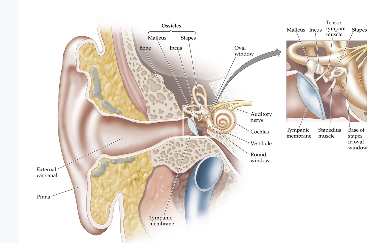
Structures of the human ear
The three parts of the outer ear are the pinna, external ear canal, and tympanic membrane. The middle ear is composed of the oval window and three tiny bones (the ossicles) that connect it to the tympanic membrane (eardrum). Note that the tympanic membrane has about 18 times as much surface area as the oval window beneath the stapes. The cochlea is the major structure of the inner ear, where sound waves (vibrations) are transduced into neural signals that are then sent to the auditory cortex of the brain
Middle ear
An air-filled chamber containing the middle bones, or ossicles. The middle ear conveys and amplifies vibration from the tympanic membrane to the oval window.
Ossicles
Any of three tiny bones of the middle ear: malleus, incus, and stapes.
Malleus
The most exterior of the three ossicles. The malleus receives vibration from the tympanic membrane and is attached to the incus.
Incus
The middle of the three ossicles, connecting the malleus and the stapes.
Stapes
The most interior of the three ossicles. Connected to the incus on one end, the stapes presses against the oval window of the cochlea on the other end.
Oval window
The flexible opening to the cochlea through which the stapes transmits vibration to the fluid inside.
Inner ear
A hollow cavity in the temporal bone of the skull and the structures within this cavity: the cochlea and the semicircular canals of the vestibular system.
tensor tympani
The muscle attached to the malleus. Tensing the tensor tympani decreases vibration.
stapedius
The muscle attached to the stapes. Tensing the stapedius decreases vibration.
acoustic reflex
A reflex that protects the ear from intense sounds via contraction of the stapedius and tensor tympani muscles.
Cochlea
A spiral structure of the inner ear containing the organ of Corti.
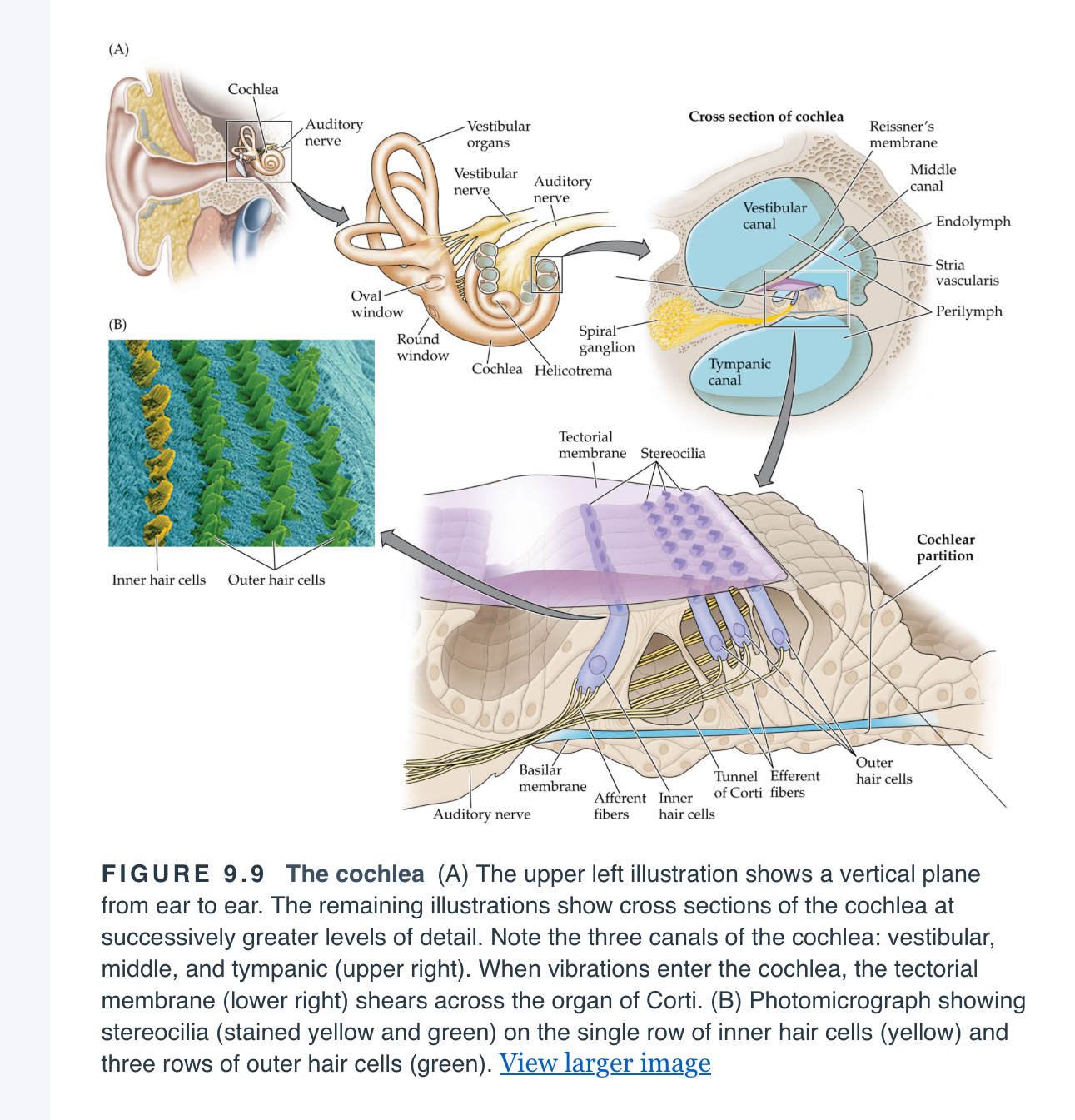
tympanic canal
One of three fluid-filled passages in the cochlea. The tympanic canal extends from the round window at the base of the cochlea to the helicotrema at the apex. Also called scala tympani.
vestibular canal
One of three fluid-filled passages in the cochlea. The vestibular canal extends from the oval window at the base of the cochlea to the helicotrema at the apex. Also called scala vestibuli.
Middle canal
One of three fluid-filled passages in the cochlea. The middle canal is sandwiched between the tympanic and vestibular canals and contains the cochlear partition. Also called scala media.
helicotrema
The opening that connects the tympanic and vestibular canals at the apex of the cochlea.
stria vascularis
Specialized tissue lines one side of the middle canal and maintains the right balance of charged ions in the endolymph to keep hair cells working at their best.
Reissner’s membrane
A thin sheath of tissue separating the vestibular and middle canals in the cochlea
basilar membrane
A plate of fibers that forms the base of the cochlear partition and separates the middle and tympanic canals in the cochlea.
cochlear partition
The combined basilar membrane, tectorial membrane, and organ of Corti, which are together responsible for the transduction of sound waves into neural signals.
Round window
A soft area of tissue at the base of the tympanic canal that releases excess pressure remaining from extremely intense sounds.
Organ of corti
A structure on the basilar membrane of the cochlea that is composed of hair cells and dendrites of auditory nerve fibers.
Hair cells
Any cell that has stereocilia for transducing mechanical movement in the inner ear into neural activity sent to the brain. Some hair cells also receive inputs from the brain.
auditory nerve (AN)
A collection of neurons that convey information from hair cells in the cochlea to the brainstem (afferent neurons) and from the brainstem to the hair cells (efferent neurons).
stereocilia
Any of the hairlike extensions on the tips of hair cells in the cochlea that, when flexed, initiate the release of neurotransmitters.
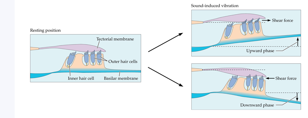
tectorial membrane
A gelatinous structure, attached on one end, that extends into the middle canal of the cochlea, floating above inner hair cells and touching outer hair cells.
Tectorial membrane shear
When vibration causes a displacement along the cochlear partition the tectorial membrane and hair cells move in opposite directions (i.e., they experience shear), and the deflection of stereocilia during this action results in the release of neurotransmitters
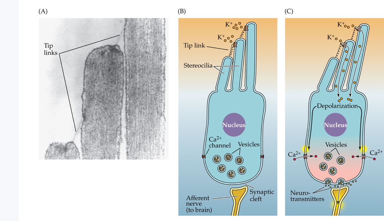
Tip link
A tiny filament that stretches from the tip of a stereocilium to the side of its neighbor.
Mechanoelectrical transduction in hair cells
Stereocilia regulate the flow of ions into and out of hair cells. (A) This photomicrograph shows the threadlike tip links that connect the tip of each shorter stereocilium to its taller neighbor. (B, C) Bending the stereocilia atop a hair cell opens the ion pores, permitting a rapid influx of potassium ions (K+) into the hair cell. This depolarization opens channels that allow calcium ions (Ca2+) to enter the base of the hair cell, causing the release of neurotransmitters into the synaptic cleft between the hair cell and an afferent auditory nerve (AN) fiber, resulting in the nerve fiber firing, sending a signal to the brain. Between 5 and 30 AN fibers synapse with each inner hair cell; only one is shown here.
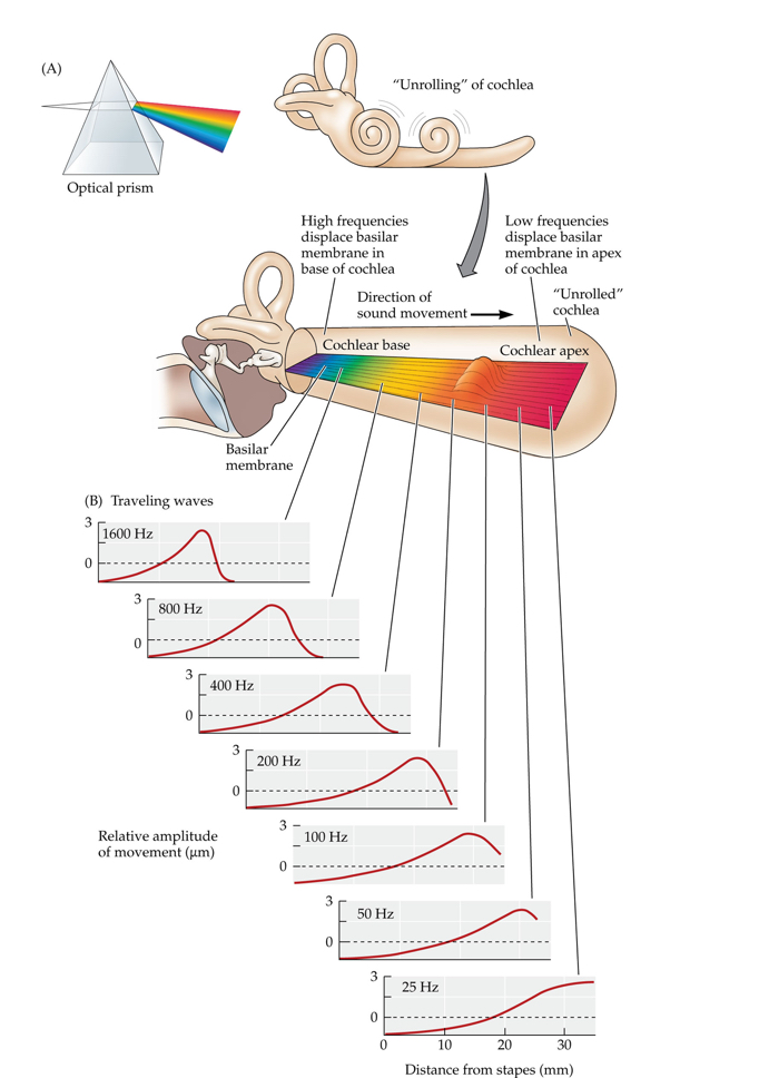
Place code
Tuning of different parts of the cochlea to different frequencies, in which information about the particular frequency of an incoming sound wave is coded by the place along the cochlear partition that has the greatest mechanical displacement.
Frequencies along the cochlea
The cochlea is like an acoustic prism in that its sensitivity spreads across different sound frequencies along its length. (A) The cochlea is illustrated as if it were uncoiled. The narrower end of the basilar membrane toward the base is stiffer and most sensitive to higher frequencies. The wider, more flexible end toward the apex is most sensitive to lower frequencies. (B) The shapes of the traveling waves for different frequencies of vibration are shown.
Afferent fibers
A neuron that carries sensory information to the central nervous system.
efferent fibers
A neuron that carries information from the central nervous system to the periphery.
threshold tuning curves
A graph plotting the thresholds of a neuron in response to sine waves with varying frequencies at the lowest intensity that will give rise to a response.
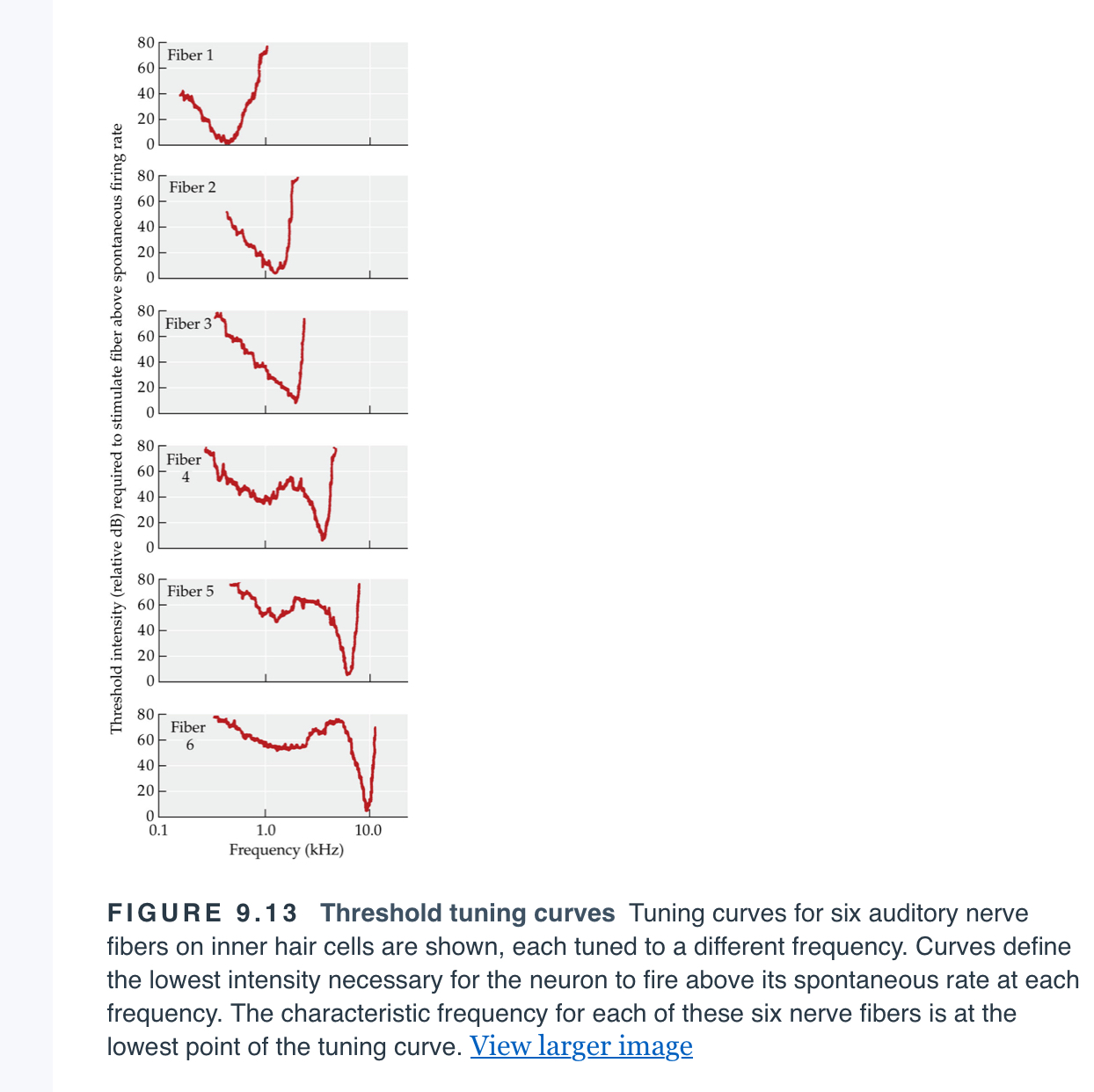
characteristic frequency (CF)
The frequency to which a particular auditory nerve fiber is most sensitive.
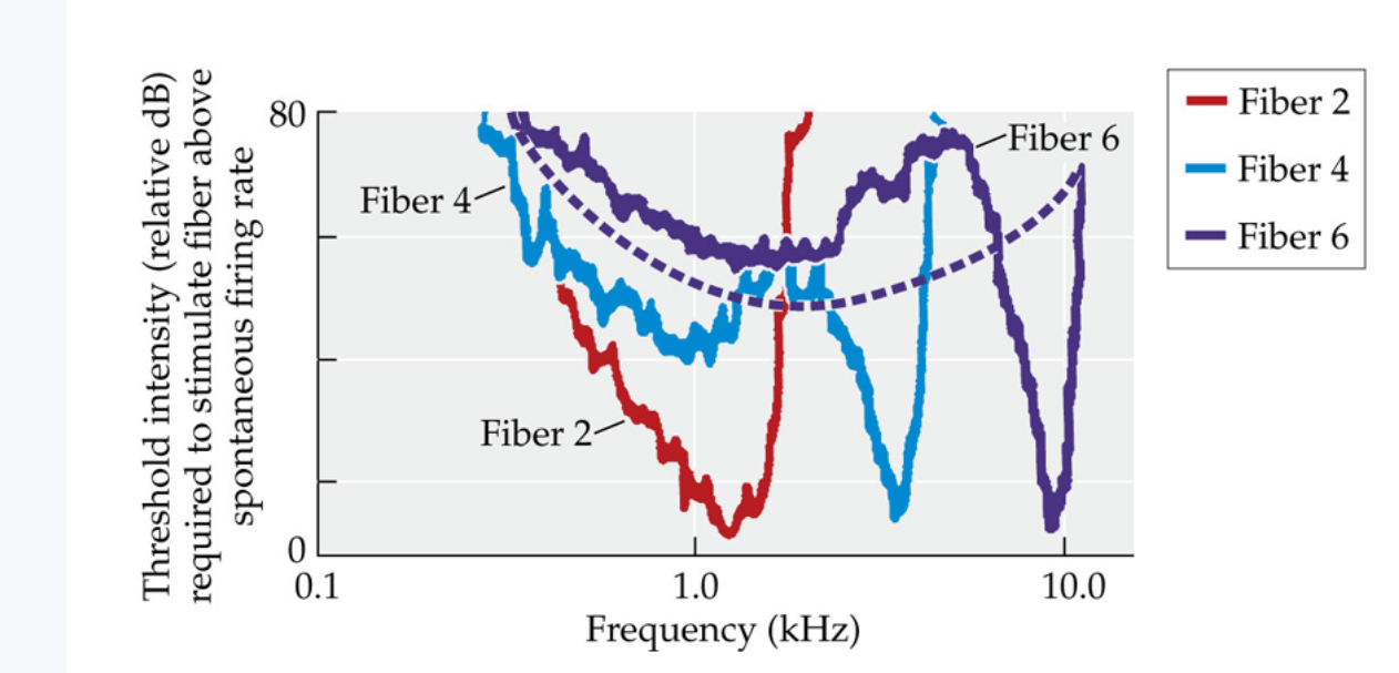
electromotility
The ability of outer hair cells to extend and contract, which changes the stiffness and sensitivity of the cochlear partition
Function of outer hair cells
Outer hair cells improve both sensitivity and frequency selectivity. Threshold tuning curves for three of the nerve fibers show responses when outer hair cells are active. The dashed line shows what the rightmost tuning curve (for fiber 6) would look like if outer hair cells were not active. Higher-intensity tones would be required to excite the auditory nerve fiber, and the fiber would be less selective in frequencies to which it would fire
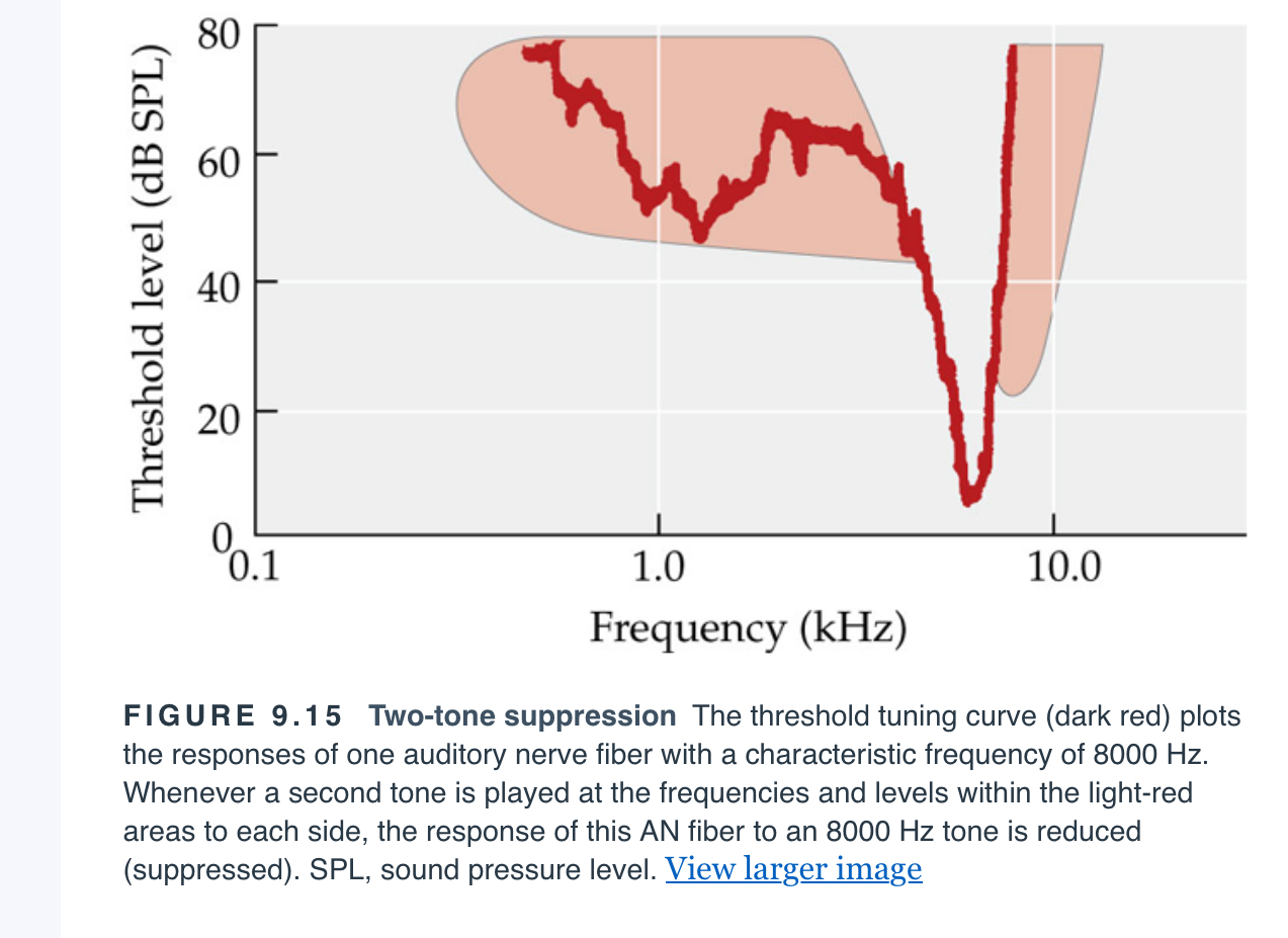
two-tone suppression
A decrease in the response (firing rate) of one auditory nerve fiber to one tone when a second tone is presented at the same time.
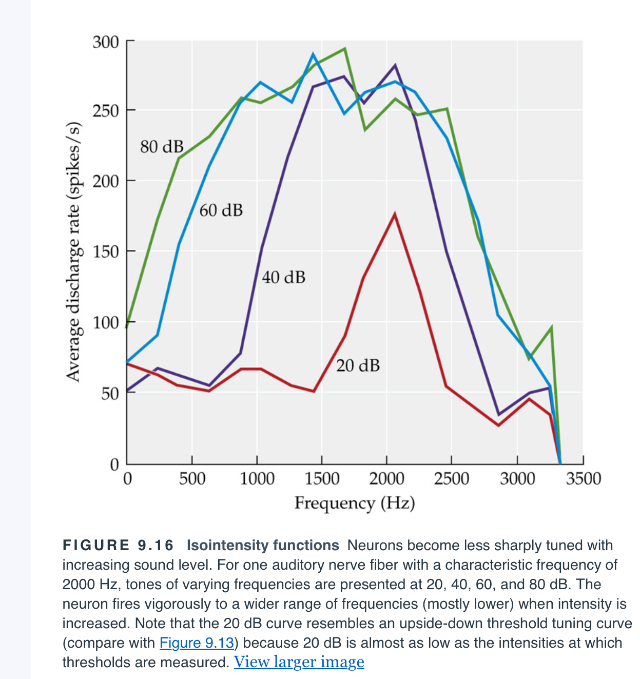
isointensity curves
A map plotting the firing rate of an auditory nerve fiber against varying frequencies at varying intensities.
rate saturation
The point at which a nerve fiber is firing as rapidly as possible and further stimulation is incapable of increasing the firing rate.
rate-intensity functions
A graph plotting the firing rate of an auditory nerve fiber in response to a sound of constant frequency at increasing intensities.
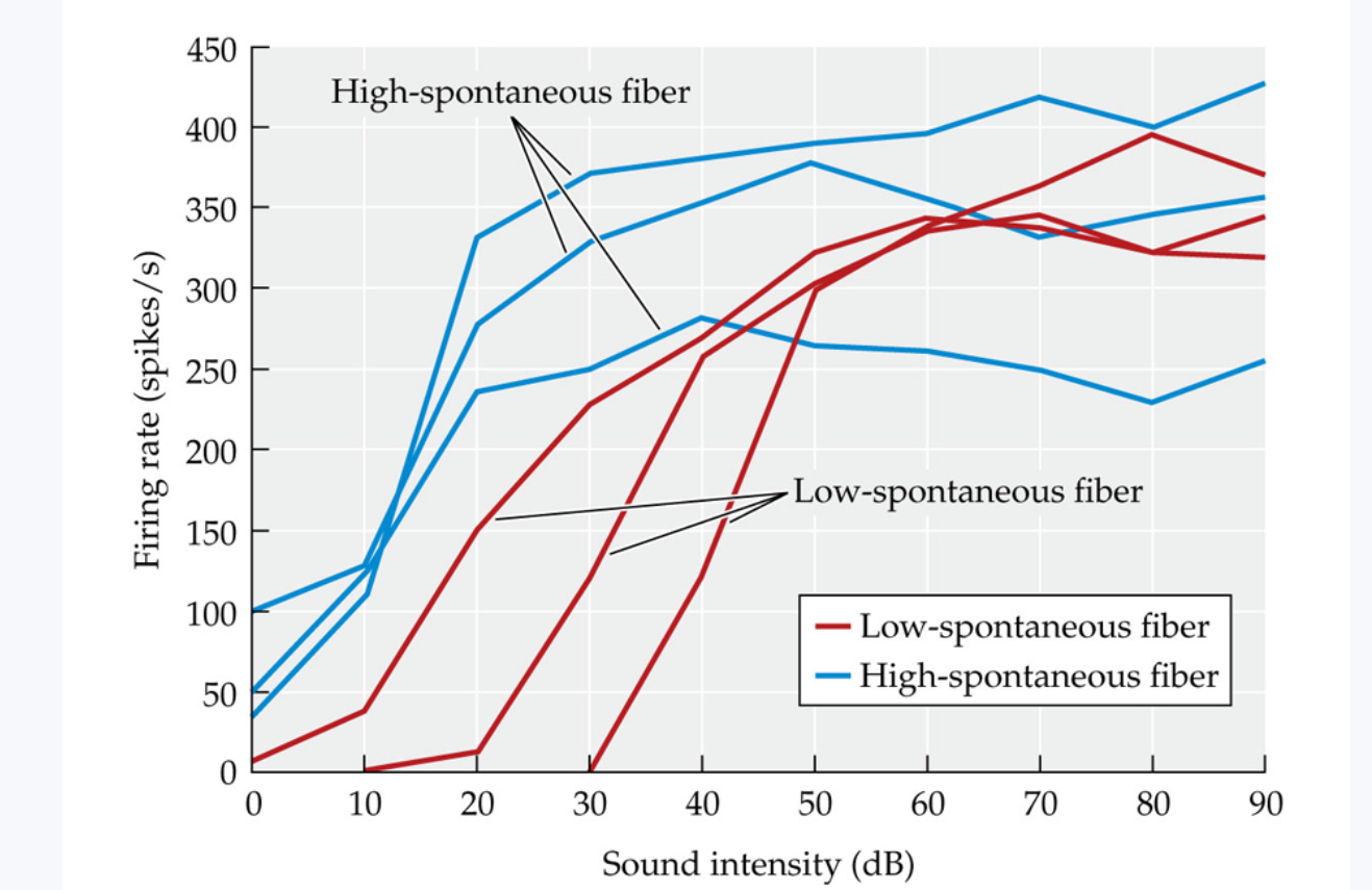
low-spontaneous fibers
An auditory nerve fiber that has a low rate (less than 10 spikes per second) of spontaneous firing. Low-spontaneous fibers require relatively intense sound before they will fire at higher rates.
high-spontaneous fibers
An auditory nerve fiber that has a high rate (more than 30 spikes per second) of spontaneous firing. High-spontaneous fibers increase their firing rate in response to relatively low levels of sound.
Mid-spontaneous fibers
An auditory nerve fiber that has a medium rate (10–30 spikes per second) of spontaneous firing. The characteristics of mid-spontaneous fibers are intermediate between those of low- and high-spontaneous fibers.
Firing rate across intensities
Neural firing rate is shown at increasing sound intensities for six auditory nerve fibers (three low-spontaneous fibers and three high-spontaneous fibers). Firing rates for all six neurons increase with increasing sound level. Low-spontaneous neurons require higher-intensity sounds before they begin to fire, and they continue to increase firing rate to higher sound levels.
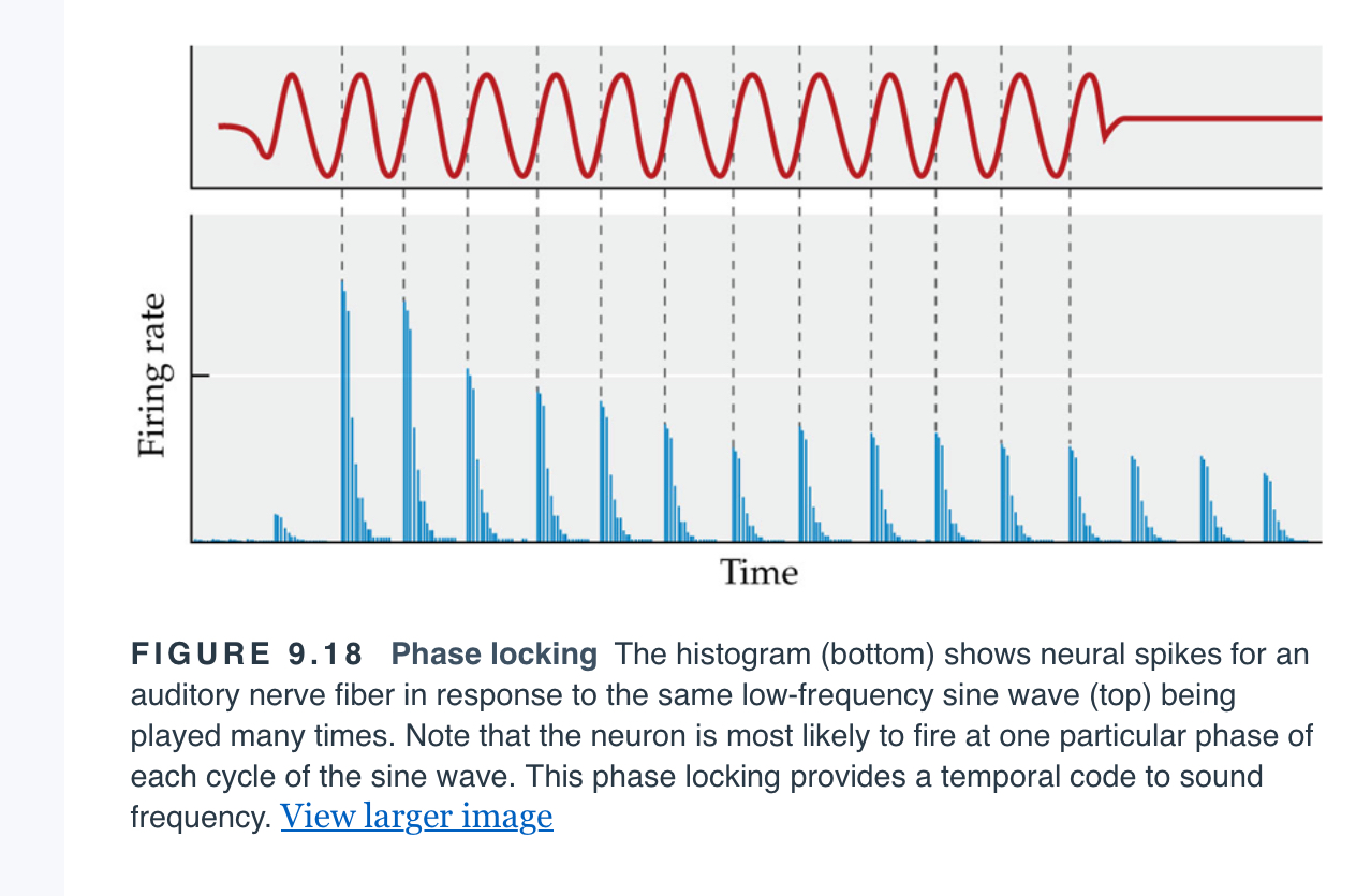
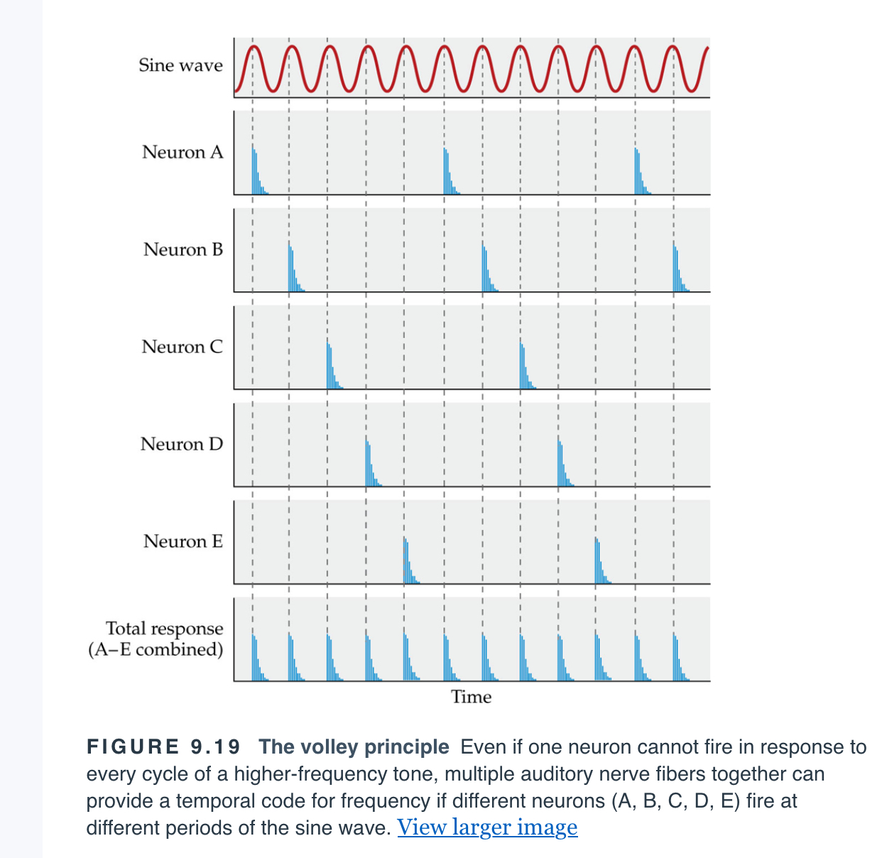
Phase locking
Firing of a single neuron at one distinct point in the period (cycle) of a sound wave at a given frequency. (The neuron need not fire on every cycle, but each firing will occur at the same point in the cycle.)
Temporal code
Tuning of different parts of the cochlea to different frequencies, in which information about the particular frequency of an incoming sound wave is coded by the timing of neural firing as it relates to the period of the sound.
Volley principle
The idea that multiple neurons can provide a temporal code for frequency if each neuron fires at a distinct point in the period of a sound wave but does not fire on every period.
Cochlear nucleus
The first brainstem nucleus at which afferent auditory nerve fibers synapse.
Pathways in the auditory system
Only a few auditory pathways are shown here. Red and blue pathways indicate input from the left and right ears, respectively, and purple indicates integration of input from both ears. Although there are two parallel pathways, information from both ears comes together very early in the auditory system, at the superior olives. Cerebral processing of auditory information begins in the primary auditory cortex, or A1
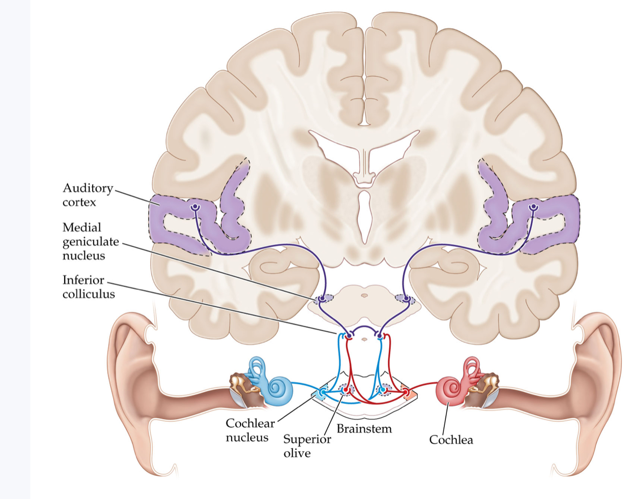
Superior olives
An early brainstem region in the auditory pathway where inputs from both ears converge.
inferior colliculus
A midbrain nucleus in the auditory pathway.
medial geniculate nucleus
The part of the thalamus that relays auditory signals to the temporal cortex and receives input from the auditory cortex.
tonotopic organization
An arrangement in which neurons that respond to different frequencies are organized anatomically in order of frequency.
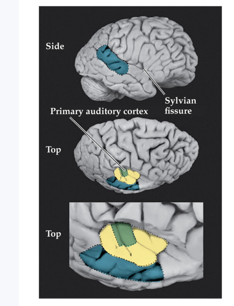
Primary auditory cortex Or A1
The first area within the temporal lobes of the brain responsible for processing acoustic information.
Belt area
A region of cortex, directly adjacent to the primary auditory cortex (A1), with inputs from A1, where neurons respond to more complex characteristics of sounds.
parabelt area
A region of cortex, lateral and adjacent to the belt area, where neurons respond to more complex characteristics of sounds, as well as to input from other senses.
Primary auditory cortex
The first stages of auditory processing begin in the temporal lobe of the cerebral cortex within the Sylvian fissure. The top picture is a side view of the brain’s right hemisphere (the front of the brain faces right). The lower two pictures are looking down at the interior of the right hemisphere, with the parietal cortex cut away. Primary auditory cortex (A1) is surrounded by belt regions, and parabelt regions extend past the belt to the front and side. These belt regions are sometimes called secondary or associational auditory areas
What structures make up the outer ear, and what are their functions?
The outer ear consists of the pinna and the external ear canal. The pinna collects sound waves from the environment and funnels them into the ear canal, which enhances frequencies between about 2000–6000 Hz and protects the tympanic membrane at its end.
What is the function of the tympanic membrane?
It vibrates in response to incoming sound waves, converting air pressure changes into mechanical motion.
What are the three ossicles of the middle ear, and what is their collective function?
The malleus, incus, and stapes amplify sound vibrations and transmit them from the tympanic membrane to the oval window of the inner ear.
How do the ossicles amplify sound vibrations in the middle ear?
They use lever-like joints to increase pressure changes and concentrate energy from the large tympanic membrane onto the much smaller oval window, magnifying pressure about 18 times.
What is the role of the tensor tympani and stapedius muscles in the middle ear?
These small muscles tense in response to loud sounds, restricting ossicle movement to protect the inner ear from potential damage.