Physiological Psychology - Quiz 2
 Call Kai
 Learn
Learn Practice Test
 Spaced Repetition
 Match
Match Flashcards
 Knowt Play
1/56

There's no tags or description
Looks like no tags are added yet.
Name | Mastery | Learn | Test | Matching | Spaced | Call with Kai | Chat |
|---|
No analytics yet
Send a link to your students to track their progress
57 Terms
What’s an action potential? (really basic)
The reversing of the sodium and the potassium is what an action potential is
The resting potential of a neuron provides a baseline level of polarization
But neurons routinely undergo a brief but radical change in polarization, sending an electrical signal from one end of the axon to the other
What are the primary inhibitory and excitatory neurotransmitters?
Primary excitatory: glutamate
Primary inhibitory: GABA
Batrachotoxin
Interfered with Voltage gated sodium channels → role in action potentials (it keeps them open!)
If you cannot create action potentials you get paralysis
*Essentially, at first the cell fires uncontrollably, and then it cannot reset to RMP so it cannot make new action potentials anymore
Death, its poison
Produced by South American poison arrow frogs, has the reverse effect and forces Na+ channels to stay open, triggering continuous action potentials, with equally lethal results
Scorpions are a rich source of channel-specific toxins
Flow of communication in neurons
Neurons communicate through the generation and propagation of action potentials (APs)
→ Travels down the length of the neuron, continues neuron to neuron

Where it begins: a neuron is either stimulated or inhibited by other neurons at the dendrites
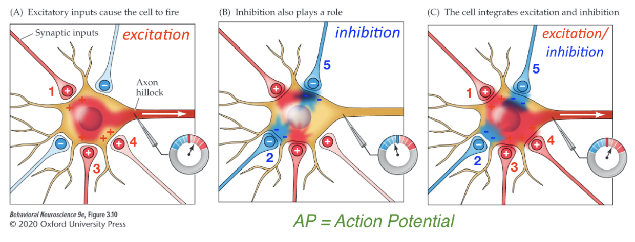
Panel on the left:
Neuron and axon and then there's another and you can see the terminal
+ and - signs, excitation with + sign and - for inhibitory
At any given time, a neuron is summing excitation and subtracting inhibition
Excitatory inputs are firing 1,2,3,4 are excited and releasing glutamate, which is causing excitation in the postsynaptic neuron
If it’s high enough it reaches threshold(~55), and if you get to the voltage point, you get an action potential formed in the axon hillock and travels down the axon
What determines whether a synapse excites or inhibits the postsynaptic cell?
One factor is the particular neurotransmitter released by the presynaptic cell.
Some transmitters usually generate an EPSP in the postsynaptic cell, others usually generate an IPSP
The same neurotransmitter may be excitatory at one synapse and inhibitory at another, depending on the receptors present in the postsynaptic cell
Whether a neuron fires an action potential at any given moment is decided by the balance between the number of excitatory and inhibitory signals that it is receiving
Action potentials
Very brief but large changes in neuronal polarization that arise in the initial segment of the axon and are propagated at high speed along the axon’s length
The propagated electrical message a neuron sends along its axon to the presynaptic axon terminals
The information that a neuron sends to its postsynaptic targets is encoded in patterns of these action potentials, so we need to understand their properties (where they come from, how they race down the axon and how they communicate information across synapses to other cells. Let’s turn first to the creation of the action potential
The point of an action potential
To transmit information to other cells
The action potential is re-created down every branch of the axon until it reaches every terminal
At each axon terminal, the electrical signal of the action potential is converted into a chemical signal as the axon releases a neurotransmitter
A substance released from a presynaptic terminal that serves to communicate with the postsynaptic cell.
Synapses can be excitatory or inhibitory
Neurotransmitters released into synapses briefly alter the membrane potential of the postsynaptic cell
These brief changes are called postsynaptic potentials
A local potential that is initiated by simulation at a synapse, can vary in amplitude, and spreads passively across the cell membrane, decreasing in strength with time and distance
A neuron, receiving synapses from hundreds of other cells, is subject to hundreds or thousands of postsynaptic potentials
When integrated, this massive array of local potentials determines whether the neuron will reach threshold and therefore generate an action potential of its own
*It is important to note that excitatory and inhibitory neurons get their names from their actions on postsynaptic neurons, not from their effects on behavior
When an excitatory presynaptic neuron (red) fires, it shows a normal action potential and causes depolarization (EPSP) in the postsynaptic neuron (yellow)
When an inhibitory presynaptic neuron (blue) fires, it also shows a normal action potential, but it causes hyperpolarization (IPSP) in the post-synaptic neuron

The stimulation of a presynaptic neuron (red) causes it to produce an all-or-none action potential that spreads to the end of the axon, releasing transmitter.
After a brief delay, the postsynaptic cell (yellow) displays a small local depolarization, as channels open to let cations in. This local postsynaptic membrane depolarization is known as an excitatory postsynaptic potential (EPSP) because it pushes the postsynaptic cell a little closer to the threshold for an action potential, thus the red neuron forms an excitatory synapse upon the yellow target neuron
The combined effect of many excitatory synapses is needed to elicit an action potential in a postsynaptic neuron
If EPSPs are elicited by many neurons that converge on the postsynaptic cell, these potentials can produce a depolarization large enough to reach threshold and can trigger an action potential
There is a delay: in the fastest cases, the postsynaptic depolarization begins about half a millisecond after the action potential arrives at the presynaptic terminal
This synaptic delay reflects the time needed for the neurotransmitter to be released and diffuse across the synapse
When the blue neuron fires, the postsynaptic effect is an increase of the resting membrane potential
This hyperpolarization moves the cell membrane potential away from threshold, decreasing the probability that the neuron will fire an action potential, so it is called an inhibitory postsynaptic potential (IPSP)
Usually IPSPs result from the opening of channels that permit chloride ions (Cl-) to enter the cell
Because Cl- ion are much more concentrated outside the cell than inside, they rush into the cell, making it even more negative
Just as driving a car requires brakes as well as an accelerator, neural circuits must be turned off as well as on
The nervous system treads a narrow path between overexcitation, which leads to seizures such as those that plagued Deidre, and underexcitation, which leads to coma and death
So excitatory and inhibitory presynaptic neurons both work in the same way with one exception: they have opposite effects on the postsynaptic cell
Excitation that diffuses down from the dendrites
Some inputs are “excitatory” which excites the neuron, depolarized (value of membrane potential is getting closer to 0), may cause the neuron to fire an AP (depolarized)
Other inputs are “inhibitory”, which makes neurons less likely to fire an action potential (hyperpolarized)
The neuron simultaneously “adds up” all of the excitatory inputs and “subtracts” all the inhibitory inputs; if there’s enough excitation to reach “threshold” (-55mV), the neuron generates an AP
The AP starts at the axon hillock and travels down the axon to cause neurotransmitter release at the synaptic terminal
Cooling effect, hyperpolarization, even less likely to fire an action potential
Middle panel is being inhibited by GABA no firing
At any given time there are excitatory inputs and inhibitory inputs at one time
If there is enough to get to the threshold, it fires
Neuron simultaneously receives excitatory and inhibitory
Neurons can be stimulated in a few different ways: Spatial vs. Temporal Summation

3 presynaptic neurons coming from different places (so they are different neurons) are simultaneously firing action potentials coming from different points in space.
Spatial summation:
Excitatory potentials from multiple presynaptic inputs arrive at the same time to stimulate a neuron, causing an action potential in the post-synaptic neuron (left) if the stimulation is sufficient for the neuron to reach “threshold” If the action potential from A and then B and then C arrives all three at the same time, they overreact the neuron, excitation is added up in the nervous system
Usually the convergence of excitatory messages from many presynaptic neurons is required for a neuron to fire an action potential. This summation of potentials from different physical locations across the neuron is called spatial summation
Temporal summation

Excitatory potentials from the same presynaptic input arrive close in time to one another (“train” of APs), and build on one another to potentially cause an action potential in the post-synaptic neuron (right)
Neuron A is being excited, firing a lot of action potentials
Postsynaptic neuron will add up the excitation
First excitation causes neurotransmitter release
Neurons do both of those at the same time
2 ways dendrites can input/smth
Even if postsynaptic effects/potentials are not absolutely occurring at the same exact time, each potential lasts a few milliseconds before fading away. The closer they are in time, the greater the overlap and the more complete is the summation
Temporal summation = sum of postsynaptic potentials that reach the axon hillock at different times, the closer in time they occur, the more complete the summation
Summation
Complex behavior requires neurons to integrate and transform the messages they receive, in other words, they perform information processing, using a sort of neural algebra, each neuron adds and subtracts the myriad inputs it receives from other neurons.
Postsynaptic potentials are caused by transmitter chemicals that can be either depolarizing (excitatory) or hyperpolarizing (inhibitory)
From their points of origin on the dendrites and cell body, these graded EPSPs and IPSPs spread passively over the neuron, decreasing in strength over time and distance
Whether the postsynaptic neuron will fire an action potential is determined by whether a depolarization exceeding threshold reaches the portion of the axon just beyond the hillock, where action potentials begin
The presynaptic terminals provide excitatory (depolarizing) or inhibitory (hyperpolarizing) stimulation to the postsynaptic cell membrane
If the membrane potential rises (depolarizes) above a threshold level, an action potential is fired
So lets say for example that two excitatory terminals are activated in the neuron, which cause local depolarizations of the cell body. These depolarizations spread out over the neuron, dissipating as they spread, so only a fraction of the original depolarization reaches the axon hillock. Taken alone, neither would be sufficient to cause threshold depolarization, but when they both arrive at about the same time, the two depolarizations sum to push the membrane potential of the hillock region to threshold.
*the image shows what happens when inhibitory synapses are also active, creating postsynaptic hyperpolarizations. These polarizations also spread passively, dissipating as they travel. Because some potentials excite and others inhibit the hillock, these effects partially cancel each other. Thus, the net effect is the difference between the two: the neuron subtracts the IPSPs from the EPSPs and no action potential arises. Simple arithmetic, right?
When summed, EPSPs and IPSPs do tend to cancel each other out. But because postsynaptic potentials spread passively and dissipate as they cross the cell membrane, the resulting sum is also influenced by distance.
For example, simultaneous EPSPs from two synapses close to the hillock will produce a larger sum there than will two EPSPs from farther away. Only if the overall sum of all the potentials—both EPSPs and IPSPs—is sufficient to depolarize the cell to threshold at the axon hillock is an action potential triggered
If EPSPs arrive one right after the other, they sum and the postsynaptic cell eventually reaches threshold and produces an action potential. If too much time passes between EPSPs, each will fade away before the next occurs, and the neuron will never fire.
Likewise, EPSPs from two different synapses may push the target to fire if they arrive at nearly the same time (they sum temporally) but will not if the second arrives after the first EPSP has faded away.
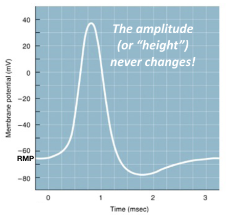
The “All or Nothing” Action potential (nerve impulse)
Time on Y axis
Action potentials are super short
The neuron depolarizes , the inside become more positive
Undershoots the membrane potentials
They either get an action potential or they don’t
No big or little action potentials, heights never changes,
The frequency increases, stimulus intensity
Action potential anatomy

Start at the resting potential 65mV
Threshold: Threshold is the critical level of depolarization that must be reached to trigger an action potential (-55mV)
If excitation is insufficient to get to that value, no action
Rising Phase: Rapid depolarization of the membrane (membrane becomes more positive)
Overshoot: Inside of the neuron is positively charged relatively to the outside
Falling phase: Repolarizing of the membrane (membrane becomes more negative)
Undershoot: The inside of the cell is briefly more negative than the resting potential (RMP)
Absolute refractory period: A period of time (1 millisecond) where another action potential cannot be generated
You can't get action potentials so close to each other that they combine.
*amplitude does not change
Rising phase is going up falling phase is going down
Generating multiple action potentials
If a large enough electrical current is injected into a neuron, the membrane is depolarized sufficiently to fire multiple (a “train”) action potentials (right)
In the nervous system, the intensity/strength of a signal is most often encoded by firing frequency
You don’t get bigger action potentials, you get more of them
Pervieving loud noises is repeated
If not excited, slows down
The intensity of a signal/stimulus is encoded by action potential firing frequency (larger depolarization = more action potentials)
If injected current does not depolarize the membrane to threshold, no action potentials will be generated
If injected current depolarizes the membrane beyond threshold, action potentials will be generated
The action potential firing rate increases as the depolarizing current increases
But how do we record this? Axons are tiny!
If you're depolarizing and stimulating the neuron more the (more about number not how strongly they are signaled)

Recording the first action potentials → the squid giant axon
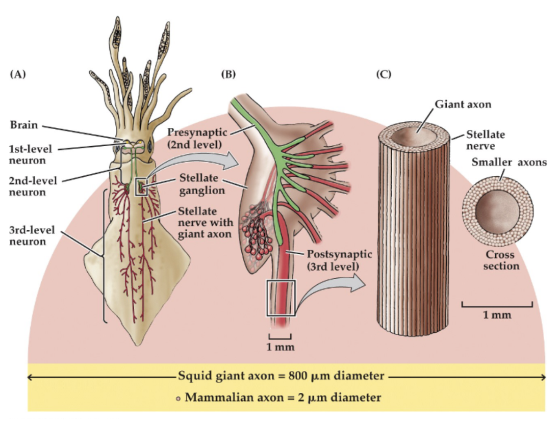
The squid giant axon is 400x larger in diameter than the typical mammalian axon
Vertebrates have not evolved myelin? Idk something about the axon size
Big diameter
The squid’s giant axon has an unusually high conduction rate for invertebrates
The giant squid axon
A neuron involved in the animal’s emergency escape behavior. The squid’s giant axon is readily apparent to the naked eye and is much better suited to experimentation than mammal’s axons which are very small in size.
Ionic basis of the axon potential
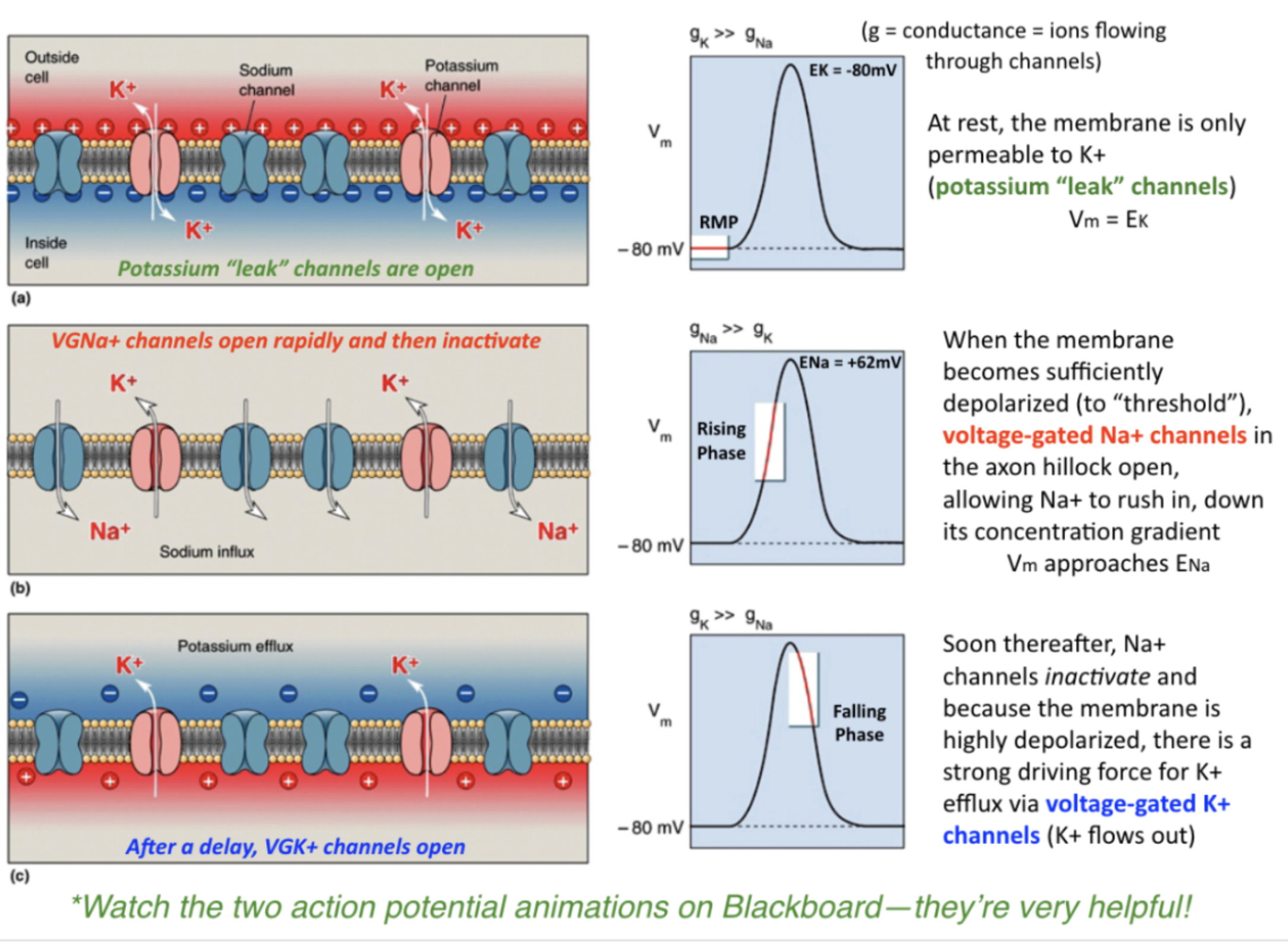
White box is where a neuron at rest is, and on the left what the channels are doing at rest, membrane is only permeable to potassium here. Potassium is mostly responsible for the channel
“Leak channels” cuz potassium is leaking out
During the rising phase, voltage gated channels open and sodium flows down its conc. Gradient
Sodium will try to go down the gradient and go in
Coming close to reaching the equilibrium Pot. for sodium reaches equilibrium
During action potential, sodium goes in, then potassium flows out (takes a positive charge with it so that's why the inside is negative) pot. Flows out during the second phase sodium first phase
Some potassium is always leaking out of the cell
Don’t eat me!
Both the pufferfish (TTX - Tetrodotoxin) and the frog block voltage-gated sodium channels
If those channels are blocked, you don't get sodium potentials, and you don't get axon potentials
Selectively block voltage-gated sodium channels, preventing the production of action potentials; paralysis and death rapidly follow. Tetrodotoxin is found in the ovaries of puffer fish, which is a delicacy in Japan. If the ovaries of puffer fish aren’t removed properly, and the fish isn’t cleaned right, and people eat it, they may be poisoned by TTX. Saxitoxin is a seafood threat: it comes from a bloom of algae that can affect shellfish.
Batrachotoxin
Produced by South American poison arrow frogs, has the reverse effect and forces Na+ channels to stay open, triggering continuous action potentials, with equally lethal results
Scorpions are a rich source of channel-specific toxins
*Tiny quantities of any of these toxins can dramatically impede the ability of neurons to function
Although, the same specificity that makes channel toxins so deadly also makes them useful tools in the laboratory. For example, the venom of the tarantula spider contains toxins that very specifically target the voltage sensor of certain voltage-gated ion channels. In the lab, these toxins provided important clues about how these channels work.
Ionic basis of the action potential (greater detail)
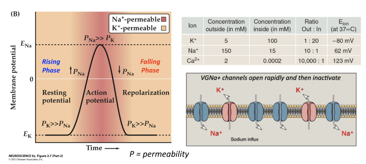
Ek = resting potential, very close to the equilibrium of the potassium, because its channels are open so it can reach its potential, far greater than sodium
At rest sodium channels are not open, high potassium permeability because K+ leak channels are open (Vm = EK)
During an action potential:
Rising phase: strong sodium influx because gated channels open (1-2ms) (inside becomes more positive)
Falling phase: with delay, voltage gated channels open, allowing K+ efflux out of the neuron (inside starts to get more negative)
During an action potential, sodium flows in first, and then potassium flows out
Then sodium channels start to open, brings positive charge with it
Gets close to the equilibrium potential for sodium (peak) ~60mV and with a little delay, potassium channels open and (theres still more K in) so then it goes down conc. Gradient → causes repolarization
Sodium channels close within a millisecond
Sodium flows in first → potassium out → these are responsible for rising(sodium in) and falling phase (potassium out) they are trying to reach their equilibrium
This is quickly reset by the ATPase, reset so that another action potential can happen
Potassium leak channels are always open but fewer of them
Voltage gated
The process:
RMP: Some K+ channels are open, and Na+ channels are closed, which creates the resting potential
THRESHOLD: When a stimulus occurs: any depolarizing force will bring the membrane potential closer to threshold (-55mV)
RISING PHASE: At threshold, the voltage gated Na+ channels stay open for only a millisecond, and then close again (only a few Na+ ions need to enter to change the membrane potential) (step 4) before the neuron can reach the Na+ equilibrium potential of about +65mV, this causes a rapid change of polarity (the action potential) —> inside becomes more positive
*Most of the change in ion concentration taking place during an action potential is happening right next to the membrane, leaving the ionic concentrations in the interior of the axon relatively unaffected
*In the long run, the sodium-potassium pump enforces the low concentrations of Na+ ions inside the neuron.
*Hodgkin and Huxley detected these ion movements by manipulating the concentration of ions in and out of the axon and then monitoring current across the membrane, using voltage clamping. (Forces the axon membrane to remain at a particular potential/voltage. Uses two electrodes; one inside the axon, and the other is used as a reference electrode in the bath surrounding the axon. A voltmeter measures the potential difference. Information is sent to a voltage clamp amplifier, which compares the membrane potential with a “command” voltage that the experimenter sets. If they match, nothing happens, if there’s a difference, an electrical current is injected into the axon to force the membrane potential to match.
OVERSHOOT: The sodium builds up inside the cell (positive)
FALLING PHASE: Na+ channels automatically close again, gated K+ channels open and they flow out, repolarizing and even hyperpolarizing the cell (afterpotential)
Now the positive charge inside the nerve cell (the overshoot) pushes K+ ions out of the channels that were always open, plus voltage-gated K+ channels open as well, making the membrane even more permeable to K+, so the resting potential is quickly restored (step 5)
REFRACTORY PHASE: The neuron cannot fire again
Applying very strong stimuli reveals another important property of axonal membranes
The upper limit to the frequency of action potentials is at about 1200 spikes per second (many neurons have even slower maximum rates of response)
Applying pairs of stimuli that are spaced closer together reveals a related phenomenon: beyond a certain point, only the first stimulus is able to elicit an action potential
The axonal membrane is said to be refractory (unresponsive) to the second stimulus
ATPase (sodium-potassium pump) restores the gradients to RMP (more K+ inside and Na+ out!!)
Ligand-gated ion channels
Some kind of ligand/neurotransmitter binds to the receptor/channel and causes it to open
—> This is the reciever at the dendrites, once it recieves the neurotransmitter, it allows ions to go through (for excitatory, positive ones) and this creates a positive electrical wave that goes down to the hillock and then thats where those voltage gated sodium channels are!
Ligand
A substance that binds to receptor molecules, (like a key and a lock), it (correct shape) can fit into a receptor protein and activate or block it
For example, at synapses where acetylcholine (ACh), is the transmitter, it fits into ligand-binding sites in receptor molecules (a protein that binds and reacts to molecules of a neurotransmitter, hormone, or drug) located in the postsynaptic membrane
The nature of the postsynaptic receptors at a given synapse determines the action of the transmitter
For example, acetylcholine can function as either an inhibitory or an excitatory neurotransmitter, at different synapses
At excitatory synapses, binding of acetylcholine opens channels for Na+ and K+ ions
This way, acetylcholine released onto muscle fibers excites them to contract
At inhibitory synapses, acetylcholine typically acts on a different type of receptor to open channels that allow Cl ions to enter, hyperpolarizing the membrane (making it more negative and less likely to create an action potential)
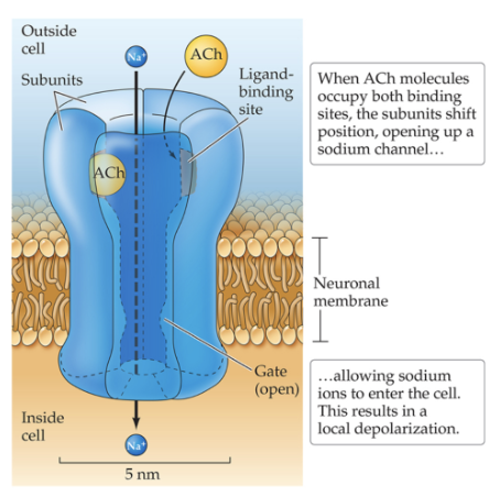
The gated part means that the neurotransmitter is responsible for opening the channel otherwise no

Ionotropic receptors/Ligand-gated ion channels
Chemically gated ion channels
When bound by the transmitter, the ion channel opens and ions flow across the membrane
Ligand
A substance that binds to receptor molecules, (like a key and a lock), it (correct shape) can fit into a receptor protein and activate or block it
Voltage gated sodium channel
*crucial for generating action potentials
Channels are in the axons in the axon hillock
Depolarization/excitation is what causes these channels to open
Dendrites summate excitation
Diffuses its way down the axon
What's different about this channel:

Protein, amino acids, it has positively charged inside residues called “voltage sensor”
Allows the protein channel to twist open when a depolarization invades the axon hillock (like charges repel)
The twisting results in an opening of the pore that only allows the passage of sodium ions (selectivity filter)
You get a pos charge in the axon hillock when the depolarization diffuses its way down the dendrites to the hillock, the plus signs will repel the channel’s positive interior and it causes it to twist open (when there is a depolarization that reaches the channel)
An increase in voltage causes it to open
This is the physiological threshold (-55mV)**important
There is enough positive charge that causes the sodium channels to open**important), and then sodium can go in, causing the action potential
Voltage-gated sodium channel selectivity for sodium ions
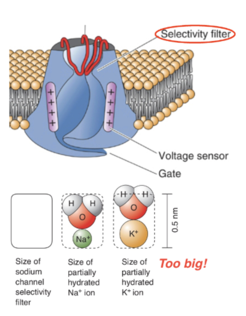
Selective for sodium, sodium mostly goes through it, potassium cannot fit (selective permeability)
Work quickly, open instantly and close (like slam the door)
Stays open for not more than a millisecond
How the channels work
Cannot be opened again by depolarization until the membrane potential resets (you cannot get action potentials on top of one another) → refractory period
THe VGNa+ channel is 12x more permeable to Na than K (pore loop selectivity filter)
Selectivity for Na+ ions is based on size, charge, and energy of hydration of the ion (basically, the hydrated K+ ion doesn’t fit through the pore)
Functional properties:
1. They open fast
2. They only stay open for 1 millisecond before they inactivate
3. They cannot be opened again by depolarization until the membrane voltage resets (absolute refractory period)
Channel Details
A cell membrane consists of two layers of fatty molecules
Because ions in body fluids are usually surrounded by clusters of water molecules, they cannot easily pass directly through neuronal membranes
They must pass through membrane-spanning ion channels, which are highly selective for particular types of ions
The inner surfaces of the K+ channel are lined with oxygen atoms that mimic water molecules
With the oxygen atoms substituting for their usual escort of water molecules, K+ ions fit exactly into this selectivity filter, Na+ does not
VGNa+ Channels start the action potential off at the Axon hillock
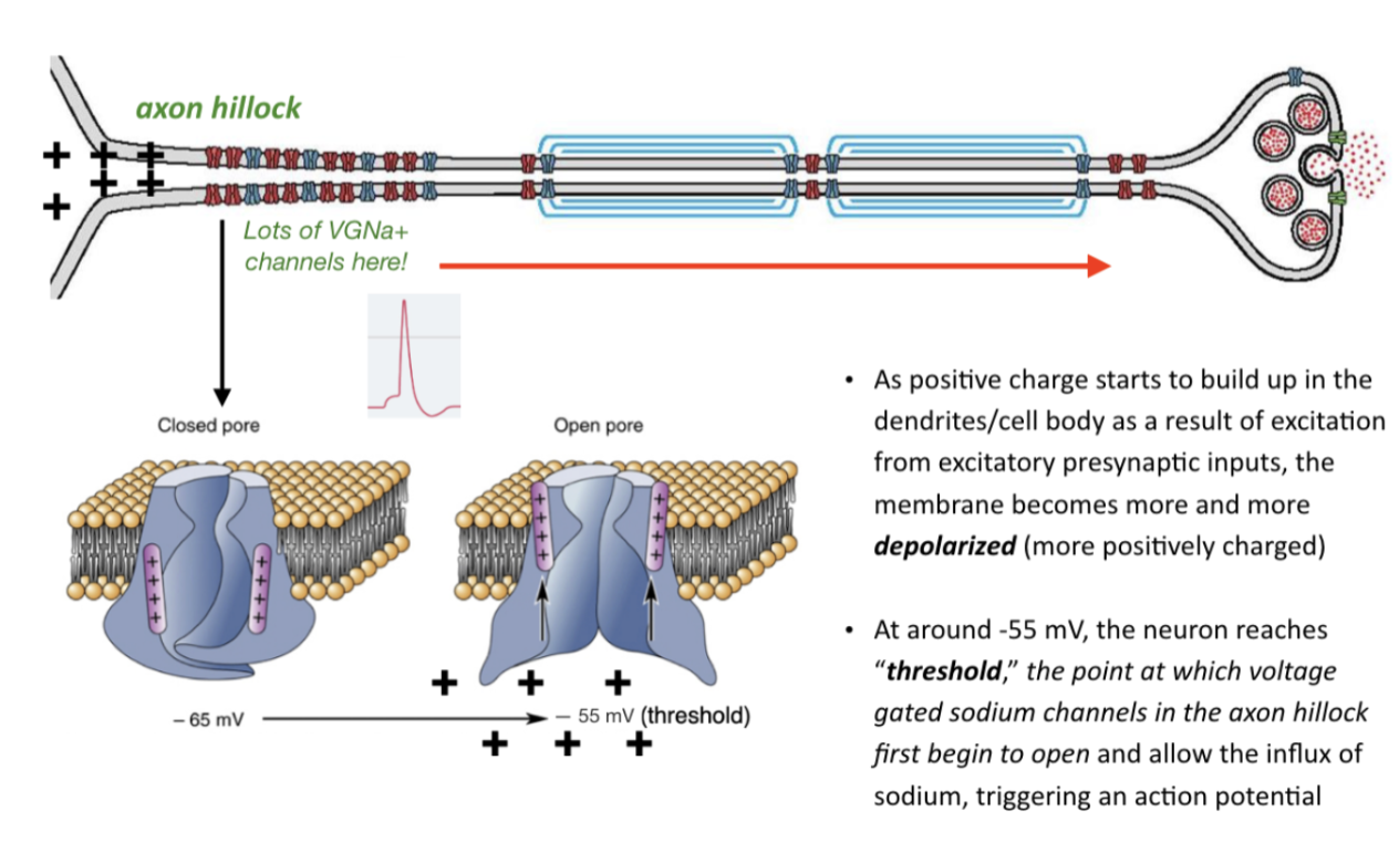
Synapses are receiving inputs
Pos charge adds up, diffuses down to the axon hillock, and then it repels the voltage sensor in the sodium channel, and then it opens and then closes quickly so sodium goes in with a positive charge and that the action potential (sodium reaching its potential)
The potential travels down the length of the axon down to the axon terminal to cause neurotransmitter release
How the action potential travels (in one direction and do not decay)
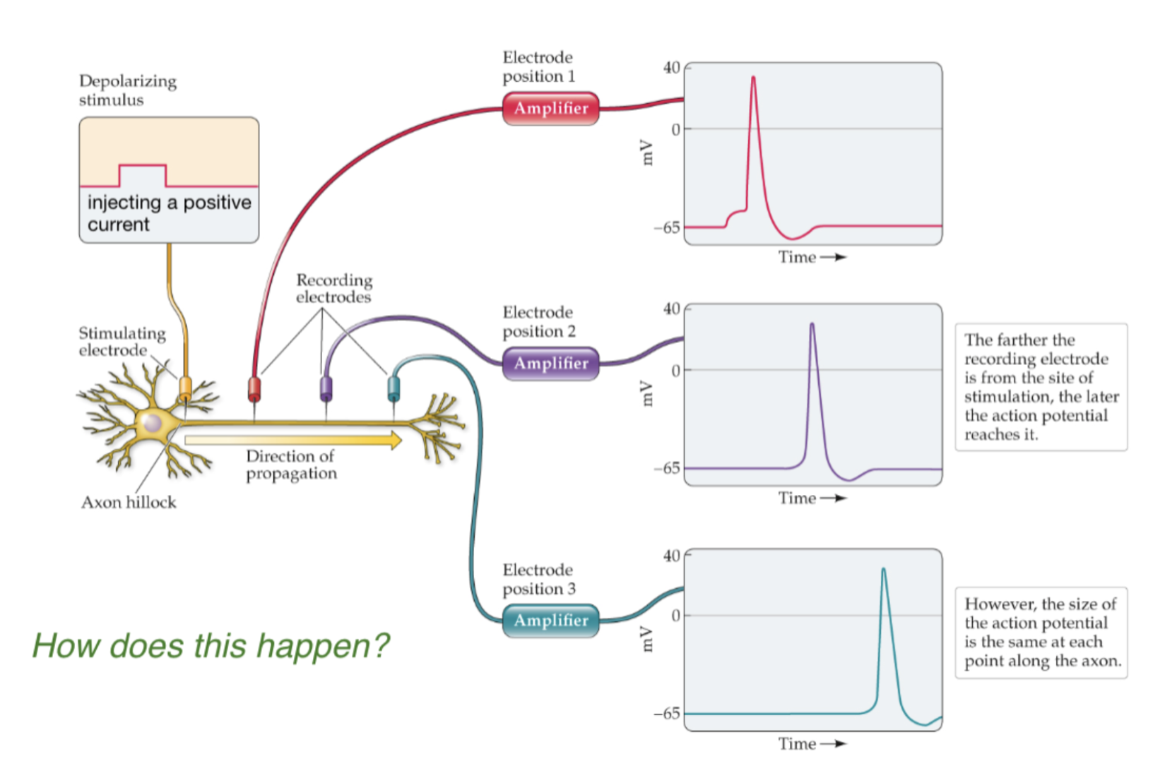
If the dendrites are being stimulated enough to cause an action potential, the height is ~40mV, and a bit down its the same amplitude, and then all the way down at the end its still the same amplitude
The signal has a high fidelity (it doesn't decrease over time) → this isn't normally how things work like in a copper wire (passive conduction)

Passive conduction decays over distance
Bidirectional, signal decays/weakens over distance from source
Not how axon’s work
In the wire, the membrane potential gets lower and lower the further it goes because current is leaking across the membrane this is why we insulate wires (cations are going away) the signal weakens
But in axons it doesn’t decay
Action potentials regenerate themselves down the entire length of the axon
The voltage gated sodium channels are expressed down the entire axon, sodium flows in, depolarization/action potential, then the charge moves and next channel, regenerated over and over again
The action potential is reborn, so the action potential regenerates
The blue is the refractory period which is why 1. You cannot get action potentials on top of each other and 2. Why the potential only goes in one direction
The channels close and slam the door behind them→ this is why the sodium can't go through the other way (one way only street for action potentials)
Important: (on slide)****Action potentials don't decay over…
Its like dominoes can only go in one direction falling (and the domino at the start and at the end is different so the action potential is regenerated not like the same (each domino))
The electrical disturbance causes the next action potential to generate (action potential propagation)
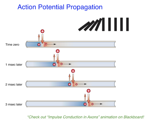
Called active conduction

Myelination for vertebrate axons
Oligodendrocytes (CNS) & Schwann Cells (PNS) produce and ensheath axons in a fatty covering called myelin similar to insulation around an electric wire
Speeds up the process of action potential conduction down the axon

Myelination, saltatory conduction, and current leakage
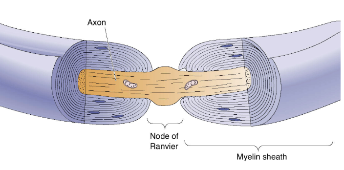
There are certain points down the axon called nodes of ranvier which are small points along the axon where there’s no myelination, and if you zoom in, at each node there's a ton/high density of voltage gated sodium channels
Insulates and allows the axon so that the charge stays and does not leak out
Myelination insulates neurons and thus dramatically reduces current/ion leakage across the membrane
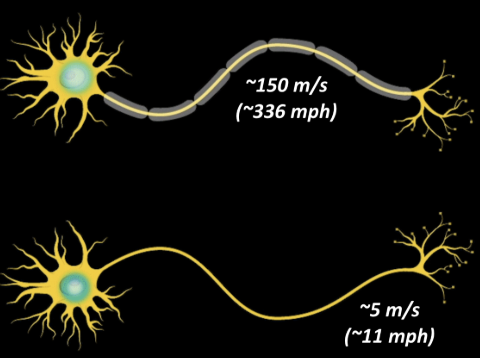
Depolarization “jumps” from node to node, increasing conduction velocity
Increases conduction velocity 15x faster
Action potential can jump from one node to the next
The channels will open, and myelin shunts the charge down to the next one because there's enough depolarization that isn't lost of current down to the next set of channels (the nodes are skipped → saltatory conduction)
Channels have to be expressed the entire length of the axon
Falling dominos
For myelinated axons, it's like an airport walkway, you walk on one, get off for a few steps and then get on the next one
5m/s for unmyelinated 500m/s for myelinated
The myelin sheath and conduction velocity
The myelin sheath that encases some axons is interrupted by nodes of ranvier, small gaps spaced out along the axon
Because the myelin insulation offers resistance to the flow of ionic currents across the membrane, the action potential jumps from node to node
This process is called saltatory conduction, which means to jump/leap
Axons are unmyelinated and mostly small in diameter and slower in conduction
Many invertebrates(animal without a spinal cord) have a few giant axons that mediate essential motor responses, such as escape behavior
The squid’s giant axon has an unusually high conduction rate for invertebrates
To conduct action potentials as swiftly as a myelinated vertebrate axon does, an unmyelinated invertebrate axon would have to be 100 times larger in volume
Estimated at least 10% of the volume of the human brain is occupied by myelinated axons
Myelination is important for the developing nervous system
Purpose of the nodes: you need space for the sodium to flow in
Having large diameter axons (squid giant axon for example) is another way to increase conduction velocity (fire hose vs. garden hose) in the absence of myelination


Multiple sclerosis
Demyelinating disease, oligodendrocytes get attacked and die, and myelin is lost, so you have slow conduction, and it might not even be able to activate the next potential
The extent of MS depends on which part of the brain is attacked
MS does not only affect the motor system
Emotional processing
I think theres more…?
MS associated ataxia (loss of control of body movements), particularly with cerebellar involvement, affects gait:
Irregular, unpredictable step lengths and foot placement
Jerky, hesitant movements
Lack of deceleration and trunk control
Often requires use of upper extremities for support
Decreased safety
A disorder that happens when a person’s immune system makes antibodies that attack myelin, disrupting the conduction of action potentials
The symptoms vary, affecting sensory and/or motor systems, depending on which axons are attacked
The risk of developing MS is much greater in those who have been exposed to the Epstein-Barr virus

But wait! What about times when we do not want to conduct action potentials? Like when we are getting teeth pulled?
In this situation, we would turn to using drugs that block Na+ channels to stop the action potential from propagating pain signals to the brain.
Scientists have discovered a host of drugs and toxins that block specific channels which have helped to understand neural transmission
Two concepts are central to understanding how action potentials are triggered:
Hyperpolarization
An increasing negativity of the membrane potential (the interior of the neuron becomes more negative on the inside relative to the outside)
So if the neuron already has a resting membrane potential of -65mV, hyperpolarization makes it even farther from zero, maybe -70mV
Applying a hyperpolarizing stimulus to the membrane produces an immediate response that passively follows the stimulus pulse
The distortions at the beginning and end of the neuron’s response are caused by the membrane’s ability to store electrical charge known as capacitance
The greater the stimulus, the greater the response, so the neuron’s change in potential is called a graded response
Depolarization
A decreased polarization of the cell membrane
The depolarization of a neuron from a resting potential of -65mV to -60mV makes the inside of the neuron more like the outside.
In other words, the depolarization of a neuron brings its membrane potential closer to zero
Larger depolarizations produce more action potentials, not larger action potentials
The size/amplitude of the action potential is independent of the strength of the stimulation
This characteristic is referred to as the all-or-none property of the action potential; either it fires at full amplitude, or it doesn’t fire at all. It turns out that information is encoded by changes in the frequency of action potentials rather than in their amplitude
Axons exhibit electrical oscillations immediately following the spike; these changes are called afterpotentials, which are also related to the movement of ions in and out of the cell
If we measured the membrane response at locations successively farther and farther away from the stimulus location, we would see another way in which the membrane response seems passive and graded
Like ripples spreading from a pebble dropped in a pond, the potentials produced by stimulation of the membrane diminish as they spread away from the point of stimulation
This is described by a physics law phenomenon: as the potential spreads across the membrane, its size decays as a function of the square of the distance
Local potentials
An electrical potential that is initiated by stimulation at a specific site, which is a graded response that spreads passively across the cell membrane, decreasing in strength with time and distance
Also arise at synapses in response to other neurons
The application of depolarizing pulses to the membrane follows the same pattern as for hyperpolarizing stimuli, producing local, graded responses.
At this point, known as the threshold, a sudden and brief response (the action potential) is provoked
The action potential (also referred to as a spike), is a rapid reversal of the membrane potential that momentarily makes the inside of the membrane positive with respect to the outside
That brief period when the neuron interior is positive with respect to the outside is called an overshoot
Unlike the previous passive spread of graded potentials, the action potential is actively propagated/regenerated down the axon, through ionic mechanisms
Connexons
Made of proteins, arranged to allow ions to flow from one neuron directly into the other. As a consequence, the electrical current that is associated with neural activity in one neuron can flow directly across the gap junction to affect the other neuron
Transmission at these synapses closely resembles action potential conduction along the axon.
Electrical synapses work with practically no time delay, in contrast to chemical synapses, where the delay is on the order of a millisecond, slow in terms of neurons
Because of the speed of their transmission, electrical synapses are frequently found in neural circuits that mediate escape behaviors in invertebrates. They are also found where many fibers must be activated synchronously, as in the system for moving our eyes
Electrical synapses may contribute to the spread of synchronized seizure discharges in epileps
Convergence & Divergence
Convergence:
Many cells send signals to a single cell
Divergences:
One cell sends signals to many other cells
In many parts of the nervous system, the axons from large numbers of neurons converge on a small number of cells. In each human eye, about 100 million receptor cells concentrate their information on 1 million axons that carry the information from the eye to the brain
Higher in the visual system there’s a lot of divergence, the 1 million axons of the optic nerve communicate to billions of neurons in several different specialized regions of the cerebral cortex
Two kinds of synapses:
Electrical synapses
Chemical synapses
Electrical and Chemical Synapses Fundamentally Differ in Their Transmission Mechanisms
Chemical

In the synaptic vesicle is neurotransmitter
Release neurotransmitters into the synaptic cleft which then the neurotransmitters bind to receptors in the postsynaptic receptors
Electrical

No vesicles, no neurotransmitters
The synaptic cleft is super close together, there is an alignment between the pre and post neuron’s ion channels
Structure of electrical synapses

Electrical Synapses
Directly connected (called Gap junctions), which contain aligned, paired channels called connexons
The presynaptic membrane comes even closer to the postsynaptic membrane than it does at a chemical synapse. The gap at an electrical synapse is very small compared to the synaptic cleft of a chemical synapse
Presynaptic connexons pair with post-synaptic connexons to form a pore (gap junction) through which ions and other small molecules directly travel
Depolarization (cations) travel from the presynaptic neuron to postsynaptic neuron; it is essentially instantaneous!

Each one of the proteins is a “connexon”
They connect to the postsynaptic connexon, forms a bridge that connects the neurons together, called a “gap junction” where the depolarization moves from the presynaptic side of the membrane, through the channels that make up the gap junction and into the postsynaptic cell. They travel from the presynaptic neuron to the postsynaptic neuron
Each one of those channels forms a gap junction (two stacked = 1 gap junction) and the pores of these channels can open and close. No neurotransmitters involved in the electrical synapses.
Connexons
Made of proteins, arranged to allow ions to flow from one neuron directly into the other. As a consequence, the electrical current that is associated with neural activity in one neuron can flow directly across the gap junction to affect the other neuron
Transmission at these synapses closely resembles action potential conduction along the axon.
Electrical synapses work with practically no time delay, in contrast to chemical synapses, where the delay is on the order of a millisecond, slow in terms of neurons
Because of the speed of their transmission, electrical synapses are frequently found in neural circuits that mediate escape behaviors in invertebrates. They are also found where many fibers must be activated synchronously, as in the system for moving our eyes
Electrical synapses may contribute to the spread of synchronized seizure discharges in epilepsy

Convergence vs. divergence
Each one of the proteins is a “connexon”
They connect to the postsynaptic connexon, forms a bridge that connects the neurons together, called a “gap junction” where the depolarization moves from the presynaptic side of the membrane, through the channels that make up the gap junction and into the postsynaptic cell. They travel from the presynaptic neuron to the postsynaptic neuron
Each one of those channels forms a gap junction (two stacked = 1 gap junction) and the pores of these channels can open and close. No neurotransmitters involved in the electrical synapses.
Function of electrical synapses - what are they for?
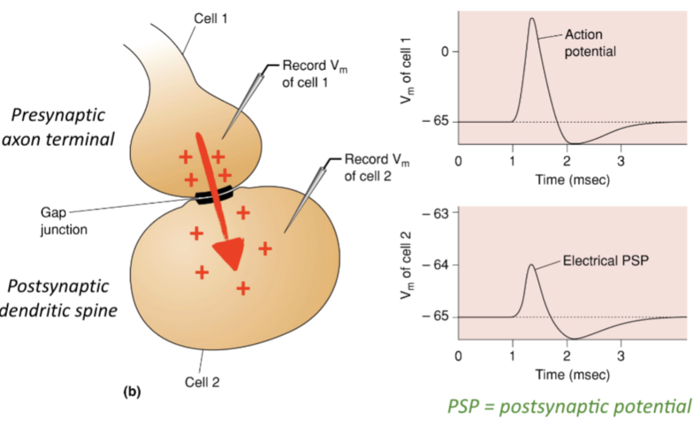
Depolarization on the presynaptic terminal can go straight through and impact the postsynaptic terminal
EPSP
PSP = postsynaptic potential
Instantaneous = small delay (<0.1ms), the two graphs align with each other. Electrical synchronization of the neurons
Action potentials (cations) from the presynaptic neuron directly invade the postsynaptic neuron
Mostly good for synchronizing
Important for really fast behavioral responses (Good for escape behaviors (especially in fish and amphibians) and synchronizing electrical activity among populations of neurons:
Synchronous movements of eye muscles, generation of rhythmic brainstem activity important for breathing and sleep
Eyes have to move at the same time, all at once firing, also fast movements
There is no delay from the presynaptic to the postsynaptic side, ions are just flowing from one side to the other
Chemical synapses
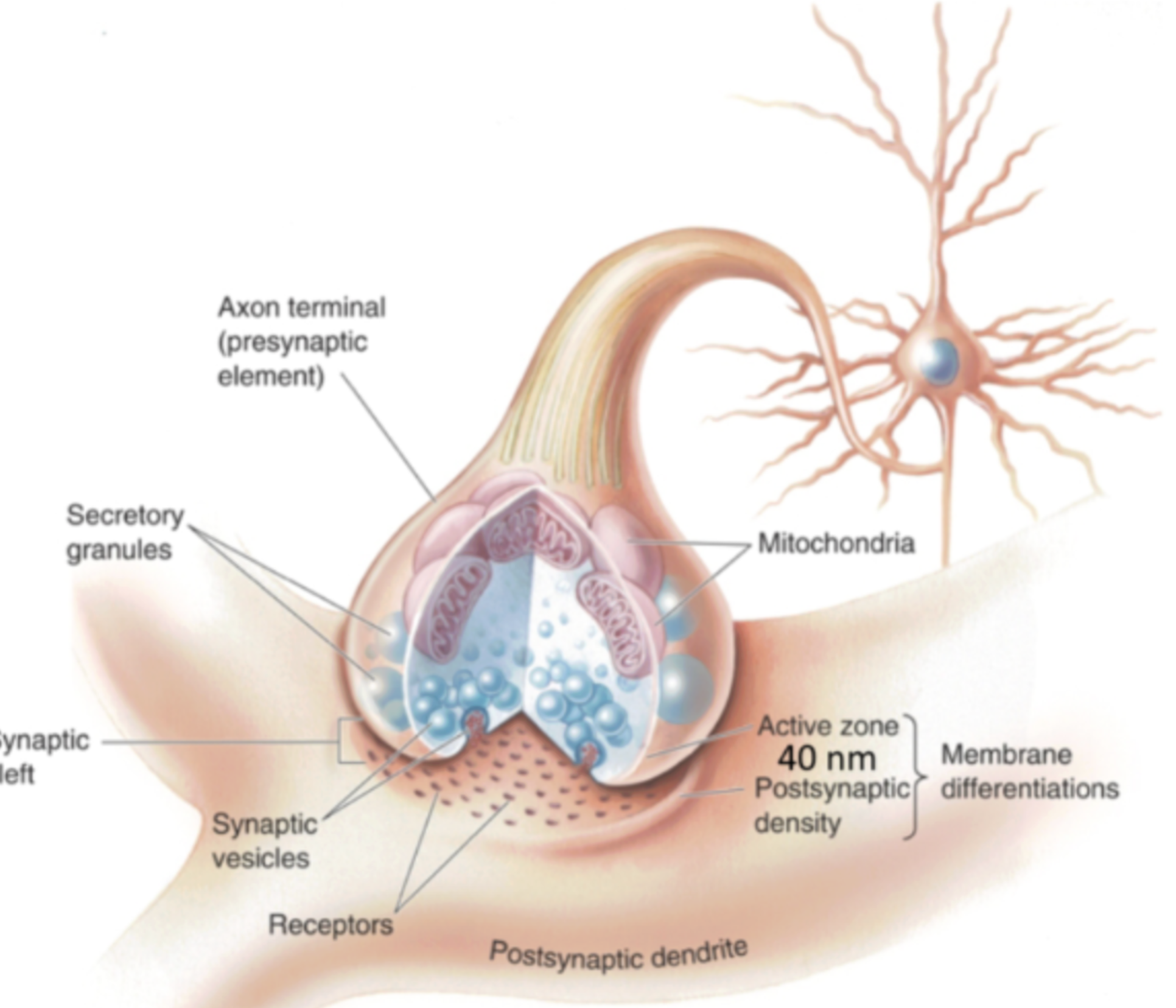
Amplification of signal
If you translate the electrical signal into a chemical one, and just one neuron can result in the activation of a bunch of them
Responsible for the majority of synaptic transmission in the nervous system
Chemical synapses use neurotransmitters to transfer information
Neurotransmitters

You can take the frog out and keep it in this liquid thing and it will keep beating
He found out that there were smth from the brain
He stimulated the vagus nerve of an isolated frog heart, and looked at what effect it had on the heart
He observed that heart rate was reduced, and the contractile force/contraction was lowered, more spread out heart beating
Vagus nerve has an inhibitory effect on the heart
Then he was like maybe there's a chemical that is responsible for slowing the heart rate, he took the perfusion fluid and put it into another heart beaker, maybe it'll have an effect on the second heart without stimulating the nerve. And he realized it worked, there was a reduced heartrate, just when he took the liquid, which means it was a chemical signal, not electrical that made the difference with the second heart, so acetylcholine was discovered
Loewi reasoned that the stimulation of the first frog’s nerve must have caused the release of a chemical, Vagusstoff (substance from the vagus) into the fluid
This is how they learned that the nervous system, long known to use electrical signals, also uses chemical signals
He discovered that Vagusstoff was Acetylcholine = excitatory, but heart is the exception, it had an inhibitory effect in the heart
Neurotransmistteres contr.
Chemicals released by brain cells that affect neighboring cells
Designations:
Classical neurotransmitters:
Are released due to an action potential
Presynaptic releasing, postsynaptic binding to receptor, acts at synapse where it is released
Either neurotransmitter gets released or it doesn’t
Allows signals to be transmitted from neuron to neuron
Classical neurotransmitters include:
Gaba (inhibitory), glutamate, acetylcholine, norepinephrine, serotonin, dopamine
Dopamine acts like a neurotransmitter in one system but in prefrontal cortex acts like a neuromodulator, so these neurotransmitters can fall into both categories depending on context
Neuromodulators:
Alter the effectiveness of signal transmission
May be released at various times and act more distantly
Neuromodulator release might affect other neurotransmitters?
They can turn up the volume
If there's a circuit of neurotransmitters communicating, and the neuromodulator might turn it up, do double the amount of transmission and stuff
Criteria that define a neruotransmitter
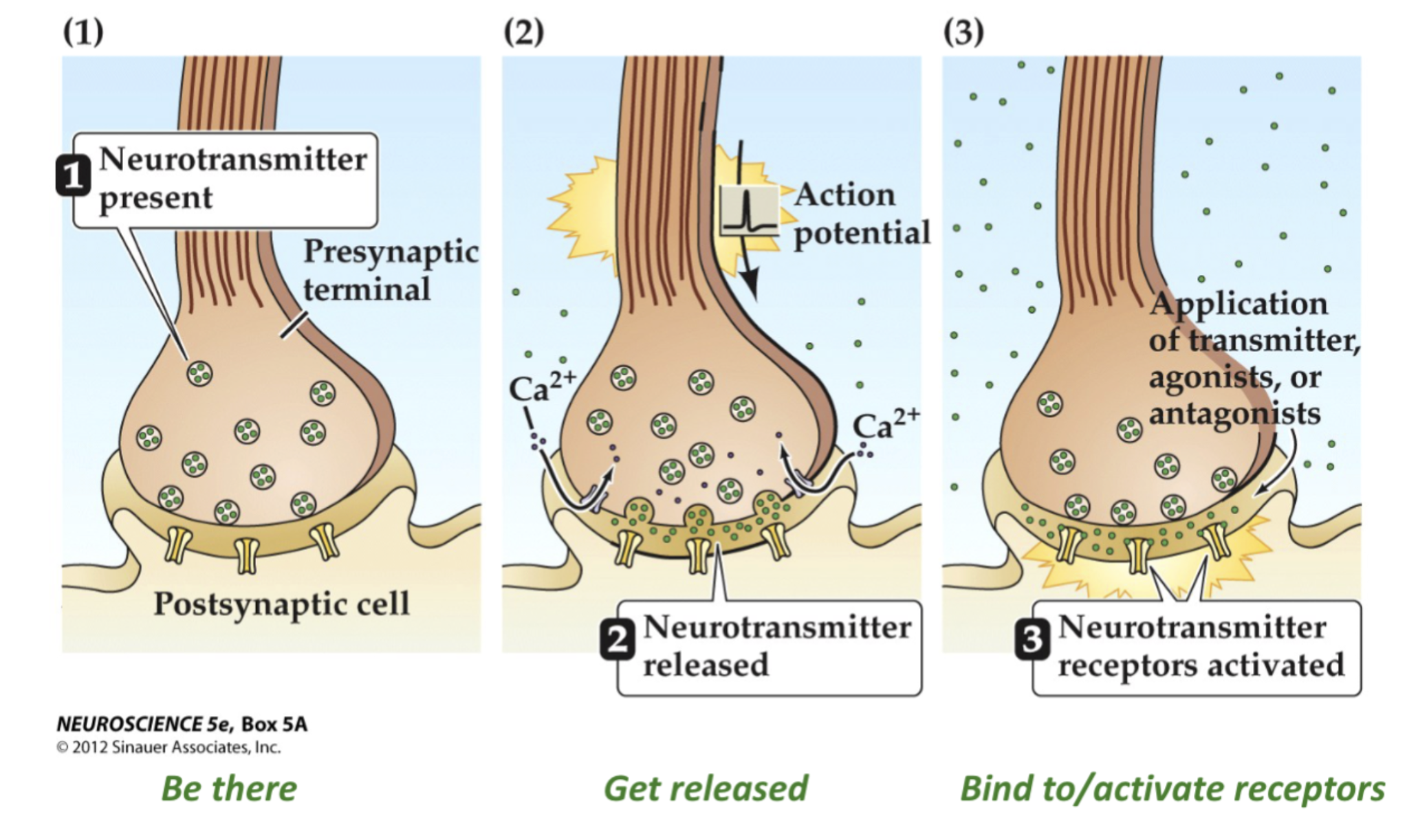
It has to be in the synaptic terminal
It has to get released upon stimulation, when the action potential gets down to the axon? it has to get released
It has to do smth, it has to bind to receptors in the postsynaptic cell
Some synaptic transmitters and families
Amino acids (tiny)
Building blocks of proteins
Ex. histamine, gaba, ,glutamate, glycine, histamine
Synthesized and packaged in the synaptic terminal
Amines (act like classical neurotransmitters with exceptions)
Acetylcholine, norepinephrine, epinephrine (adrenaline), dopamine, serotonin, melatonin
Synthesized and packaged in the synaptic terminal
Amines are put into vesicles right at the synaptic
Neuropeptides (long strings of amino acids)
Proteins
Ex. endorphins (reduce sensation of pain), exercise, euphoric effect, mood enhancer, modulatory influence
Synthesized and packaged into vesicles in the cell body
Oxytocin, neuropeptides are not “classical neurotransmitters”, and are usually neuromodulators
Neuropeptides are made in the cell body and have to be transported down, they are modulators
Gases
Carbon monoxide, nitric oxide
Gaba is major inhibitory, glutamate is major excitatory neurotransmitter
Neurotransmitters are packaged and stored in synaptic vesicles
Usually a lot of neurotransmitter contained in the vesicles
Release of 100,000s of molecules
Slower than electrical synapses but more powerful
At the neuromuscular junction (NMJ), each presynaptic vesicle contains up to 10,000 molecules of acetylcholine (ACh)

Vesicular proteins
Synaptogagamin
Voltage sensor, senses when there is depolarization in the membrane
What happens when the action potential makes and reaches its way all the way down the synaptic terminal?

In the presynaptic terminal, Ca is important (without it no neurotransmitter release)
Neurotransmitter release (via Exocytosis) is initiated by Voltage-Gated Calcium channels
The action potential is depolarization, positive charge invading the presynaptic terminal (AP invades terminal)
Once it gets there, there’s one more type of channel 9calculm channel that when it opens, calcium flows in (VGCa2 channels open
Equilibrium potential is 123mV, so when they open the Ca really wants to get in (high concentration gradient)
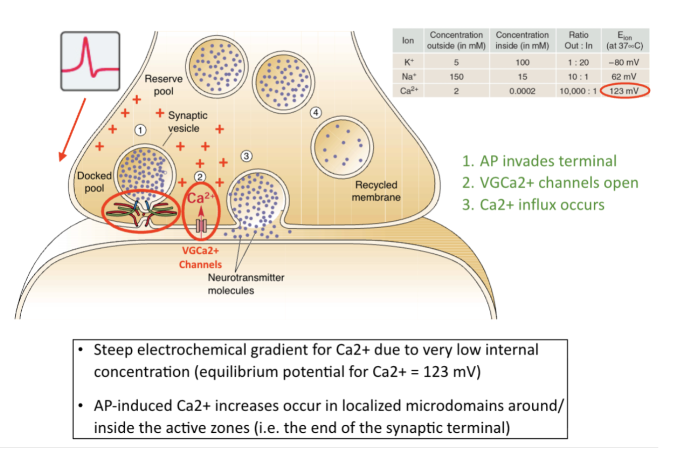
*the squiggly proteins = SNARE proteins (they are tethered together)
SNARE proteins mediate vesicle fusion with the synaptic membrane

Synaptotagrin, a protein on the vesicle that binds to calcium (red dots) which mediates the fusion of the vesicles with the membrane “voltage sensor"
On membrane, t-SNAREs, part of the axon terminal itself
It's like docking a boat, that's like what the proteins do. Once the vesicle is packaged with neurotransmitters they tether together (docking), vesicle is docking with the membrane (don't need calcium for this process), when action potentials are going
Binding sites on the blue protein, calcium binds there, when it binds it causes the protein to become so tightly bound together that it causes the two membranes to fuse, causes the formation of a pore and neurotransmitter goes through, causes the activation of VGC channels, causes vesicular fusion of the two membranes, causing neurotransmitter release
More about Synaptotagrin
Another protein attached to the vesicle, responds to calcium ions to trigger vesicular exocytosis
When the action potential arrives at the axon terminal, the incoming Ca2+ ions bind and activate synaptotagmin, which then triggers the final fusion of the vesicular and presynaptic membranes, allowing the neurotransmitter molecules to enter the synaptic cleft
Botox and tetanus toxin both silence synapses by cutting up SNARE proteins, disabling exocytosis and synaptic transmission
The vesicles are filled with around the same amount of neurotransmitters so when they rupture at the membrane to release their contents, they produce about the same amount of change in postsynaptic potential
The presynaptic terminal normally produces and stores enough transmitter to ensure that it is ready for activity
The rate of production of transmitter is governed by enzymes that are manufactured in the neuronal cell body and transported down the axons to the terminals
Intense activity of the neuron reduces the number of available vesicles
But then more vesicles are produced to replace those that were discharged
Random fact: There's a process called kiss and run in which a vesicle fuses with the presynaptic membrane just long enough to release neurotransmitter into the cleft, and then pinches off again to return to the pool of vesicles

Botox & SNARE Proteins
Cuts up SNARE proteins, and and they cannot tether together, no vesicle fusion, no neurotransmitter release, no wrinkles, paralyzes the neuromuscular junction
Synaptic transmission steps

The action potential traveling down the axon arrives at the axon terminal
This depolarization opens voltage-gated calcium channels in the membrane of the axon terminal, allowing Ca+ ions to enter the terminal
The Ca+ causes synaptic vesicles filled with neurotransmitter to fuse with the presynaptic membrane and rupture, releasing the transmitter molecules into the synaptic cleft
Transmitter molecules cross the cleft to bind to special receptor molecules in the postsynaptic membrane, leading to the opening of ion channels in the postsynaptic membrane
This ion flow creates a local EPSP (Excitatory postsynaptic potential) or IPSP (Inhibitory postsynaptic potential) in the postsynaptic neuron
The IPSPs and EPSPs in the postsynaptic cell spread throughout its interior. If the integration of all the EPSPs and IPSPs depolarizes the axon hillock enough, the postsynaptic neuron will fire an action potential of its own.
Synaptic transmitter is either a) inactivated (degraded) by enzymes or b) removed from the synaptic cleft by transporters, so the transmission is brief and accurately reflects the activity of the presynaptic cell
Synaptic transmitter may also activate presynaptic autoreceptors, regulating future transmitter release
Review
A typical neuron integrates a variety of inputs, and if sufficiently excited (depolarized), it fires a distinctive brief electrical impulse, called an action potential, which rapidly sweeps down the length of the axon toward the axon terminals
Events upon arrival of the action potential:
Because the action potential strongly depolarizes the axon terminal, voltage-gated calcium channels in the terminal membrane are induced to open
The resulting influx of calcium ions triggers the SNAREs, large proteins on the presynaptic membrane and on the synaptic vesicles that cause the vesicles to fuse to the presynaptic membrane and release their cargo of neurotransmitter molecules into the synaptic cleft (a process called exocytosis)
Following their diffusion across the synaptic cleft, neurotransmitter molecules briefly bind to their corresponding receptors, which then mediate a response on the postsynaptic side
Receptors are protein molecules embedded in the postsynaptic membrane that recognize a specific transmitter
The transmitter molecule binds to the receptor, either changing its shape to open an ion channel, as with fast ionotropic receptors (ligand-gated and chemically gated ion channel that is opened when the receptor is bound by a neurotransmitter molecule), or altering chemical reactions within the target cell, as with slow metabotropic receptors (a receptor protein that does not contain an ion channel but may, when activated, use a second-messenger system to alter the functioning of the postsynaptic cell)
Detailed steps of synaptic transmission
When an action potential reaches a presynaptic terminal, it opens voltage-gated calcium channels which allow an influx of Ca+ ions, rather than K+ or Na+, into the axon terminal. These Ca2+ ions activate enzymes that cause vesicles near the presynaptic membrane to fuse with the membrane and discharge their contents into the synaptic cleft
The higher the frequency of action potentials arriving at the terminal, the greater the influx of Ca2+, and the more vesicles that dump transmitter into the synapse
Most synaptic delay is caused by the time needed for Ca2+ to enter the terminal and the vesicles to fuse
Both the diffusion of the transmitter across the cleft and the interaction of transmitter molecules with their receptors also take some time
Lecture 6 - Learning Objectives
Learn how neurons are stimulated and/or inhibited at the dendrites and the different ways that stimulation can simultaneously occur (e.g. spatial vs. temporal summation)
Understand the “anatomy” of an action potential (“nerve impulse”) and how it’s generated
Use the principles from our last lecture to understand why membrane voltage changes as a function of K+/Na+ permeability to produce action potentials (the ionic basis of APs—more on this on Friday)
Lecture 6 - Thought Questions
What do the terms, “depolarization” and “hyperpolarization” mean? What is spatial summation, what is temporal summation, an how do the two forms of summation differ? What does it mean for a neuron to be “excitatory?” What is an “inhibitory” neuron?
Describe the “anatomy” of an action potential. Are there such things as “big” and “little” action potentials? If not, what is one way that signal intensity is encoded in the nervous system?
What role do voltage-gated sodium channels play in the generation of action potentials? How does their activity relate to the concept of “threshold,” and what is the approximate value of “threshold” in millivolts (mV)? Which ions are flowing in and/or out of the neuron during the “rising phase” of the action potential? What about during the “falling phase” of the action potential?
Lecture 7 - Learning Objectives
Understand the role of voltage-gated sodium channels in the generation of the action potential (e.g. function, kinetics, location on neurons)
Learn why action potentials 1) propagate down the axon in one direction and 2) do not decay (same amplitude all the way down)
Discover the role myelin plays in insulating neurons and thus speeding up action potential propagation down the length of the axon, and its implications for demyelinating diseases (e.g. Multiple Sclerosis)
Lecture 7 - Thought Questions
What are voltage-gated sodium channels, where on the neuron are they concentrated, how do they open (or “activate”), and how does their activity relate to the concept of action potential “threshold?”
Explain how action potentials propagate down the axon. What role does myelination play in this process? What are “Nodes of Ranvier?” Why is it that action potentials do not “decay” as they propagate down the axon? Under normal physiological conditions, why can’t action potentials travel backwards, back up the axon toward the cell body? Hint: think about the properties of the voltage-gated sodium channel.
Lecture 8 - Learning Objectives
Learning Objectives:
Appreciate what synapses are—provide an overview of electrical vs. chemical synaptic communication
What are electrical synapses?
What are chemical synapses and what are the “chemicals?”
2. Learn about neurotransmitters—review the major types, how they were first discovered, how a neurotransmitter is defined, etc.
3. Understand how AP arrival in the synaptic terminal leads to neurotransmitter release (synaptic transmission)
Recognize the critical roles of Ca2+, VGCa2+ channels, synaptotagmin, and SNARE proteins in NT release
Lecture 8 - Thought Questions
Compare and contrast chemical and electrical synapses. Which synapse type uses vesicles and releases neurotransmitters? Which synapse type has a smaller synaptic cleft? Which synapse type is the most common in the central nervous system? Which is good for synchronizing neural activity? Why?
List some of the major neurotransmitters of the brain and give an example o an excitatory and inhibitory neurotransmitter. What are the three main criteria that define a neurotransmitter? How do neuromodulators (e.g. neuropeptides) differ from classical neurotransmitters?
What are voltage-gated calcium channels and what role do they play in neurons? What role does calcium play in neurotransmitter release? What role do synaptotagmin and SNARE proteins play in vesicular fusion? List (from memory) the sequence of events underlying synaptic transmission starting with an action potential invading the presynaptic terminal.