rdcs prep
 Call Kai
 Learn
Learn Practice Test
 Spaced Repetition
 Match
Match Flashcards
 Knowt Play
1/131

Earn XP
Description and Tags
thx jack :)
Name | Mastery | Learn | Test | Matching | Spaced | Call with Kai | Chat |
|---|
No analytics yet
Send a link to your students to track their progress
132 Terms
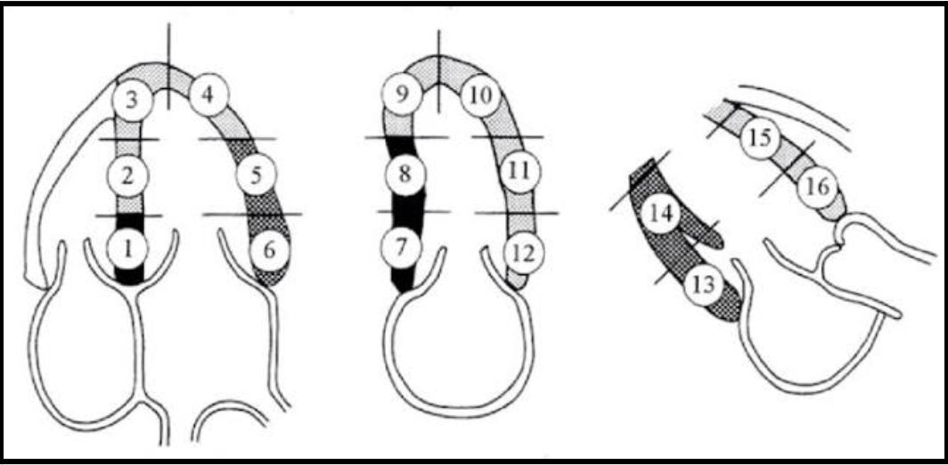
1
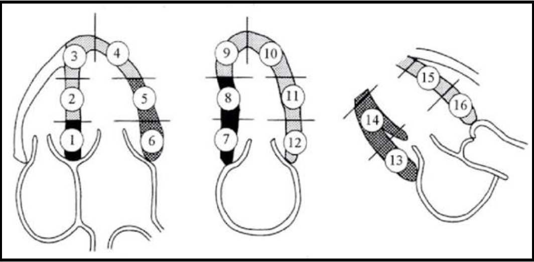
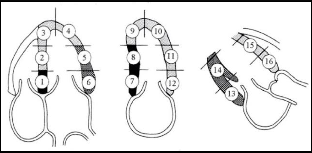
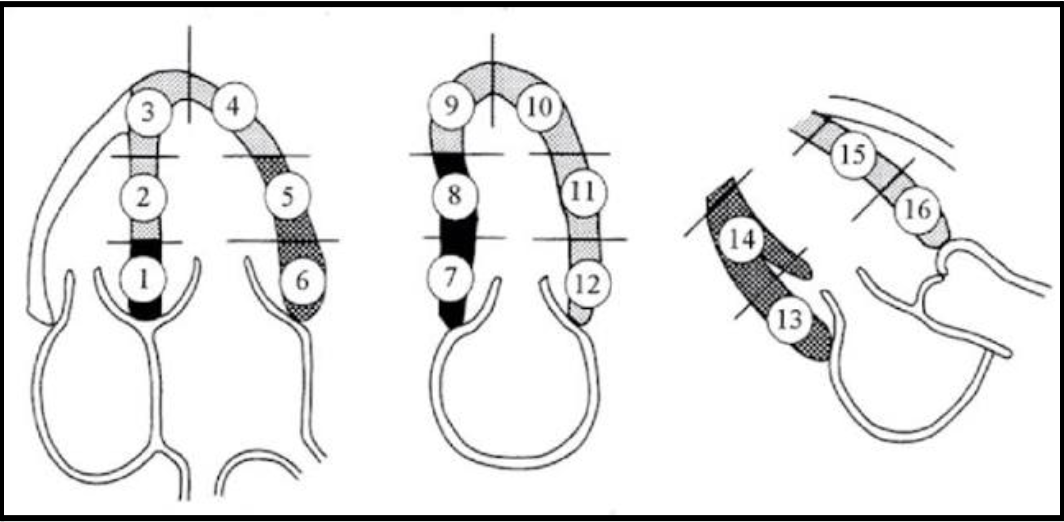
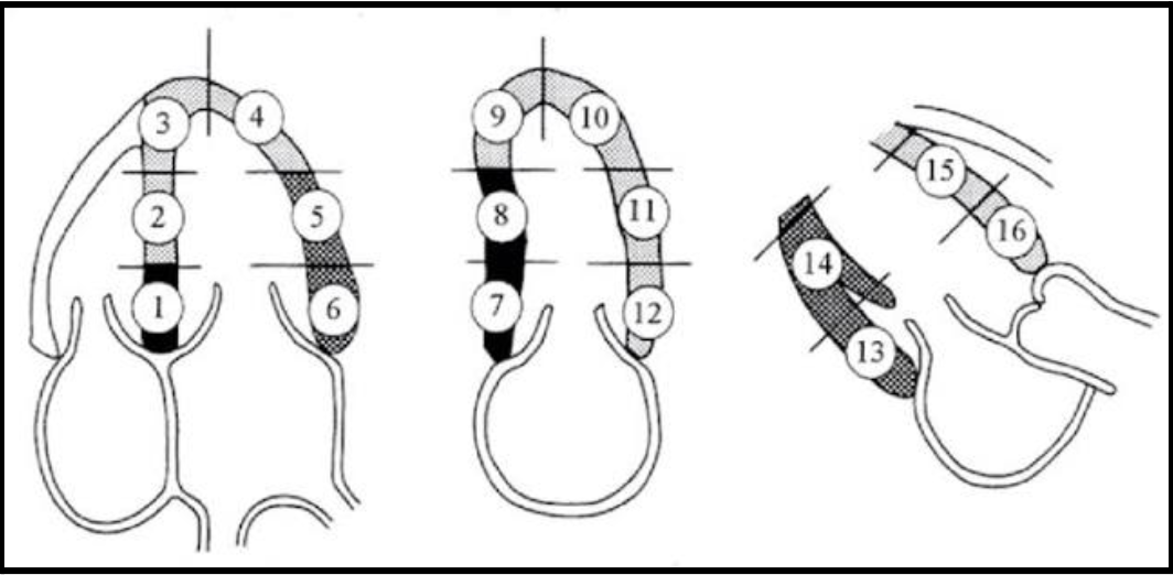
basal inferoseptal
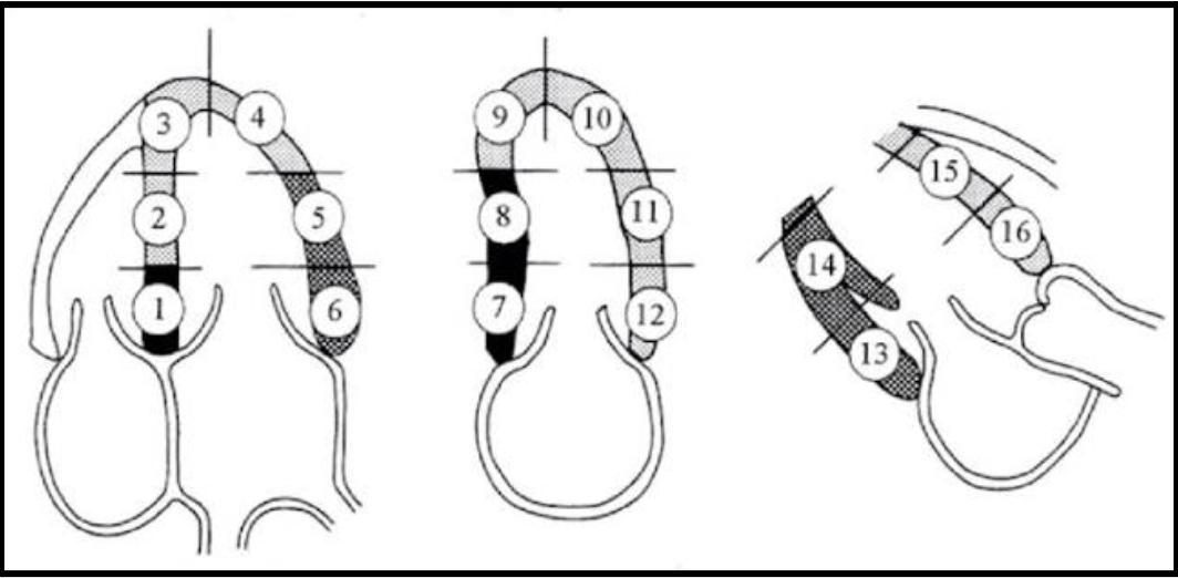
2
mid inferoseptal

3
apical septal

4
apical lateral
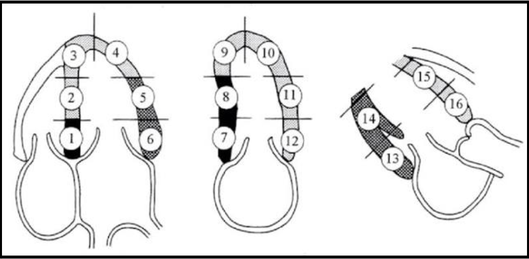
5
mid anterolateral
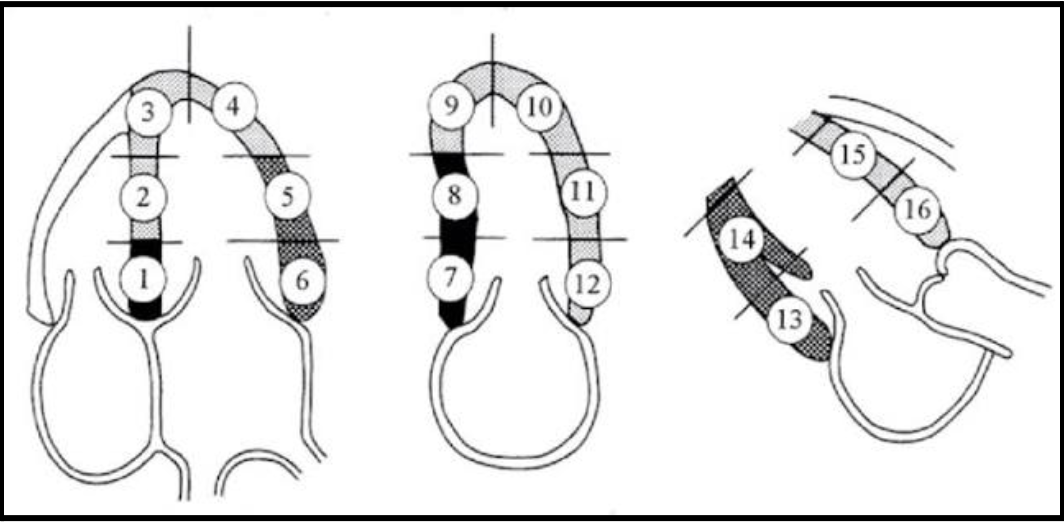
6
basal anterolateral
7
basal inferior
8
mid inferior
9
apical inferior

10
apical anterior

11
mid anterior
12
basal anterior
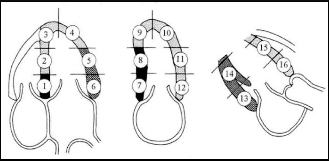
13
basal inferolateral

14
mid inferolateral
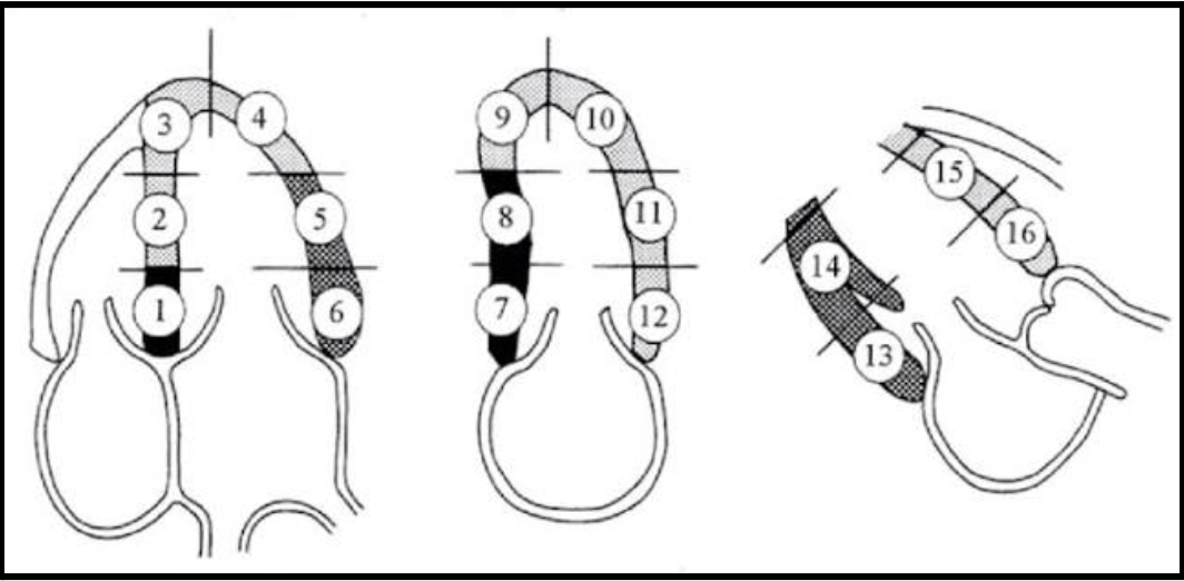
15
mid anteroseptal

16
basal anteroseptal
what structures does the RCA feed?
inferior, inferoseptal, RV apex, RV free wall, IAS
what structures does the LCX feed?
lateral segments
what structures does the LAD feed?
anterior, anteroseptal, LV apex, generally the apical segments
what does TOF cause?
severe cyanosis
TOF characteristics
VSD
overriding AO
pulmonary/infundibular stenosis
RVH
what heart defects are common with trisomy 21/down syndrome
AVSD, CHD (in >50% of cases)
what structures don’t form correctly in trisomy 21 patients?
endocardial cushions (MV, TV, & AV septum)
in trisomy 21, if there is a complete AVSD…
primum ASD + inlet VSD + common AV valve
in trisomy 21, if there is a partial AVSD…
primum ASD + abnormal MV + NO VSD
why would trisomy 21 result in pulmonary HTN?
left to right shunting occurs
what is wolf-parkinson-white syndrome?
abnormal electrical pathways in the heart
causes disruption of normal rhythym
arrhythmia & changes to wall motion are common
what is commonly seen with WPW?
early systolic notching of the posterior wall or IVS
wall motion changes
arrhythmias
characteristics of Ebsteins anomaly
apical displacement of the TV
what is the s wave on hepatic vein PW?
ventricular systole (normal = below baseline)
what is the d wave on hepatic vein PW?
ventricular diastole (normal = below baseline)
what is the a wave on hepatic vein PW?
atrial systole (normal = above baseline)
should the s or d wave of the hepatic veins be a higher velocity on PW?
s wave
what characteristic on PW of the pulmonary veins will severe MR demonstrate?
systolic flow reversal
what is the s wave on pulmonary vein PW?
systole
what is the d wave on pulmonary vein PW?
early diastole
what is the a wave on pulmonary vein PW?
late diastole
hypertrophic cardiomyopathy
⇩LV dimension
⇧A, E/A < 0.8
Lower E, Higher A
Mitral regurgitation
restrictive cardiomyopathy
Endocarditis, infection(amyloidosis)
grade 3 DD, increased stiffness
⇧atrial size
⇩LV size
⇧E, E/A > 2
dilated cardiomyopathy
⇩EF%
⇧Ventricles AND Atria
Thrombus formation
common with diabetics & alcohol use
average E/e’ diastolic dysfunction
>14
average e’ diastolic dysfunction
<6.5 cm/s
septal e’ diastolic dysfunction
<6 cm/s
lateral e’ diastolic dysfunction
<7 cm/s
TR velocity diastolic dysfunction
>2.8 m/s
PASP diastolic dysfunction
>35 mmHg
LAVI diastolic dysfunction
>34 mL/m²
LARS diastolic dysfunction
</= 18%
if E/A is </= 0.8…
grade I DD
if E/A is <2…
grade II DD
if E/A is >2…
grade III DD
MVP murmur
mid-systolic click and late systolic murmur at the apex
MR murmur
high pitched blowing holosystolic murmur with radiation to the axilla
MS murmur
low pitched diastolic rumble with opening snap/click at apex
SEVERE AI/AR murmur
AUSTIN FLINT!!!!
low pitched, mid-diastolic rumble at apex
AS murmur
high pitched systolic crescendo-decrescendo murmur with systolic ejection click at RSB
TR murmur
holosystolic high pitched blowing murmur that may be intensified with inspiration at SSN
TS murmur
diastolic rumble with an opening snap that varies with respiration at mid sternum
PR murmur
low bitched diastolic rumble that may be intensified with inspiration at the left of the sternum
PS murmur
harsh systolic ejection click that may be intensified with expiration at the right of the sternum
PDA murmur
continuous throughout cardiac cycle
VSD murmur
harsh holosystolic murmur that does not radiate or change with respiration located at the left sternal border
general stenotic murmur
crescendo/decrescendo
what does tamponade look like on m-mode & PW?
RV collapse
flow changes vary with respiration
if a patient has an aortic coarctation, what other abnormality are they likely to have?
bicuspid AV (most people have both)
MVP
leaflet bows into LA during systole (>2mm displacement)
flail MV
leaflet TIPS go into LA during systole from ruptured chordae (BIG MR JET)
cleft MV
actual split in the leaflet
constrictive pericarditis can look similar to…
RCM
has similar E/A pattern
what is the key difference in constrictive pericarditis and RCM?
CP has respiratory variation
constrictive pericarditis:
e’ is preserved or increased
myocardium is intact
eggshell pericardium
RCM:
e’ is reduced due to myocardial stiffening
inhalation with tamponade
right side E velocity increases
left side E velocity decreases
exhalation with tamponade
right side E velocity decreases
left side E velocity increases
E wave
early passive LV filling
A wave
atrial kick/contraction
normal E/A value
>1 (E bigger than A)
low E/A
impaired relaxation
high E/A
>/= 2
restrictive filling
E/e’ >14
elevated filling pressure
which is a cardiac emergency: LV pseudoaneurysm or true aneurysm?
PSEUDO: high risk of rupture
when is the septal TV leaflet seen?
when the IVS or coronary sinus ostium is visible
when is the posterior TV leaflet visible?
when the probe is angled more inferiorly towards the right so the septum leaves the image
what percentage of adults have a PFO?
30%
myxoma
MC benign cardiac tumor
MC associated with systolic embolic events
MC in LA with grape cluster appearance
rhabdomyoma
MC benign tumor in peds
MC in RV
adults (18) with tuberous sclerosis
90% of patients have multiple tumors
papillary fibroelastoma
MC valvular tumor
attached to surface of aortic or MV leaflets
small and mobile (can mimic vegetation)
angiosarcoma
MC primary malignancy of the heart
RCC infiltration
finger like projection
MC in IVC that extends into the RA
what percentage of cardiac tumors are benign?
80%
are metastatic malignancies or primary malignancies more common in the heart?
metastatic malignancies (up to 40% more common)
impaired relaxation grade I E/A
< 0.8
pseudonormal grade 2 E/A
<2 + elevated LAP
restrictive grade 3 E/A
>/= 2 + elevated LAP
VSD
MC defect in peds
perimembranous VSD
MC type - upper portion of the septum by semilunar valves
muscular VSD
muscular portion (mid) of IVS
outlet VSD
in RVOT by pulmonic valve
inlet VSD
posterior septum near posterior leaflet of TV
septum secundum
MC form of congenital heart disease in adults
lower septum by IVS