MRIQUIZ TOTAL SET
1/1453
Earn XP
Description and Tags
All of MRIQUIZ questions -> updated 12/5/25 *corrected Q w/ hepatic portal vein
Name | Mastery | Learn | Test | Matching | Spaced |
|---|
No study sessions yet.
1454 Terms

Letter A in Image 16 is responsible for blood supply to the:
A. Anterior brain
B. Posterior brain
C. Face
D. Upper extremities
C. Face

Letter G in Image 12 is pointing to:
A. Lens
B. Lateral rectus muscle
C. Medial rectus muscle
D. Internal carotid artery
E. Globe
B. Lateral rectus muscle

Letter C in image 8 is pointing to:
A. Tentorium
B. Corpus callosum
C. Thalamus
D. Fourth Ventricle
E. Medulla oblongata
C. Thalamus

Letter F in Image 5 is pointing to:
A. Anterior cerebral artery
B. Internal carotid artery
C. Basilar artery
D. Posterior cerebral artery
E. Middle cerebral artery
E. Middle cerebral artery
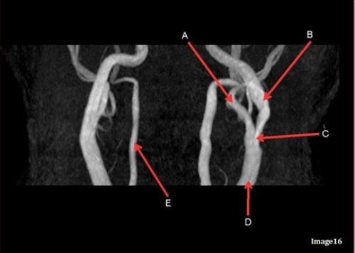
Letter D in Image 16 is pointing to:
A. Internal carotid artery
B. External carotid artery
C. Vertebral artery
D. Common carotid artery
E. Common carotid bifurcation
D. Common carotid artery

Image 16 is an example of what type of MR image?
A. MR Spectroscopy
B. MRA Circle of Willis
C. MRV intracranial circulation
D. MRA extracranial circulation
E. MRA intracranial circulation
D. MRA extracranial circulation

Image 9 is an example of a _____ weighted sequence acquired in the ______ scan plane.
A. T1; Axial
B. T2 FLAIR; sagittal
C. T2; Axial
D. T2; Coronal
C. T2; Axial
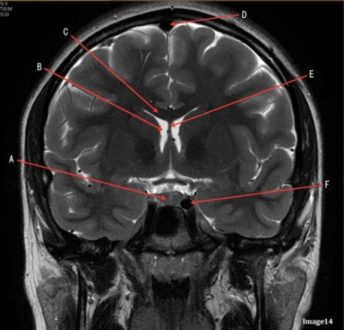
Letter A in image 14 is pointing to:
A. Corpus callosum
B. Third ventricle
C. Lateral Ventricle
D. Pituitary gland
E. Fornix
D. Pituitary gland

Letter C in image 12 is pointing to:
A. Left optic nerve
B. Lateral rectus muscle
C. Medial rectus muscle
D. Lens
E. Midbrain
A. Left optic nerve
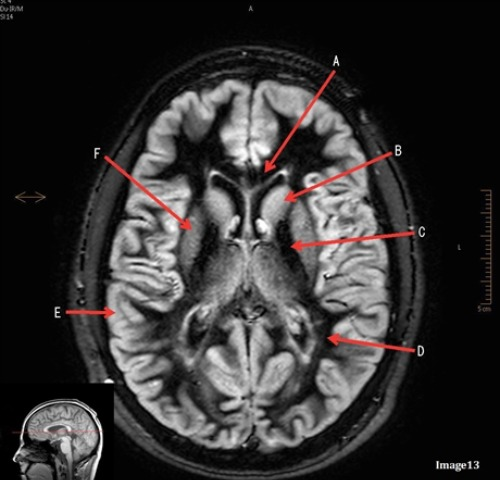
Letter B in image 13 is pointing to:
A. Splenium of the corpus callosum
B. Genu of the corpus callosum
C. Lentiform Nucleus
D. Caudate nucleus
E. Internal capsule
D. Caudate nucleus

Letter D in Image 13 is pointing to:
A. Grey matter
B. White matter
C. Lentiform nucleus
D. Caudate nucleus
E. Internal capsule
B. White matter

Letter C in Image 6 is pointing to:
A. 7th cranial nerve
B. Cochlea
C. Trigeminal nerve
D. Semicircular canal
A. 7th cranial nerve

Letter D in Image 7 is pointing to:
A. Pons
B. Cerebellum
C. Hypothalamus
D. Genu of the corpus callosum
E. Splenium of the corpus callosum
B. Cerebellum

Letter F in image 14 is pointing to:
A. Third ventricle
B. Pituitary gland
C. Hypothalamus
D. Internal carotid artery
D. Internal carotid artery

Letter D in Image 12 is pointing to:
A. Optic nerve
B. Pituitary gland
C. Globe
D. Lens
E. Midbrain
B. Pituitary gland

Letter D in Image 8 is pointing to:
A. Tentorium
B. Cerebellum
C. Thalamus
D. Fourth Ventricle
E. Medulla oblongata
A. Tentorium

Letter C in Image 10 is pointing to:
A. Third ventricle
B. Basal ganglia
C. Anterior horn lateral ventricle
D. Posterior horn lateral ventricle
C. Anterior horn lateral ventricle

Letter E in Image 12 is pointing to:
A. Optic nerve
B. Pituitary gland
C. Globe
D. Pons
E. Left lens
D. Pons

Letter B in Image 6 is pointing to:
A. Maxillary sinus
B. Sphenoid sinus
C. Vertebral artery
D. Internal carotid artery
D. Internal carotid artery
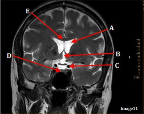
Image 11 is an example of a _______ weighted sequence acquired in the _______ scan plane.
A. T1; Axial
B. T1; Coronal
C. T2; Axial
D. T2; Coronal
E. STIR; Axial
D. T2; Coronal
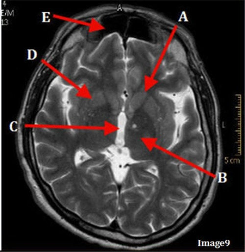
Letter E in Image 9 is pointing to:
A. Maxillary sinus
B. Sphenoid sinus
C. Frontal sinus
D. Ethmoid sinus
C. Frontal Sinus

Letter E in Image 16 is responsible for blood supply to the:
A. Anterior brain
B. Posterior brain
C. Face
D. Upper extremities
B. Posterior brain

Letter A in Image 8 is pointing to:
A. Tentorium
B. Corpus callosum
C. Hypothalamus
D. Fourth ventricle
E. Lateral Ventricle
E. Lateral Ventricle
The medial and lateral rectus muscles are located in the:
A. Ears
B. Gluteus maximus
C. Eyes
D. Outer neck regions
C. Eyes

Letter D in Image 6 is pointing to:
A. 7th cranial nerve
B. Cochlea
C. Trigeminal Nerve
D. Semicircular canal
D. Semicircular canal

Letter B in Image 10 is pointing to:
A. Third ventricle
B. Basal ganglia
C. Anterior horn lateral ventricle
D. Posterior horn lateral ventricle
D. Posterior horn lateral ventricle

Letter B in Image 16 is responsible for blood supply to the:
A. Anterior brain
B. Posterior brain
C. Face
D. Upper extremities
A. Anterior brain

Letter E in Image 14 is pointing to:
A. Corpus callosum
B. Third ventricle
C. Lateral ventricle
D. Internal carotid artery
E. Fornix
E. Fornix

Letter E in Image 6 is pointing to:
A. 7th cranial nerve
B. Cochlea
C. Trigeminal nerve
D. Semicircular canal
B. Cochlea
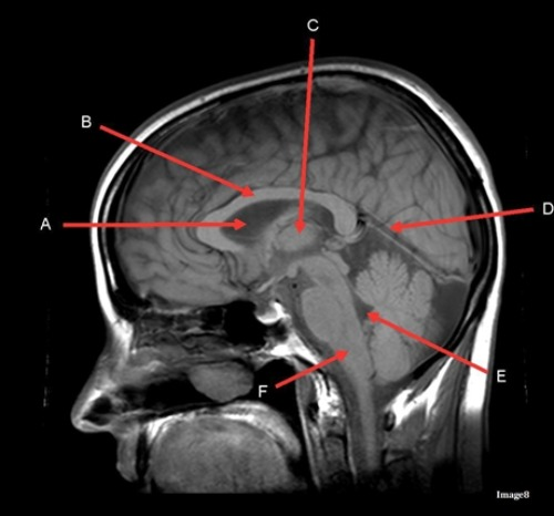
Image 8 is an example of a ____ weighted sequence acquired in the ____ scan plane.
A. T1; Axial
B. T1; Sagittal
C. T2; Axial
D. T2; Sagittal
B. T1; Sagittal

Letter C in Image 5 is pointing to:
A. Vertebral artery
B. Internal carotid artery
C. Basilar artery
D. Posterior cerebral artery
E. Middle cerebral artery
A. Vertebral artery

Letter C in Image 13 is pointing to:
A. Splenium of the corpus callosum
B. Genu of the corpus callosum
C. Lentiform nucleus
D. Caudate nucleus
E. Internal capsule
E. Internal capsule

Letter A in Image 7 is pointing to:
A. Pons
B. Cerebellum
C. Hypothalamus
D. Genu of the corpus callosum
E. Splenium of the corpus callosum
D. Genu of the corpus callosum

Letter F in Image 6 is pointing to:
A. Maxillary sinus
B. Sphenoid sinus
C. Frontal sinus
D. Optic chiasm
A. Maxillary sinus
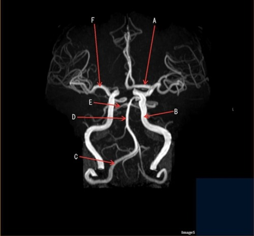
Letter D in Image 5 is pointing to:
A. Anterior cerebral artery
B. Internal carotid artery
C. Basilar artery
D. Posterior cerebral artery
E. Middle cerebral artery
C. Basilar artery

Letter A in image 12 is pointing to:
A. Left optic nerve
B. Lateral rectus muscle
C. Medial rectus muscle
D. Lens
E. Midbrain
C. Medial rectus muscle
The right and left optic nerve join at the:
A. Optic chiasm
B. Foramen magnum
C. Transverse sinus
D. Internal optic canals
A. Optic Chiasm

Letter B in Image 7 is pointing to:
A. Pons
B. Cerebellum
C. Hypothalamus
D. Genu of the corpus callosum
E. Splenium of the corpus callosum
E. Splenium of the corpus callosum

Letter D in Image 14 is pointing to:
A. Tentorium
B. Sphenoid sinus
C. Frontal Sinus
D. Sagittal sinus
E. Fornix
D. Sagittal sinus
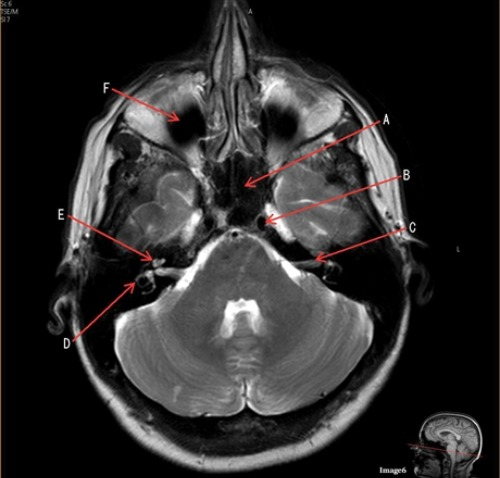
Letter A in Image 6 is pointing to:
A. Maxillary sinus
B. Sphenoid Sinus
C. Frontal Sinus
D. Internal Carotid artery
B. Sphenoid sinus

Letter B in Image 14 is pointing to:
A. Corpus callosum
B. Third ventricle
C. Lateral ventricle
D. Pituitary gland
E. Fornix
C. Lateral ventricle

Letter A in Image 5 is pointing to:
A. Anterior cerebral artery
B. Internal carotid artery
C. Basilar artery
D. Posterior cerebral artery
E. Middle cerebral artery
A. Anterior cerebral artery

Letter C in Image 7 is pointing to:
A. Pons
B. Cerebellum
C. Hypothalamus
D. Genu of the corpus callosum
E. Splenium of the corpus callosum
A. Pons
Which intracranial artery passes through the sylvian fissure?
A. Basilar
B. Internal carotid
C. External mesenteric
D. Middle cerebral
D. Middle cerebral


Letter C in Image 9 is pointing to:
A. Third ventricle
B. Thalamus
C. Lentiform nucleus
D. Caudate nucleus
A. Third ventricle

Letter A in Image 16 is pointing to:
A. Internal carotid artery
B. External carotid artery
C. Vertebral artery
D. Common carotid artery
E. Common carotid bifurcation
B. External carotid artery

Letter C in Image 14 is pointing to:
A. Corpus callosum
B. Third ventricle
C. Lateral ventricle
D. Pituitary gland
E. Fornix
A. Corpus callosum

Letter H in Image 12 is pointing to:
A. Lens
B. Lateral rectus muscle
C. Medial rectus muscle
D. Internal carotid artery
E. Globe
E. Globe

Letter E in Image 13 is pointing to:
A. Grey matter
B. White matter
C. Lentiform nucleus
D. Caudate nucleus
E. Internal capsule
A. Grey matter

Image 6 is an example of a ____ weighted sequence acquired in the _____ imaging plane.
A. T1; Axial
B. T1; Coronal
C. T2; Axial
D. T2; Coronal
E. STIR; Axial
C. T2; Axial
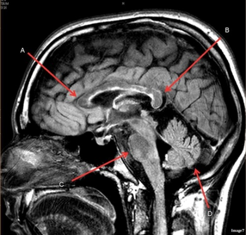
Image 7 is an example of a _____ weighted sequence acquired in the ______ scan plane.
A. T1; Axial
B. T2 FLAIR; Sagittal
C. T2; Axial
D. T2; Sagittal
B. T2 FLAIR; Sagittal

Letter B in Image 8 is pointing to:
A. Tentorium
B. Corpus callosum
C. Hypothalamus
D. Fourth ventricle
E. Medulla oblongata
B. Corpus callosum
Which arteries join together to form the basilar artery?
A. Vertebral arteries
B. External carotids
C. Internal carotids
D. Iliacs
E. None of the above
A. Vertebral arteries

Letter A in Image 9 is pointing to:
A. Third ventricle
B. Thalamus
C. Lentiform nucleus
D. Caudate nucleus
D. Caudate nucleus

Letter F in Image 13 is pointing to:
A. Grey matter
B. White matter
C. Lentiform nucleus
D. Caudate nucleus
E. Internal capsule
C. Lentiform nucleus

Letter D in Image 9 is pointing to:
A. Third ventricle
B. Thalamus
C. Lentiform nucleus
D. Caudate nucleus
C. Lentiform nucleus

Letter B in Image 16 is pointing to:
A. Internal carotid artery
B. External carotid artery
C. Vertebral artery
D. Common carotid artery
E. Common carotid bifurcation
A. Internal carotid artery

Letter E in Image 8 is pointing to:
A. Tentorium
B. Cerebellum
C. Thalamus
D. Fourth ventricle
E. Medulla oblongata
D. Fourth ventricle

Letter B in Image 12 is pointing to:
A. Left optic nerve
B. Lateral rectus muscle
C. Medial rectus muscle
D. Lens
E. Midbrain
D. Lens

Letter E in Image 5 is pointing to:
A. Anterior cerebral artery
B. Internal carotid artery
C. Basilar artery
D. Posterior cerebral artery
E. Middle cerebral artery
D. Posterior cerebral artery
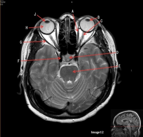
Letter J in Image 12 is pointing to:
A. Globe
B. Lateral rectus muscle
C. Medial rectus muscle
D. Right lens
E. Left lens
D. Right lens
The pituitary stalk is also known as:
A. Hypothalamus
B. Internal capsule
C. Lentiform nucleus
D. Infundibulum
D. Infundibulum

Letter C in Image 16 is pointing to:
A. Internal carotid artery
B. External carotid artery
C. Vertebral artery
D. Common carotid artery
E. Common carotid bifurcation
D. Common carotid artery
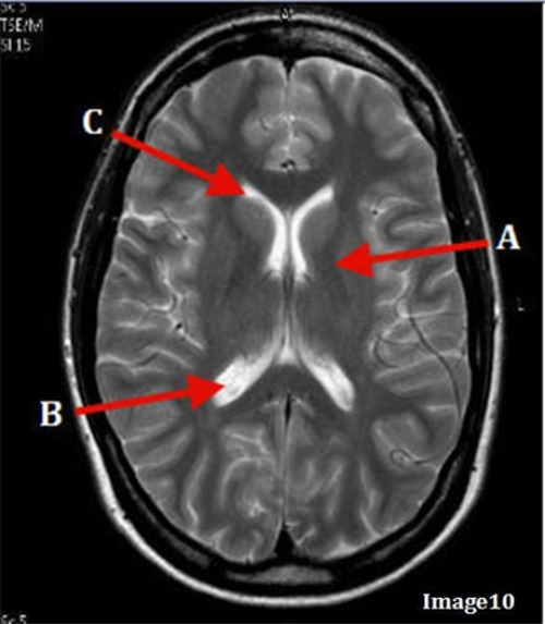
Letter A in Image 10 is pointing to:
A. Third Ventricle
B. Basal ganglia
C. Anterior horn Lateral Ventricle
D. Posterior horn lateral ventricle
B. Basal ganglia

Letter E in Image 16 is pointing to:
A. Internal carotid artery
B. External carotid artery
C. Vertebral artery
D. Common carotid artery
C. Vertebral artery

Image 5 is an example of an:
A. MRI brain
B. MRV sagittal sinus
C. MRS single Voxel
D. MRA Circle of Willis
D. MRA Circle of Willis

Letter F in Image 8 is pointing to:
A. Tentorium
B. Cerebellum
C. Thalamus
D. Fourth Ventricle
E. Medulla oblongata
E. Medulla oblongata

Letter F in Image 12 is pointing to:
A. Lens
B. Lateral rectus muscle
C. Medial rectus muscle
D. Internal carotid artery
E. Globe
D. Internal Carotid artery

Letter B in Image 5 is pointing to:
A. Anterior cerebral artery
B. Internal carotid artery
C. Basilar artery
D. Posterior cerebral artery
E. Middle cerebral artery
B Internal carotid artery

Letter B in Image 9 is pointing to:
A. Third ventricle
B. Thalamus
C. Lentiform nucleus
D. Caudate nucleus
B. Thalamus

Letter A in Image 16 is responsible for blood supply to the:
A. Anterior brain
B. Posterior brain
C. Face
D. Upper extremities
C. Face

Letter C in Image 136 is pointing to:
A. Anterior frontal veins
B. Posterior frontal veins
C. Parietal veins
D. Internal cerebral veins
E. Vein of trolard
C. Parietal veins
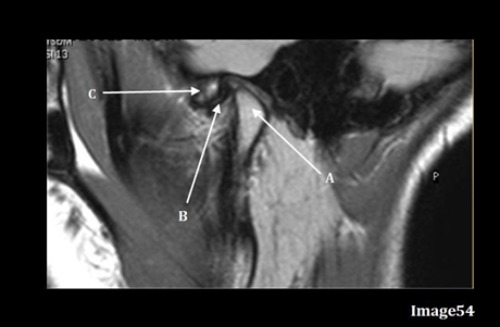
Letter A in Image 54 is pointing to:
A. Articular disk
B. Articular tubercle
C. Intervertebral disk
D. Mandibular condyle
E. Lateral epicondyle
D. Mandibular condyle

Letter C in Image 133 is pointing to:
A. Middle cerebral artery
B. Common carotid bifurcation
C. VertebroBasilar junction
D. Internal carotid artery
E. External carotid artery
F. Abdominal aorta
B. Common carotid bifurcation

Letter C in Image 54 is pointing to:
A. Articular disk
B. Articular tubercle
C. Intervertebral disk
D. Mandibular condyle
E. Lateral epicondyle
B. Articular tubercle

Letter R in Image 137 is pointing to:
A. Pons
B. Cerebral peduncle
C. Thalamus
D. Caudate nucleus
E. Cerebellum
F. Basal vein
A. Pons

Letter Q in Image 136 is pointing to:
A. Right transverse sinus
B. Left transverse sinus
C. Right parietal veins
D. Left parietal veins
E. Right sigmoid sinus
F. Left sigmoid sinus
A. Right transverse sinus

Letter N in Image 137 is pointing to:
A. Pons
B. Cerebral peduncle
C. Genu of corpus callosum
D. Body of corpus callosum
E. Splenium of corpus callosum
D. Body of corpus callosum

Letter J in Image 137 is pointing to:
A. Aqueduct of Sylvius
B. Third ventricle
C. Sulcus
D. Fourth ventricle
E. Superior sagittal sinus
F. Lateral ventricle
D. Fourth ventricle

Letter H in Image 137 is pointing to:
A. Grey matter
B. White matter
C. Sulcus
D. Fourth ventricle
E. Superior sagittal sinus
C. Sulcus

Letter F in Image 136 is pointing to:
A. Anterior frontal vein
B. Posterior frontal vein
C. Parietal vein
D. Internal jugular vein
E. Internal cerebral vein
D. Internal jugular vein

Letter D in Image 136 is pointing to:
A. Right transverse sinus
B. Left transverse sinus
C. Sigmoid sinus
D. Superior sagittal sinus
E. Vein of trolard
D. Superior sagittal sinus

Letter J in Image 136 is pointing to:
A. Right transverse sinus
B. Left transverse sinus
C. Right parietal veins
D. Left parietal veins
E. Right sigmoid sinus
F. Left sigmoid sinus
C. Right parietal veins

Letter M in Image 136 is pointing to:
A. Right transverse sinus
B. Left transverse sinus
C. Right parietal veins
D. Left parietal veins
E. Right sigmoid sinus
F. Left sigmoid sinus
F. Left sigmoid sinus

Letter F in Image 133 is pointing to:
A. Middle cerebral artery
B. Common carotid bifurcation
C. Brachiocephalic artery
D. Thoracic aorta
E. Subclavian artery
D. Thoracic aorta

Letter O in Image 137 is pointing to:
A. Pons
B. Cerebral peduncle
C. Genu of corpus callosum
D. Body of corpus callosum
E. Splenium of corpus callosum
F. Basal vein
E. Splenium of corpus callosum

Image 54 is an MRI of the ______.
A. Shoulder joint
B. Hip Joint
C. TMJ
D. Pituitary gland
C. TMJ

Letter R in Image 136 is pointing to:
A. Rosenthal vein
B. Posterior frontal vein
C. Basal vein
D. Internal cerebral vein
E. Vein of trolard
F. A and / or C
F. A and / or C

Letter P in Image 136 is pointing to:
A. Right transverse sinus
B. Left transverse sinus
C. Right parietal veins
D. Left parietal veins
E. Right sigmoid sinus
F. Left sigmoid sinus
B. Left transverse sinus

Letter G in Image 136 is pointing to:
A. Superior sagittal sinus
B. Right sigmoid sinus
C. Left sigmoid sinus
D. Right transverse sinus
E. Left transverse sinus
D. Right transverse sinus

Letter K in Image 133 is pointing to:
A. Vertebral artery
B. Common carotid bifurcation
C. Brachiocephalic artery
D. Internal carotid artery
E. External carotid artery
F. Abdominal aorta
D. Internal carotid artery
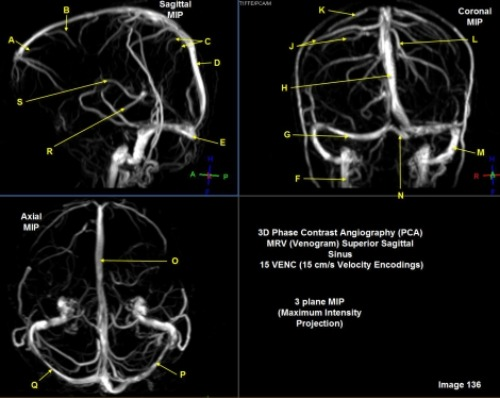
Letter K in Image 136 is pointing to:
A. Anterior frontal vein
B. Posterior frontal vein
C. Parietal vein
D. Internal cerebral vein
E. Vein of trolard
E. Vein of trolard
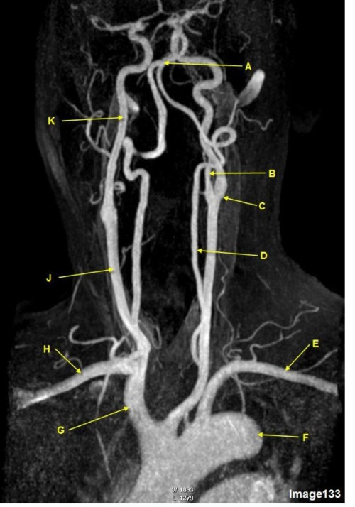
Letter E in Image 133 is pointing to:
A. Right subclavian artery
B. Left subclavian artery
C. Brachiocephalic artery
D. Innominate artery
E. Aortic arch
B. Left subclavian artery

Letter E in Image 136 is pointing to:
A. Anterior frontal vein
B. Vein of trolard
C. Torcular herophili
D. Internal cerebral vein
E. Posterior sigmoid sinus
C. Torcular herophili

Letter M in Image 137 is pointing to:
A. Pons
B. Cerebral peduncle
C. Thalamus
D. Caudate nucleus
E. Cerebellum
C. Thalamus

Letter D in Image 133 is pointing to:
A. Vertebral artery
B. Common carotid bifurcation
C. Brachiocephalic artery
D. Internal carotid artery
E. External carotid artery
A. Vertebral artery

Letter X in Image 137 is pointing to:
A. Anterior frontal vein
B. Cerebral peduncle
C. Sulcus
D. Vein of galen
E. Superior sagittal sinus
D. Vein of galen

Letter B in Image 136 is pointing to:
A. Anterior frontal vein
B. Posterior frontal vein
C. Parietal vein
D. Internal cerebral vein
E. Vein of trolard
B. Posterior frontal vein

Letter B in Image 137 is pointing to:
A. Pons
B. Cerebral peduncle
C. Thalamus
D. Caudate nucleus
E. Cerebellum
D. Caudate nucleus
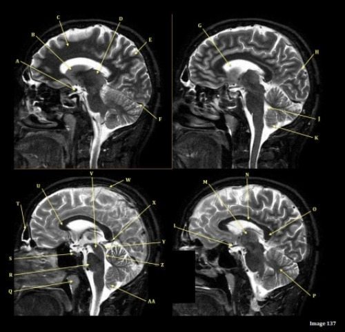
Letter S in Image 137 is pointing to:
A. Optic chiasm
B. Pituitary gland
C. Inferior colliculus of midbrain
D. Internal carotid artery
E. Vein of galen
F. Straight sinus
B. Pituitary gland