Biology chapter 4
 Call Kai
 Learn
Learn Practice Test
 Spaced Repetition
 Match
Match Flashcards
 Knowt Play
1/54

Earn XP
Description and Tags
Cell structure
Name | Mastery | Learn | Test | Matching | Spaced | Call with Kai | Chat |
|---|
No analytics yet
Send a link to your students to track their progress
55 Terms
Organisms are organized in a hierarchy
Cells
⬇
Tissues
⬇
Organ
⬇
Organ system
⬇
Organism
Cells
The basic structural, functional, and biological unit of all known living organisms
The basic unit
Tissues
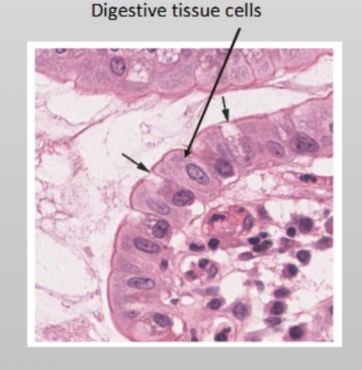
An intermediate level of biological organization consisting of a group of structurally and functionally similar cells working together to perform a specific task
Are made of cells with a common function
Organ
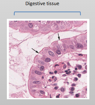
A specialized structure composed of two or more different types of tissues that work together to perform a specific, complex function for the organism
Formed by several tissues

Organ system
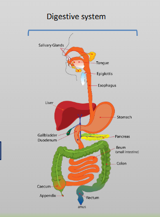
A group of organs that work together to perform vital, complex body functions
Made up of organs working together
Organism
An individual living system—such as a plant, animal, fungus, or microorganism—that can carry out all basic life processes independently
Composed of multiple organ systems
Microscopes
Cells are too small to be seen with the naked eye
These make it possible to see small cells
Ex: light microscope, electron microscope
Light microscope
An optical instrument that uses visible light and glass lenses to magnify and view small biological specimens like cells and tissues

Magnification
The process of enlarging an object in appearance
The ratio of an object's image size to its real, actual size, making a specimen appear larger than it is

Resolution/Resolving power
The ability of a microscope to distinguish two adjacent structures as seperate; the higher the resolution, the better the clarity and detail of the the image

Stains
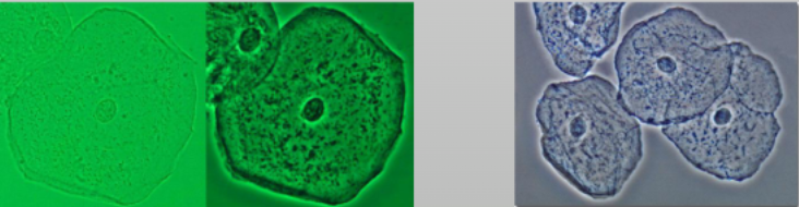
Transparent objects (like cells) are treated with chemical stains to
distinguish different parts
Chemical: a dye or reagent used to color microscopic structures, cells, or tissues to increase visual contrast and highlight specific components
Fluorescent: specialized chemical dyes or tagged molecules that bind to specific cellular components or macromolecules and emit visible light when excited by a specific wavelength of light
Electron microscope
An imaging tool that uses a beam of electrons instead of visible light to view ultra-small specimens at a much higher resolution and magnification

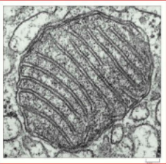
Transmission electron microscope
An advanced imaging tool that passes a beam of electrons through an ultra-thinly sliced specimen to view its internal structures at a nanometer or atomic scale
show fine detail within cells
Scanning electron microscope
An instrument that uses a focused beam of electrons to scan the surface of a specimen, creating detailed, three-dimensional surface images
provide 3-D exterior views

Cell theory
A significant principle of biology proposed by Schleiden and Schwann in the 1830’s
Cells are basic units of life
All living organisms are made of cells
All cells come from pre-existing cells
Cells have 4 components
Plasma membrane: separates the cell’s interior from the outside
Cytoplasm: inside of cell (cytosol) where organelles are found
DNA: the genetic material
Ribosomes: synthesize proteins
Characteristics of prokaryotes
No nucleus or organelles
Have a cell wall made of peptidoglycan
Prokaryotes are similar to the the first cells
Prokaryotes divided into domains: Archaea & Bacteria

General structure of prokaryotic cells
One chromosome
Ribosomes
The cell membrane surrounded by a cell wall (made of peptidoglycan)

Prokaryotic cells are smaller than eukaryotic cells
Reasons for small size of prokaryotic cells:
Surface area to volume ratio is more favorable for moving material in and out of the cell
They lack organelles found in eukaryotes
Surface area–to–volume ratio
A mathematical comparison of a cell's outer membrane area to its internal volume, determining how efficiently it can exchange materials with its environment
As cells get bigger, volume increases faster than surface
areaHigh SA:V Ratio (Small Cells)
Low SA:V Ratio (Large Cells)

Eukaryotic cells
Have a nucleus
More complex than prokaryotic
Highly compartmentalized (membrane-bound organelles and endomembrane system)
Have a cytoskeleton for support and to maintain cellular structure

Plasma membrane of Eukaryotes
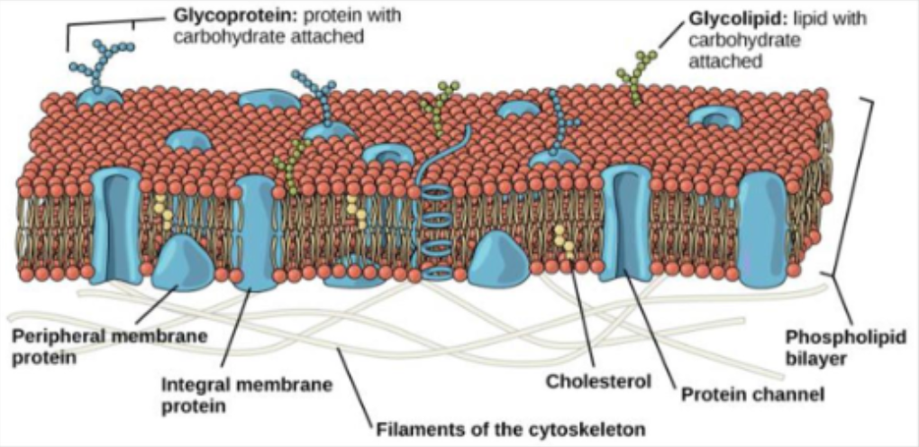
A selectively permeable phospholipid bilayer that surrounds the cell, separates internal processes from the outside environment, and regulates the movement of substances in and out
Phospholipid bilayer with embedded proteins
Cytoplasm
The entire region of a cell between the plasma membrane and the nuclear envelope, consisting of the gel-like cytosol, suspended organelles, and the cytoskeleton
70% of the cytoplasm is water but it has semi-solid consistency due because of proteins within

Nucleus
The membrane-bound organelle in eukaryotic cells that houses the cell's genetic material (DNA) and serves as the cell's control center
Only one per cell
The largest organelle (site of genetic information)

Ribosomes
Non-membrane-bound cellular structures composed of ribosomal RNA (rRNA) and proteins that function as the sites of protein synthesis in all living cells
Made of two different subunits
Made of RNA (rRNA) and proteins
During protein synthesis, ribosomes assemble amino acids into proteins

Mitochondrion
Membrane-bound organelles in eukaryotic cells that produce adenosine triphosphate (ATP) through cellular respiration
Site where energy is converted to ATP (cell energy)
Inner membrane is folded: area inside is the mitochondrial matrix
Extract energy from nutrients; convert energy into ATP

Peroxisomes
A small, single membrane-bound organelles in eukaryotic cells that specialize in lipid metabolism and the detoxification of harmful reactive oxygen species
Small round organelles
Break down fatty acids and amino acids
Peroxisomes detoxify poisons

Comparing animal/plant cells
Animal cells have (plant cells do not):
A centrosome
Lysosomes
Plant cells have (animal cells do not):
Cell wall
Chloroplasts
Large central vacuole
Plant cell walls
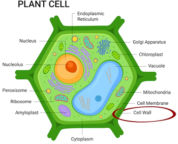
The protective structure outside the plasma membrane
A tough, rigid extracellular matrix located outside the plasma membrane that provides structural support, shape, and protection against osmotic lysis
Plant cell walls different than bacteria because they are made up of cellulose rather than peptidoglycan
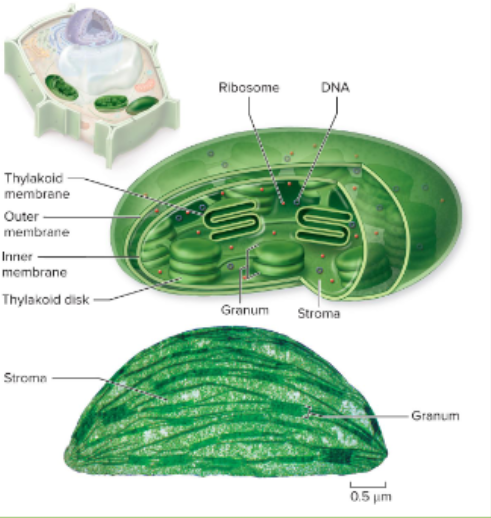
Chloroplasts
A double-membrane organelle found in plants and algae that carries out photosynthesis, converting light energy, water, and carbon dioxide into sugars
Double-membrane organelles; have their own ribosomes
and DNA like mitochondriaSite of photosynthesis
Vacuole
Plant cells have a large central vacuole
Regulates water concentration and contributes to cell
expansiona large central vacuole maintains turgor pressure in plants; in animal cells, vacuoles are smaller and more numerous
a membrane-bound sac inside eukaryotic cells that handles storage, waste disposal, and structural support

Endosymbiotic theory
Theory states that eukaryotic organelles—specifically mitochondria and chloroplasts—evolved from free-living prokaryotic cells that were engulfed by a larger host cell
It is hypothesized that mitochondria and chloroplasts originated from prokaryotes
Evidence for endosymbiotic theory
Mitochondria have their own DNA and ribosomes. The DNA is similar to prokaryotic DNA
The size of these organelles is similar to that of prokaryotes
Enzymes and transport mechanisms also similar to prokaryotic cells
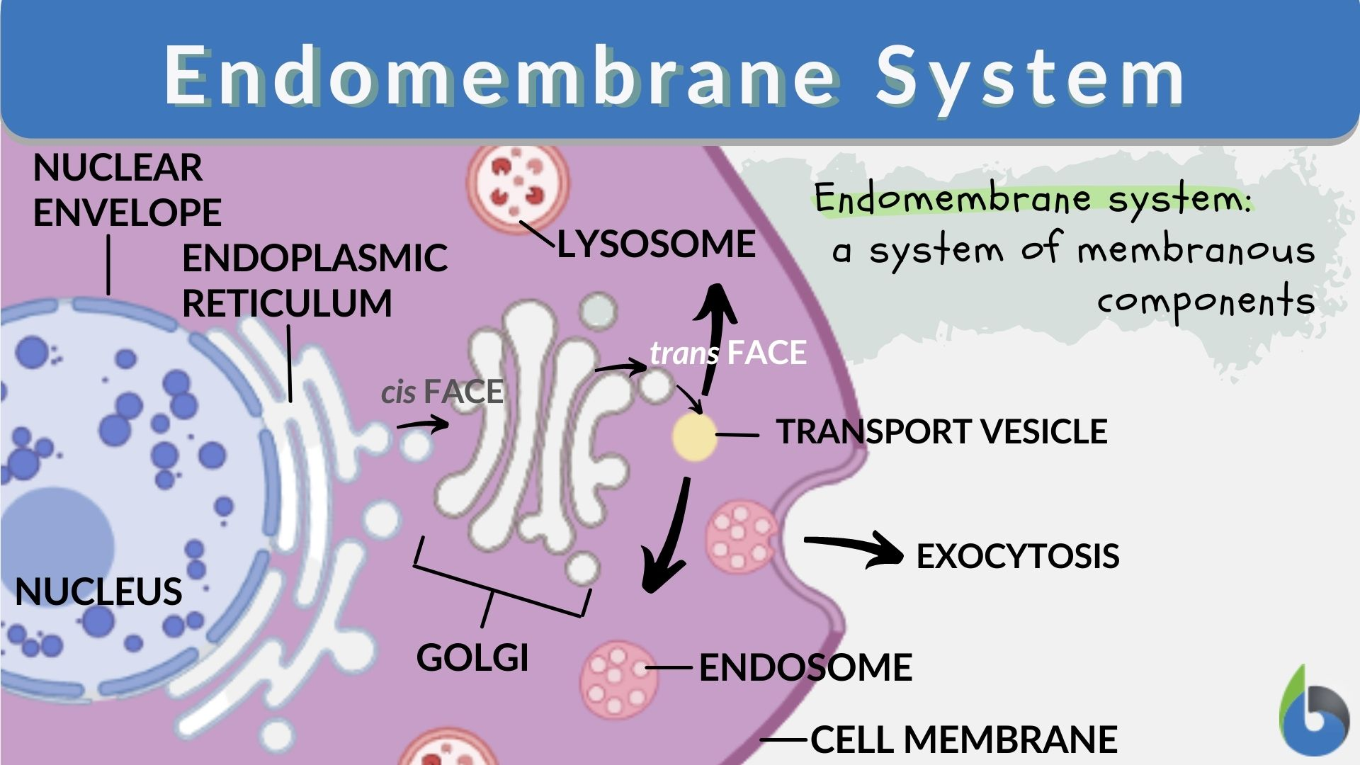
The endomembrane system
A network of interacting membranes and organelles in eukaryotic cells that work together to modify, package, and transport lipids and proteins
Includes:
Nuclear membrane
Lysosomes
Vesicles
Endoplasmic reticulum
Golgi apparatus
Lysosomes
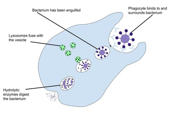
In animal cells contain digestive enzymes
The enzymes breakdown biomolecules and old organelles
Membrane-bound organelles packed with hydrolytic enzymes that function as the cell’s recycling and waste disposal system

Phagocytosis
Aspecific type of endocytosis where a cell engulfs large, solid particles—such as bacteria, cell debris, or whole cells—by extending its plasma membrane around them
Endoplasmic reticulum (ER)
Connected membranous sacs
Makes proteins (rough ER) and synthesizes lipids (smooth ER)
The membrane of the ER is continous with the nuclear envelope
A continuous network of membrane-enclosed sacs and tubules within eukaryotic cells that manufactures, folds, and transports proteins and lipids

Rough endoplasmic reticulum
Ribosomes in the ER manufacture proteins
New proteins are modified (folding or adding side chains) in the lumen of the rER
Modified proteins are incorporated into cellular membranes or secreted from the cell (example: protein hormones, enzymes)

Rough endoplasmic reticulum (rER)
The rER also makes phospholipids for cells membranes
Proteins that do stay in the rER travel to their destinations via transport vesicles that bud from the rER’s membrane
A membrane-bound eukaryotic organelle studded with ribosomes that synthesizes and folds proteins destined for export or membrane insertion

Smooth endoplasmic reticulum (sER)
Is continuous with the rER but doesn’t have any ribosomes on its
surface
A membrane-bound organelle made of a network of tubular sacs that lacks ribosomes on its surface
Synthesizes lipids, phospholipids, and steroid hormones, carbohydrates

Functions of the sER
Synthesis of:
Carbohydrates
Lipids
Steroid hormones
Detoxification of medications and poison
Storage of Ca++

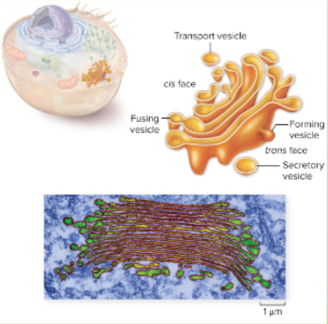
Golgi apparatus
Lipids or proteins within vesicles are sorted, packaged, and tagged to send to the right place
A membrane-bound organelle in eukaryotic cells that modifies, sorts, and packages proteins and lipids received from the endoplasmic reticulum (ER) for transport
Golgi apparatus features
The receiving side of the Golgi apparatus is the cis face; the opposite side is the trans face
Vesicles from the ER fuse with the cis face and empty their contents into the lumen of the Golgi apparatus
As the proteins and lipids travel through the Golgi, they are modified and sorted
This often involves adding short chains of sugar molecules

The cytoskeleteon
A network of protein fibers with several functions:
Support against tension
Support against compression
Support organelles
Serve as transport highways for some molecules
Enables cells in organisms to move
a dynamic network of protein fibers extending throughout the cytoplasm that gives a cell its structural support, shape, and ability to move.

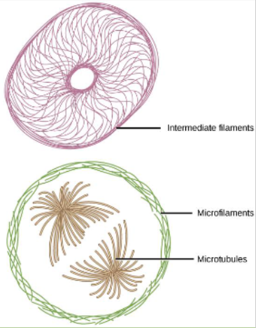
Three components of cytoskeleton
Different sizes and functions
Intermediate filaments: throughout the cell hold organelles in place
Microfilaments: around the cell's inner edge resist tension
Microtubules: maintain cell shape resisting compressive forces
Microfilaments
The thinnest components of the cell's cytoskeleton
Involved in movement
– Whole cell or internal partsDetermine & stabilize shape
Made from actin monomers

Microtubules
The largest hollow, rigid protein tubes of the cell's cytoskeleton
Form rigid internal skeleton for some cells
Provide framework for motor proteins to move within cell
Made of tubulin dimers

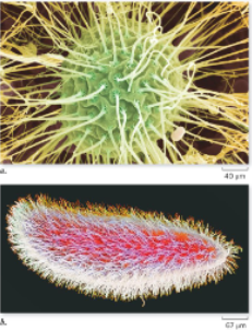
Cilia & Flagella (structures for movement)
Microtubule-based cellular projections that extend from the plasma membrane to facilitate cell motility or move fluids across the cell surface
Made of microtubules (only in eukaryotes)
Cilia shorter and more numerous
Cilia only in eukaryotes
Flagella: a long, whip-like cellular appendage primarily responsible for locomotion in various prokaryotic and eukaryotic cells
Extracellular structures - plant cell wall
A tough, rigid extracellular matrix that surrounds the plasma membrane, providing structural support, shape, and protection against osmotic lysis
Support
Barrier to infection
Plasmodesmata connect cells
Made of cellulose

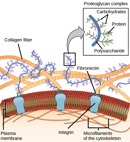
Extracellular matrix in animals
complex meshwork of proteins and carbohydrates secreted by animal cells that lies outside the plasma membrane to provide structural support and cell signaling
3 components:
Collagens & other fibrous
proteinsGlycoproteins called
proteoglycansLinking proteins
Intercellular junctions
Specialized contact points between the plasma membranes of adjacent cells that allow them to stick together, form barriers, or communicate
Provide direct channels of communication between cells
Plants and animals do this differently
Plasmodesmata
Channels that connect cells and allow materials to move from cell to cell
Microscopic channels that pass through the cell walls of adjacent plant cells, connecting their cytoplasm to allow direct communication and transport of molecules

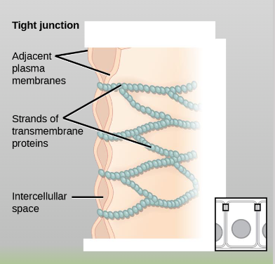
Tight junctions
Watertight, plasma-membrane seals between adjacent animal cells that prevent fluid and molecules from leaking through the intercellular space
Found in epithelial cells of internal organs and cavities
Only in animal cells
Desmosomes
Specialized cell-to-cell adhesion structures that act like spot welds to hold adjacent cells together under mechanical stress
Short proteins in the plasma membrane
Join adjacent cells in tissues that stretch (eg. heart, lungs, muscles)
Only in animal cells

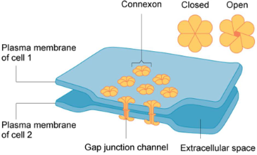
Gap junctions
Specialized protein channels that directly connect the cytoplasm of adjacent animal cells, allowing for the free passage of ions, water, and small molecules
Connect animal cells