BMA End of Semester Test
1/127
Earn XP
Description and Tags
Week 7 - Week 10
Name | Mastery | Learn | Test | Matching | Spaced | Call with Kai |
|---|
No analytics yet
Send a link to your students to track their progress
128 Terms
Cranial Nerve I - Name
Olfactory Nerve
Cranial Nerve I - Function
Conducts sensory input for olfaction
Cranial Nerve II - Name
Optic Nerve
Cranial Nerve II - Function
Conducts sensory input for vision
Cranial Nerve III - Name
Oculomotor Nerve
Cranial Nerve III - Function
Conducts motor output for:
The movement of the eyeball
Constriction of the pupils
Changes in lens shape
Cranial Nerve IV - Name
Trochlear Nerve
Cranial Nerve IV - Function
Conducts motor output for eyeball movements
Cranial Nerve V - Name
Trigeminal nerve
Cranial Nerve V - Function
Conducts sensory input for pain, touch and temperature from the face
Conducts motor output to the muscles involved in mastication (chewing)
Cranial Nerve VI - Name
Abducens nerve
Cranial Nerve VI - Function
Conducts motor output for eyeball movement (abduction)
Cranial Nerve VII - Name
Facial nerve
Cranial Nerve VII - Function
Conducts sensory input for taste
Carries motor output for facial expression, salivation and tears
Cranial Nerve VIII - Name
Vestibulocochlear nerve
Cranial Nerve VIII - Function
Conducts sensory input for hearing and balance
Cranial Nerve IX - Name
Glossopharyngeal nerve
Cranial Nerve IX - Function
Conducts sensory input for taste
Carries motor output for movements of pharynx (swallowing, speech) and salivation
Cranial Nerve X - Name
Vagus nerve
Cranial Nerve X - Function
Conducts sensory input for taste, proprioception (pharynx), blood pressure, blood gasses, visceral sensations
Conducts motor output for swallowing, breathing, cardiac function, digestive activities
Cranial Nerve XI - Name
Accessory nerve
Cranial Nerve XI - Function
Conducts motor output for movements of head, neck and shoulders
Cranial Nerve XII - Name
Hypoglossal nerve
Cranial Nerve XII - Function
Conducts motor output for tongue movements (swallowing and speech)
Fasciculus gracilis and fasciculus cunteatus tracts
Location
Conducted information
Posterior white columns
Fine touch, vibration, light pressure and conscious proprioception
Posterior column pathway
First-order neurons conduct sensory input from proprioceptors and tactile receptors into the spinal cord and to the medulla oblongata
Ascend in a fasciculus gracilis or fasciculus cuneatus tract (each does one side of the body)
Within the medulla oblongata the first-order neuron synapses with and passes information on to a second-order neuron
Second-order neuron conducts information up the brain stem and to the thalamus
Third-order neuron carries the information from the thalamus and to the primary somatosensory cortex
Anterior spinothalamic tract
Location
Conducted information
Anterior white columns
Crude touch and deep pressure
Lateral spinothalamic tract
Location
Conducted information
Lateral white columns
Pain and temperature
Spinothalamic pathway
First-order neurons conduct the sensory input detected by thermoreceptors, nociceptors or tactile receptors into the spinal cord
The sensory information is delivered to the posterior gray horn and within a sensory nucleus the first-order neuron will synapse with and pass the information onto a second-order neuron
Second-order neuron decussates and conducts the sensory information through the spinal cord and brain stem to the thalamus
If they are conducting pain or temperature information they will be located in a lateral spinothalamic tract
If they are conducting crude touch or deep pressure information they will be located in an anterior spinothalamic tract
Third-order neurons conduct the sensory information from the thalamus to the primary somatosensory cortex
Anterior and posterior spinocerebellar tracts
Location
Conducted information
Lateral white columns
Unconscious proprioception, helps to maintain smooth muscle movements and balance and posture
Spinocerebellar pathway
First-order neurons conduct sensory information into a posterior gray horn within a sensory nucleus and synapses with and passes the information onto a second-order neuron
Second-order neuron conducts the information to the cerebellum via a posterior or anterior spinocerebellar tract
Lateral corticospinal tracts
Lateral white columns
Somatic motor output that controls the skeletal muscles of the limbs
Lateral corticospinal pathway
Upper motor neurons, located in the primary motor cortex generate the somatic motor output that stimulates your skeletal muscles to contract
The axons conduct the information down the brain stem and the spinal cord through the lateral corticospinal tract
Within a nucleus of an anterior gray horn the upper motor neuron synapses with and passes the information onto a lower motor neuron
Lower motor neuron conducts the information away from the spinal cord to the skeletal muscle of a limb
Anterior corticospinal tracts
Anterior white columns
Somatic motor output that controls the skeletal muscles of the trunk/axial skeleton
Anterior corticospinal pathway
Upper motor neurons, located in the primary motor cortex generate the somatic motor output that stimulates your skeletal muscles to contract
The axons conduct the information down the brain stem and the spinal cord through the anterior corticospinal tract
Within a nucleus of an anterior gray horn the upper motor neuron synapses with and passes the information onto a lower motor neuron
Lower motor neuron conducts the information away from the spinal cord to the skeletal muscle of the trunk
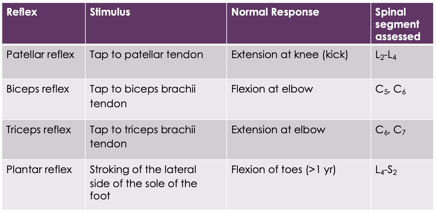
Purpose of somatic reflexes
Produced by the grey matter of the spinal cord
Since the reflex response is always the same, somatic reflexes are often tested to quickly diagnose disorder of the nervous system
Injuries or disease that affect the spinal cord segments that mediate the response of lower motor neurons can result in loss of reflex activity
Brain injuries that affect the primary motor cortex and corticospinal tract can result in abnormal reflexes
Examples of somatic reflexes

Babinski sign
Indicates damage to the primary motor cortex or corticospinal tracts
Spinal cord injuries
Damage to the posterior gray or ascending spinal cord tracts located in the posterior, lateral and anterior white columns can result in a loss of sensation
What is spastic paralysis?
Occurs when a spinal cord injury damages upper motor neurons where the somatic motor output generated by the primary motor cortex, will not be conducted down the spinal cord to lower motor neurons and therefore skeletal muscles will not be stimulated to contract voluntarily
*Reflex integration centres have not been affected within the spinal cord or lower motor neurons which
What is flaccid paralysis?
Occurs when a spinal cord injury damages lower motor neurons where somatic motor output will not be conducted away from the spinal cord to skeletal muscles and therefore our muscles will not be stimulated to contract through reflex activity
*Loss in voluntary muscle movements and reflex activity which will result in abnormal reflex response due to the primary motor cortex can no longer influence the reflex response
A complete transection of the spinal cord
Transection in the cervical region all four limbs are affected, known as quadriplegia
Transection in the thoracic or lumber region both legs will be affected, known as paraplegia
If it occurs in the thoracic region, patients will need a manual wheelchair as all leg muscles are affected
If it occurs in the lumber region, patients may require assistive devices (e.g. leg braces and crutches) as not all leg muscles are affected
Providing there is no damage to the reflex centres or lower motor neurons below the level of the transection, reflex activity will be present but abnormal as the primary motor cortex is unable to influence the reflex response
Sympathetic NS functions
Blood vessels
Lungs
Liver
Salivary glands
Kidney
Spleen
Blood vessels, vasodilation to increase blood flow
Lungs, bronchiole dilation
Liver, stimulates glucose into blood
Salivary glands, stimulates secretion of thick saliva
Kidney, reduced blood flow and urine formation
Spleen, release of stored blood
Parasympathetic NS functions
Heart
Liver
Heart, blood pressure decreases
Liver, increases glucose uptake
What organs do not have dual innovation, and only apply to the sympathetic NS?
Blood vessels, sweat glands, adrenal gland and spleen
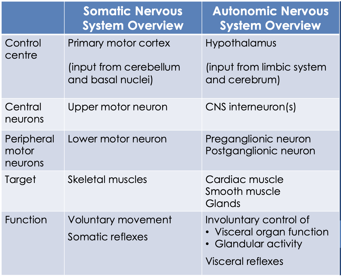
Somatic NS vs Autonomic NS Overview

Somatic pathway and neurotransmitters
Lower motor neuron releases ACh
Skeletal muscles have cholinergic, nicotinic receptors
Sympathetic NS pathway and neurotransmitter
Preganglionic neuron is short and it releases ACh which binds to the postganglionic neuron which is an excitatory response
Postganglionic has a cholinergic, nicotinic receptor type and it releases NA and is long
Targets are cardiac muscles, smooth muscles and glands muscles which have adrenergic (alpha or beta subtype) which have either an inhibitory or excitatory response
Parasympathetic NS pathway and neurotransmitter
Preganglionic neuron is long and it releases ACh which binds to the postganglionic neuron which is an excitatory response
Postganglionic has a cholinergic, nicotinic receptor type and it releases ACh and is short
Targets are cardiac muscles, smooth muscles and glands muscles which have cholinergic (muscarinic subtype) which have either an inhibitory or excitatory response
Subtypes of cholinergic receptors
Nicotinic, also bind nicotine and are always excitatory
Leads to an action potential which increases the activity of the target
Found in the cell bodies of the postganglionic neurons in the synapse
When Ach is released by the sympathetic preganglionic neuron this innervates these cells this excites the cells to release adrenaline and noradrenaline into the blood
When the lower motor neuron releases ACh it binds to skeletal muscles which leads to action potentials that increase muscle activity
Muscarinic, also bind muscarine that comes from mushrooms
Mostly excitatory
Some of them are inhibitory which means it makes the action potential less likely which reduces the activity of the target
Receptors on cardiac muscle are inhibitory which reduces the likelihood of the action potential and overall decreases the activity of the heart
Subtypes of adrenergic receptors
Alpha receptors, two types of alpha receptors
A1, when A or NA bind this will increase the activity of the target which is going to cause smooth muscle in blood vessels to constrict and visceral organs sphincters, dilates pupils
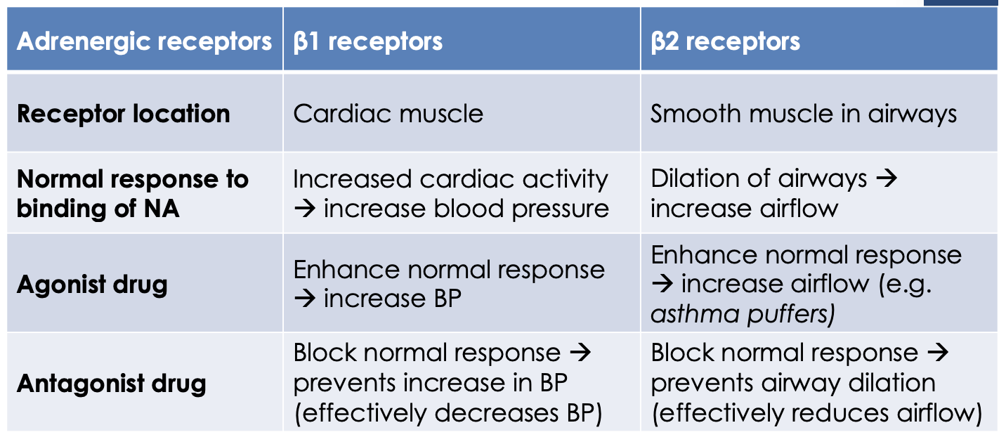
Beta receptors, three types of beta receptors
B1, when noradrenaline or adrenaline bind this increases the activity of the heart and increases blood flow
B2, found on smooth muscles and are inhibitory so when the NA or A bind this decreases the activity which relaxes the smooth muscle that is present in airways
Adrenergic receptor drugs

Acetylcholine (ACh)
In the CNS
Low levels of this neurotransmitter are found in individuals with dementia, especially Alzheimer’s disease
This is where there is a loss of neurons particularly in the prefrontal cortex and in the hippocampus which produces problems in cognition and memory
Noradrenaline
Stimulates the reward and pleasure centres in the brain and helps to see rewards and take actions to move towards those rewards ("feel good" NT)
Involved in reducing stress and enhancing attention
Dopamine
Associated with addiction which is released during pleasurable activities, therefore this NT reinforces those behaviours and helps promote addition
People with schizophrenia (a disorder that affects a person's ability to think, feel and behave clearly) have high levels of dopamine
Deficient in people with Parkinson's disease as this NT is responsible for controlling the basal nuclei which is important for helping us with the fine control of our motor activity
Serotonin
Involved in regulating mood, sleep, appetite and it can be associated with nausea and migraine
Drugs that prevent the re-uptake of serotonin are thought to help with conditions like anxiety and depression as they reduce the activity of the amygdala, which means a person becomes less emotionally reactive
Dark chocolate stimulates the release of serotonin in the brain and that’s why its thought to have a natural antidepressant kind of effect
Substance P
Produced in the periphery by damaged tissue
It stimulates local nociceptors so we can transmits pain to the CNS
Endorphins
Inhibit our perception of pain
A group of chemicals that involve endorphins and enkephalins
Termed "natural opiates" as they are chemically similar to opiate drugs
Also induce sleepiness and can promote wellbeing
What is a hormonal stimulus?
One hormone stimulates the secretion of another
What is a humoral stimulus?
Changes in ion or nutrient blood levels
What is a neural stimulus?
Signals from the nervous system
Steroid hormone
Are made from cholesterol
Are lipid-soluble and can easily diffuse across the plasma membrane
Bind to receptors inside a cell
Amino acid-based hormones
Vary in size, can be single amino acids, peptides or proteins
Are lipid insoluble and cannot easily diffuse across the plasma membrane
Bind to receptors embedded in the plasma membrane
Hypothalamus Posterior Pituitary
The cell bodies of neurons within the hypothalamus produce the hormones oxytocin and antidiuretic hormone (ADH).
The axons of these neurons form the hypothalamic-hypophyseal tract, which transports these hormones through the infundibulum to the posterior pituitary, where they are stored in the axon terminals.
When these hypothalamic neurons are stimulated the stored hormones are secreted from the posterior pituitary into the bloodstream.
Antidiuretic (ADH)
Stimulus for secretion
Target organ
Main action
When blood Na+ levels increase above the normal range or blood volume and blood pressure decrease below the normal range
Kidneys
ADH decreases urine output by stimulating the kidneys to return more water to the blood
*Can cause the vasoconstriction of arterioles which helps increase blood pressure
Increases reabsorption of water from the urine being produced and returns it to the blood stream:
Dilutes the blood plasma, restoring Na+ levels
Restores blood volume and pressure to normal levels
Maintains normal blood volume and pressue
Oxytocin
Stimulus for secretion
Target organ
Main action
Stretching of the uterus during labour
Uterus
Stimulates smooth muscle contractions during labour
Suckling action of the infant during breastfeeding
Mammary glands
Stimulates ejection of milk during breastfeeding
Hypothalamus-Anterior Pituitary
When stimulated, hypothalamic neurons secrete releasing/inhibiting hormones into the hypophyseal portal system.
Hormones travel through the infundibulum via the hypophyseal portal system to the anterior pituitary.
Releasing/inhibiting hormones stimulate/inhibit the secretion of anterior pituitary hormones into the bloodstream.
Thyrotropin-releasing hormone (TRH)
Stimulus for secretion
Main action
Corticotropin-releasing hormone (CRH)
Stimulus for secretion
Main action
Growth hormone-releasing hormone (GHRH)
Stimulus for secretion
Main action
Growth hormone-inhibiting hormone (GHIH or somatostatin)
Stimulus for secretion
Main action
Gonadotropin-releasing hormone (GnRH)
Stimulus for secretion
Main action
Prolactin-inhibiting hormone (PIH)
Stimulus for secretion
Target organ
Main action
Decreased PIH secretion leads to an increase in PRL secretion
Mammary glands
Stimulates milk production
Thyroid Glands
Lies at the base of the throat and it is composed of thyroid follicles which produce and secrete T3 and T4 (a.k.a. Thyroid hormones), and parafollicular cells which produce and secrete the hormone calcitonin.
Parathyroid Gland
Are tiny masses of glandular tissue that produce and secrete parathyroid hormone (PTH). These glands are located on the posterior surface of the thyroid gland.
Thyroid hormone (TH)
Stimulus for secretion
Target organs
Main action
Thyroid-stimulating hormone (TSH)
Every cell in the body
Main actions
Increases basal metabolic rate (BMR), amount of energy required by body cells to carry out all metabolic reactions at rest
Increases body heat production to maintain a normal body temperature
Increases HR and force of contraction by increasing the number of B-adrenergic receptors on cardiac muscle cells, regulates normal heart functioning
Promotes growth of muscles and bones
Promotes nervous system development
Calcitonin
Stimulus for secretion
Target organ
Main action
Blood Ca2+ levels increase above the normal range
Bone
Main actions
Decreases blood Ca2+ to normal levels by:
Inhibiting activity of osteoclasts, specialised bone cells that resorb/digest the extracellular matrix component to release stored calcium into the blood
Stimulating calcium uptake from the blood into bone
Parathyroid hormone (PTH)
Stimulus for secretion
Target organs
Main action
Blood Ca2+ levels decrease below the normal range
Bone, kidneys and small intestines
Main actions
Increases Ca2+ to normal levels by stimulating:
Bone-resorbing osteoclasts and the release of stored Ca2+ into the blood
Kidneys return more Ca2+ to the blood
Kidneys secrete calcitriol (the active form of vitamin D) which increases the absorption of Ca2+ from digested food in the small intestines
Erythropoietin (EPO)
Stimulus for secretion
Target organ
Main action
EPO is secreted by the kidneys when blood oxygen levels drop below their normal range (this is known as hypoxemia).
EPO targets the bone marrow
Stimulates the production of red blood cells.
The adrenal glands
The outer adrenal cortex is responsible for the production and secretion of glucocorticoids (primarily cortisol) and mineralocorticoids (primarily aldosterone). The inner adrenal medulla produces and secretes the hormones adrenaline and noradrenaline
Cortisol
Stimulus for secretion
Target organs
Main action
Anterior pituitary hormone (ACTH)
Skeletal muscle, liver and adipose tissue
Main actions
Helps the body resist stressors by increasing blood glucose, fatty acids and amino acid levels by stimulating:
Skeletal muscle to breakdown muscle proteins into amino acids
Liver to produce glucose from amino acids and glycerol
Adipose tissue to breakdown stored fat into fatty acids
This allows for the production of ATP to resist stressors, from using glucose, fatty acids and amino acids
Also suppress functions of the immune system by depressing inflammatory and immune responses
Aldosterone
Stimulus for secretion
Target organ
Main action
An increase in blood K+ levels rise above the normal range
Kidneys
Main actions
Maintains blood K+ and Na+ levels by stimulating the kidneys to:
§ Remove more K+ from the blood, increases secretion of K+ from the blood into the urine being formed
Return more Na+ to the blood, increases reabsorption of Na+ from the urine being formed into the blood
Restoring and maintaining normal blood volume and blood pressure
Adrenaline and Noradrenaline
Stimulus for secretion
Target organs
Main action
When the sympathetic nervous system is activated
Heart, bronchioles, blood vessels, pupils, sweat glands
Prolong the fight-or-flight response by binding to the alpha or beta adrenergic receptors on the target cells/tissues
The pancreas
b (beta) cells produce and secrete the hormone insulin
a (alpha) cells produce and secrete the hormone glucagon
Insulin
Stimulus for secretion
Target organs
Main action
Blood glucose levels increase above the normal range (4-8mmol/L)
Body cells, liver and skeletal muscle
Decreases blood glucose by:
Stimulating body cells to uptake glucose from the blood
Targets liver to inhibit the production of glucose from amino acids and glycerol (gluconeogenesis)
Targets liver and skeletal muscle to inhibit the breakdown of glycogen and glucose (glycogenolysis)
Glucagon
Stimulus for secretion
Target organ
Main action
Blood glucose levels decrease below normal range
Liver
Stimulates the liver to:
Breakdown of stored glycogen to glucose (glycogenolysis)
Produce glucose from amino acids and glycerol (gluconeogenesis)
Release glucose into the bloodstream
Ovaries
Produce and secrete oestrogen and progesterone
Testes
Produce and secrete testosterone
Oestrogen
Stimulus for secretion
Target organs
Main action
FHS and LH
Female reproductive organs, bones and adipose tissue
Main actions
Promote growth and maturation
Targets uterus to regulate the
menstrual cyclePromotes the development of female secondary sec characteristics
Promotes growth and enlargement of breasts
Promote growth and feminisation of the skeleton
Adipose tissue to increase fat storage
Progesterone
Stimulus for secretion
Target organs
Main action
LH stimulates the ovaries to produce this hormone
Ovaries
Main actions
Prepares the uterus for pregnancy and helps maintain the pregnancy
Helps regulate the menstrual cycle with oestrogen
Testosterone
Stimulus for secretion
Target organs
Main action
LH stimulates the testes to produce and secrete this hormone
Male reproductive organs, muscles, bone and hair follicles
Main actions
Promotes their growth and maturation and targets the testes to stimulate sperm production (spermatogenesis)
]Also promotes the development of male secondary sex characteristics
Increase muscle mass and strength
Stimulate growth
Stimulate the growth of body hair
Olfactory pathway
Chemoreceptors that are bathed in mucus capture and dissolve the odorant
Olfactory sensory neurons form the olfactory nerve or cranial nerve one where the action potentials travel to the olfactory cortex of the temporal lobe where we are made consciously aware of different odours
The information then travels to two different places:
Frontal lobe for the smell to be consciously interpreted and identified
Hypothalamus and other regions of the limbic or the emotional system for us to elicit an emotional response to an odour
Gustation pathway
Gustatory epithelial cells found in taste buds, microvilli are bathed in saliva on the surface of the tongue which is where food chemicals are dissolved
Impulses travel from the taste receptors up to the medulla via the facial nerve, glossopharyngeal and the vagus nerve where they will then travel to the gustatory cortex of the insula
Fibres then travel on the hypothalamus in the limbic system so that you can get an emotional response and an appreciation to taste is elicited
Pinna - Function
Funnels sound into external acoustic meatus
External acoustic meatus - Function
Allows sound waves to travel down and vibrate the tympanic membrane
Tympanic membrane - Function
Vibrates in response to sound waves and it then transfers this sound energy as waves into mechanical energy in bones
Auditory ossicles - Function
Includes the malleus, incus and stapes which transmit and amplifies the vibratory motion of the tympanic membrane through the middle ear to the oval window
Oval window - Function
Window in the wall of the cochlea. On one side has the stapes embedded in the window and the other side the perilymph. Movement causes pressure within the perilymph
Round window - Function
Absorbs pressure waves to wait for a new sound to come through
Cochlear - Function
Spiral shaped structure that contains the cochlear duct